. 2011 Feb;138(2):135-45.
doi: 10.1016/j.clim.2010.10.006. Epub 2010 Nov 20.

Phosphatidylserine reduces immune response against human recombinant Factor VIII in Hemophilia A mice by regulation of dendritic cell function

Affiliations

Phosphatidylserine reduces immune response against human recombinant Factor VIII in Hemophilia A mice by regulation of dendritic cell function

Puneet Gaitonde et al. Clin Immunol. 2011 Feb.

Abstract

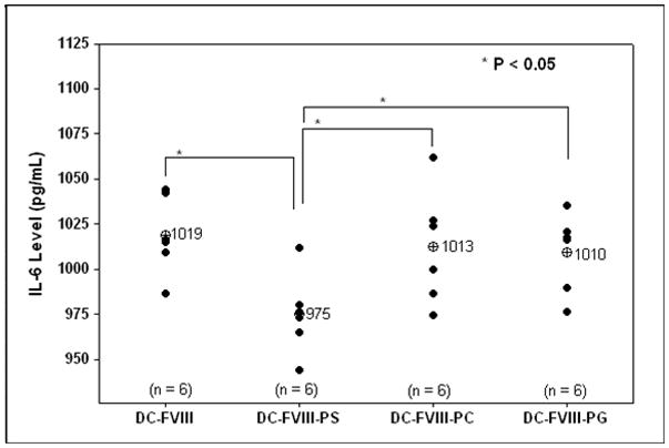
A major clinical complication in the treatment of Hemophilia A using exogenously administered recombinant Factor VIII (FVIII) is the development of neutralizing antibodies. It has been shown previously that FVIII complexed with phosphatidylserine (PS) reduces the development of total and neutralizing antibody titers in hemophilic mice. The effect of complexation of FVIII with PS upon dendritic cell (DC) uptake, maturation and processing, T-cell proliferation and cytokine secretion profiles was investigated. Flow cytometric studies of DC showed that PS inhibited the up-regulation of cell surface co-stimulatory markers (CD86 and CD40). PS reduced T-cell proliferation and significantly increased levels of TGF-β and IL-10 but reduced secretion of IL-6 and IL-17 compared to controls. The data suggest that PS reduces immunogenicity of FVIII by regulating dendritic cell maturation and subsequent T-lymphocyte activity through modulation of cytokine secretion. A possible mechanism for PS-mediated induction of FVIII tolerance is discussed.


Figures

Fig. 1
Fig. 1
Flow-cytometry analysis of the phenotypic maturation of bone marrow-derived DC following stimulation with PS, PC and PG liposomes associated with or without FVIII. The cell-surface markers were identified by FITC-MHC II antibody (1.A), PE-CD80 antibody (1.B), PE-CD86 antibody (1.C) or PE-CD40 antibody (1.D). Grey peak presents unstimulated DC (negative control).
Fig. 1
Fig. 1
Flow-cytometry analysis of the phenotypic maturation of bone marrow-derived DC following stimulation with PS, PC and PG liposomes associated with or without FVIII. The cell-surface markers were identified by FITC-MHC II antibody (1.A), PE-CD80 antibody (1.B), PE-CD86 antibody (1.C) or PE-CD40 antibody (1.D). Grey peak presents unstimulated DC (negative control).
Fig. 2
Fig. 2
DC uptake and endocytosis of HPTS-labeled PS liposomes. DC were exposed to liposomes containing the pH-indicating fluorophore for 30 min at 37°C, washed free of unbound liposomes, and examined by fluorescence microscopy. (2.A): differential interference contrast image. (2.B): violet excitation band (total cell-associated fluorescence). (2.C): blue excitation band (non-acidified compartment) of the same field as in A and B.
Fig. 2
Fig. 2
DC uptake and endocytosis of HPTS-labeled PS liposomes. DC were exposed to liposomes containing the pH-indicating fluorophore for 30 min at 37°C, washed free of unbound liposomes, and examined by fluorescence microscopy. (2.A): differential interference contrast image. (2.B): violet excitation band (total cell-associated fluorescence). (2.C): blue excitation band (non-acidified compartment) of the same field as in A and B.
Fig. 3
Fig. 3
PS mediated inhibition of T-cell proliferation as measured by 3H-thymidine incorporation. T-cell proliferation was measured for CD4+ T-cells isolated from animals immunized by subcutaneous (sc) administration of FVIII and re-stimulated in vitro with DC that were exposed to FVIII in the absence or presence of liposomes (PS or PC).
Fig. 4
Fig. 4
PS mediated modulation of cytokine secretion as measured by ELISA. Cytokine secretion of TGF-β (4.A), IL-10 (4.B), IL-6 (4.C) and IL-17 (4.D) was measured following co-culturing of CD4+ T-cells isolated from FVIII-immunized animals with naïve DC exposed to FVIII in the absence or presence of liposomes (PS, PC and PG).
Fig. 4
Fig. 4
PS mediated modulation of cytokine secretion as measured by ELISA. Cytokine secretion of TGF-β (4.A), IL-10 (4.B), IL-6 (4.C) and IL-17 (4.D) was measured following co-culturing of CD4+ T-cells isolated from FVIII-immunized animals with naïve DC exposed to FVIII in the absence or presence of liposomes (PS, PC and PG).
Fig. 5
Fig. 5
OPLS mediated modulation of cytokine secretion as measured by ELISA. Cytokine secretion of TGF-β (5.A), IL-10 (5.B), IL-6 (5.C) and IL-17 (5.D) was measured following co-culturing of CD4+ T-cells isolated from FVIII immunized animals with naïve DC exposed to FVIII in the absence or presence of liposomes (PS, PC and PG).
Fig. 5
Fig. 5
OPLS mediated modulation of cytokine secretion as measured by ELISA. Cytokine secretion of TGF-β (5.A), IL-10 (5.B), IL-6 (5.C) and IL-17 (5.D) was measured following co-culturing of CD4+ T-cells isolated from FVIII immunized animals with naïve DC exposed to FVIII in the absence or presence of liposomes (PS, PC and PG).

References

    1. Kaufman RJ, Wasley LC, Dorner AJ. Synthesis, processing, and secretion of recombinant human factor VIII expressed in mammalian cells. J Biol Chem. 1988;263(13):6352–62. - PubMed
    1. Vehar GA, et al. Structure of human factor VIII. Nature. 1984;312(5992):337–42. - PubMed
    1. Fay PJ. Factor VIII structure and function. Thromb Haemost. 1993;70(1):63–7. - PubMed
    1. Lollar P, et al. Factor VIII inhibitors. Adv Exp Med Biol. 2001;489:65–73. - PubMed
    1. Ananyeva NM, et al. Inhibitors in hemophilia A: mechanisms of inhibition, management and perspectives. Blood Coagul Fibrinolysis. 2004;15(2):109–24. - PubMed

Publication types

MeSH terms