. 2011 Jul;1813(7):1309-15.
doi: 10.1016/j.bbamcr.2010.11.005. Epub 2010 Nov 20.

Redox regulation of the mitochondrial K(ATP) channel in cardioprotection

Affiliations

Redox regulation of the mitochondrial K(ATP) channel in cardioprotection

Bruno B Queliconi et al. Biochim Biophys Acta. 2011 Jul.

Abstract

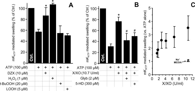
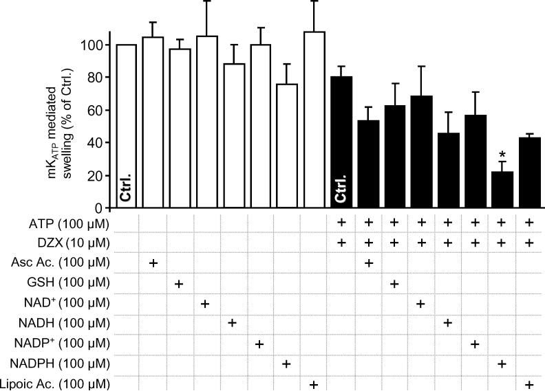
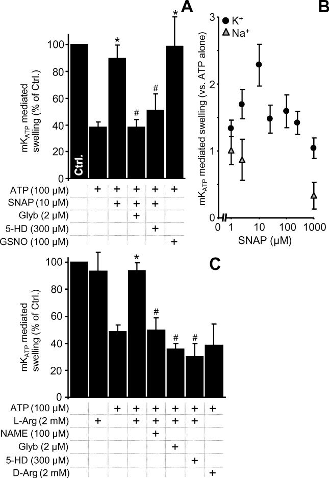
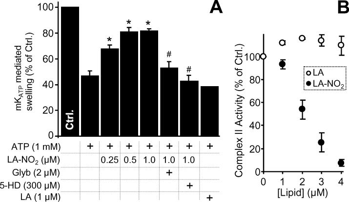
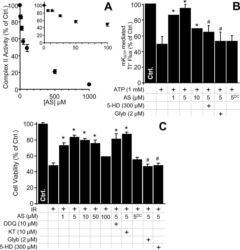
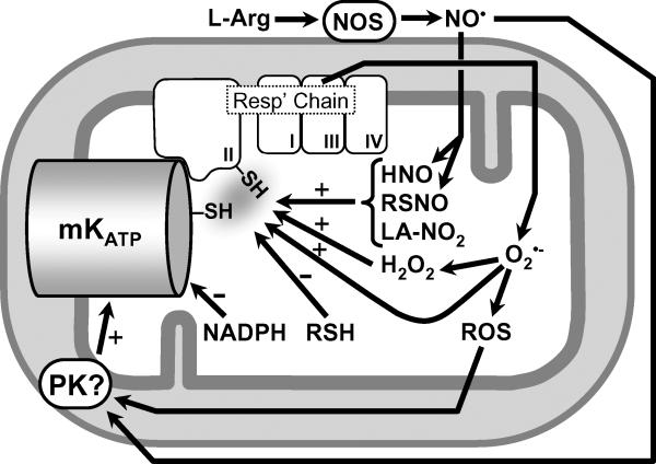
The mitochondrial ATP-sensitive potassium channel (mK(ATP)) is important in the protective mechanism of ischemic preconditioning (IPC). The channel is reportedly sensitive to reactive oxygen and nitrogen species, and the aim of this study was to compare such species in parallel, to build a more comprehensive picture of mK(ATP) regulation. mK(ATP) activity was measured by both osmotic swelling and Tl(+) flux assays, in isolated rat heart mitochondria. An isolated adult rat cardiomyocyte model of ischemia-reperfusion (IR) injury was also used to determine the role of mK(ATP) in cardioprotection by nitroxyl. Key findings were as follows: (i) mK(ATP) was activated by O(2)(-) and H(2)O(2) but not other peroxides. (ii) mK(ATP) was inhibited by NADPH. (iii) mK(ATP) was activated by S-nitrosothiols, nitroxyl, and nitrolinoleate. The latter two species also inhibited mitochondrial complex II. (iv) Nitroxyl protected cardiomyocytes against IR injury in an mK(ATP)-dependent manner. Overall, these results suggest that the mK(ATP) channel is activated by specific reactive oxygen and nitrogen species, and inhibited by NADPH. The redox modulation of mK(ATP) may be an underlying mechanism for its regulation in the context of IPC. This article is part of a Special Issue entitled: Mitochondria and Cardioprotection.


Figures

Figure 1
Figure 1. Selective mKATP Activation by ROS
mKATP activity was measured by osmotic swelling as detailed in the methods. Controls (Na+ based media) are in Figure S1. (A): mKATP is activated by H2O2 but not by t-BuOOH or LOOH. Diazoxide (DZX) opening of mKATP was used as a positive control. (B): mKATP activation by O2•- generated by the X/XO system. Glyburide (Glyb) and 5-hydroxydecanoate (5-HD) are mKATP antagonists. (C): Dose response of mKATP activation by X/XO in K+ (black circles) or Na+ (gray triangles) based media. *p<0.05 vs. ATP alone. #p<0.05 vs. ATP + H2O2 or X/XO. Experimental conditions are listed below the X axis.
Figure 2
Figure 2. Inhibition of DZX-Activated mKATP by Reductants
mKATP activity was measured by osmotic swelling as detailed in the methods. Data are shown for the baseline condition (open bars) or maximal swelling in the presence of both ATP and DZX (filled bars). Reductants were present in the media before mitochondrial addition, at the concentrations indicated. *p<0.05 vs. the appropriate control (bar marked Ctrl.) in the absence of reductant.
Figure 3
Figure 3. mKATP Activation by S-nitrosothiols and Mitochondrial-Associated NOS
mKATP activity was measured by osmotic swelling as detailed in the methods. Controls (Na+ based media) are in Figure S2. (A): mKATP was activated by S-nitrosoacetylpenacillamine (SNAP) and S-nitrosoglutathione (GSNO), at the indicated doses. (B): Dose-response of mKATP activation by SNAP in K+ (black circles) or Na+ (gray triangles) based media. *p<0.05 vs. ATP alone. #p<0.05 vs. ATP plus SNAP. (C): mKATP activation by NOS modulators. L- or D-Arginine, L-nitroarginine methyl ester (NAME), Glyb and 5-HD were present at the concentrations indicated. *p<0.05 vs. *p<0.05 vs. ATP alone. #p<0.05 vs. ATP plus L-arginine.
Figure 4
Figure 4. LA-NO2 Opens mKATP and Inhibits Complex II
(A): mKATP activity was measured by osmotic swelling as detailed in the methods. LA-NO2, native linoleate (LA), Glyb, and 5-HD were present at the indicated concentrations. *p<0.05 vs. ATP alone. #p<0.05 vs. ATP plus LA-NO2. (B): Complex II activity in the presence of LA-NO2 was determined as detailed in the methods. Values are expressed as percentage of control complex II rate (128 ± 26 nmols DCPIP . min-1 . mg protein-1).
Figure 5
Figure 5. Nitroxyl Inhibits Complex II, Opens mKATP and is Cardioprotective
(A): Complex II activity was measured, following nitroxyl exposure of mitochondria, as described in the methods. Values are expressed as percentage of control complex II rate (108 ± 8 nmols DCPIP . min-1 . mg protein-1). (B): Nitroxyl activation of mKATP was monitored using a novel Tl+ flux assay as described in the methods. Data show the magnitude of change in intra-mitochondrial Tl+ based fluorescence following Tl+ addition, relative to control. 5-HD, Glyb, the nitroxyl donor Angeli's salt (AS) and decomposed AS (ASDC) were present at the indicated concentrations. *p<0.05 vs. ATP alone. #p<0.05 vs. ATP plus 5 μM AS. (C): Nitroxyl protects against cardiomyocyte IR injury. Cell viability was measured via Trypan blue exclusion at the end of reoxygenation, as described in the methods, and expressed as percentage of control (normoxic) cell viability. 5-HD, Glyb, Angeli's salt (AS), decomposed AS (ASDC), the PKG inhibitor KT-5823 (KT) or the soluble guanylate cyclase inhibitor ODQ were present at the indicated concentrations. *p<0.05 vs. IR alone. #p<0.05 vs. IR plus 5 μM AS.
Figure 6
Figure 6. Schematic Showing Redox Regulation of mKATP
Nitroxyl (HNO), RSNO, and LA-NO2 activate the channel, possibly via thiols on the channel itself or on complex II of the respiratory chain. The ability of low molecular weight thiols (RSH) to inhibit the channel may also be mediated via thiols on the channel or on complex II. In contrast, the effects of NADPH are likely no mediated via thiols. The ability of NO to activate the channel may be mediated via the generation of secondary RNS (e.g. RSNO, LA-NO2, HNO), which can activate the channel via PKG-independent mechanisms, or via classical NO protein kinase signaling. ROS (in particular H2O2) can also activate the channel, via mechanisms that may include thiol modification or protein kinase signaling. The nature of the interaction between complex II and the subunits of the mKATP channel itself remains to be elucidated.

Similar articles

Cited by

References

    1. Murry CE, Jennings RB, Reimer KA. Preconditioning with ischemia: a delay of lethal cell injury in ischemic myocardium. Circulation. 1986;74:1124–1136. - PubMed
    1. Facundo HT, Fornazari M, Kowaltowski AJ. Tissue protection mediated by mitochondrial K+ channels. Biochim. Biophys. Acta. 2006;1762:202–212. - PubMed
    1. Garlid KD, Dos SP, Xie ZJ, Costa AD, Paucek P. Mitochondrial potassium transport: the role of the mitochondrial ATP-sensitive K(+) channel in cardiac function and cardioprotection. Biochim. Biophys. Acta. 2003;1606:1–21. - PubMed
    1. Murphy E, Steenbergen C. Preconditioning: the mitochondrial connection. Annu. Rev. Physiol. 2007;69:51–67. - PubMed
    1. Vanden Hoek TL, Becker LB, Shao Z, Li C, Schumacker PT. Reactive oxygen species released from mitochondria during brief hypoxia induce preconditioning in cardiomyocytes. J. Biol. Chem. 1998;273:18092–18098. - PubMed

Publication types

MeSH terms

LinkOut - more resources