

Hippocampal volume differences in Gulf War veterans with current versus lifetime posttraumatic stress disorder symptoms
- PMID: 21094937
- PMCID: PMC3259803
- DOI: 10.1016/j.biopsych.2010.09.044
Hippocampal volume differences in Gulf War veterans with current versus lifetime posttraumatic stress disorder symptoms
Abstract
Background: Decreased hippocampal volume is described in posttraumatic stress disorder (PTSD) and depression. However, it is not known whether it is a risk factor for the development of PTSD or a consequence of PTSD. We sought to determine the effects of PTSD and depressive symptoms on hippocampal volume.
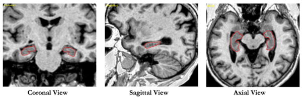
Methods: Clinical and magnetic resonance imaging data were collected in a cross sectional study of 244 Gulf War veterans. Measures included lifetime and current Clinician Administered PTSD Scale, Hamilton Depression Scale, Life Stressor Checklist, and Lifetime Drinking History. Magnetic resonance imaging data were acquired with a 1.5-T scanner and analyzed with automated and semiautomated image processing techniques.
Results: Eighty-two veterans had lifetime PTSD, 44 had current PTSD, and 38 had current depression. In the linear regression analysis, current PTSD symptoms (standardized coefficient β = -.25, p = .03) but neither lifetime PTSD symptoms nor current depression were associated with smaller hippocampal volume. Gender, age, history of early life trauma, education, lifetime and current alcohol use, current marijuana use, and treatment with antidepressants did not have independent effects. Participants with chronic PTSD had, on average, a smaller hippocampus compared with those with remitted PTSD.
Conclusions: The finding that current but not lifetime PTSD symptom severity explains hippocampal size raises two possibilities: either a small hippocampus is a risk factor for lack of recovery from PTSD (trait) or PTSD effects on hippocampal volume are reversible once PTSD symptoms remit and the patient recovers (state).
Copyright © 2011 Society of Biological Psychiatry. Published by Elsevier Inc. All rights reserved.
Conflict of interest statement
All authors report no biomedical financial interests or potential conflicts of interest.
Figures


References
-
- Karl A, Schaefer M, Malta LS, Dorfel D, Rohleder N, Werner A. A meta-analysis of structural brain abnormalities in PTSD. Neurosci Biobehav Rev. 2006;30:1004–1031. - PubMed
-
- Kitayama N, Vaccarino V, Kutner M, Weiss P, Bremner JD. Magnetic resonance imaging (MRI) measurement of hippocampal volume in posttraumatic stress disorder: A meta-analysis. J Affect Disord. 2005;88:79–86. - PubMed
-
- Smith ME. Bilateral hippocampal volume reduction in adults with post-traumatic stress disorder: A meta-analysis of structural MRI studies. Hippocampus. 2005;15:798–807. - PubMed
-
- Sala M, Perez J, Soloff P, Ucelli di Nemi S, Caverzasi E, Soares JC, Brambilla P. Stress and hippocampal abnormalities in psychiatric disorders. Eur Neuropsychopharmacol. 2004;14:393–405. - PubMed
-
- De Kloet ER, Vreugdenhil E, Oitzl MS, Joels M. Brain corticosteroid receptor balance in health and disease. Endocr Rev. 1998;19:269–301. - PubMed
Publication types
MeSH terms
Grants and funding
LinkOut - more resources
Full Text Sources
Other Literature Sources
Medical
