. 2010 Nov 24;40(4):558-70.
doi: 10.1016/j.molcel.2010.11.003.

Pervasive and cooperative deadenylation of 3'UTRs by embryonic microRNA families

Affiliations

Pervasive and cooperative deadenylation of 3'UTRs by embryonic microRNA families

Edlyn Wu et al. Mol Cell. .

Abstract

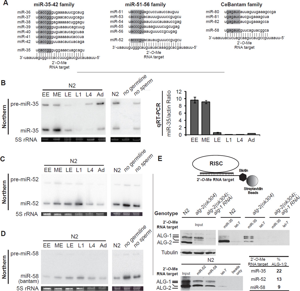
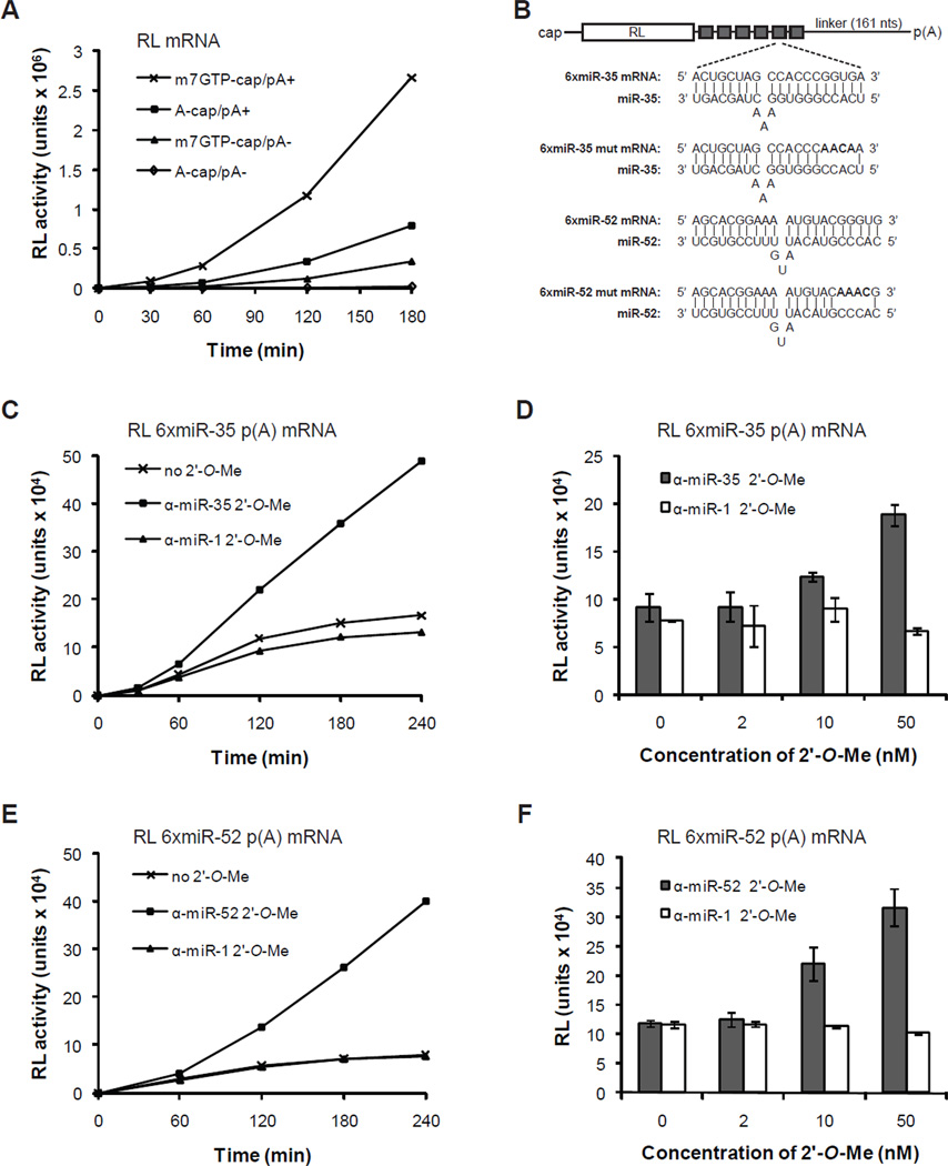
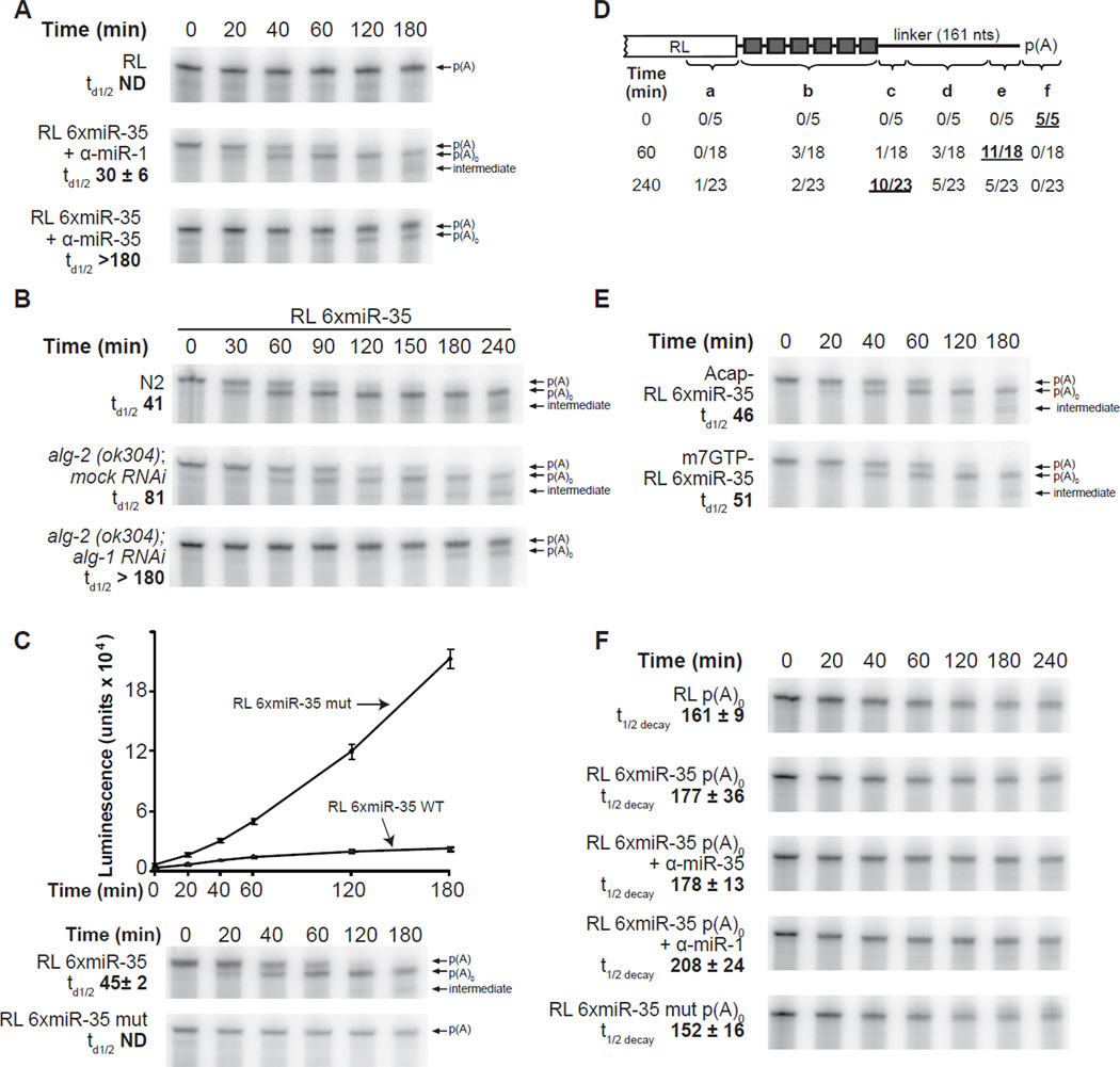
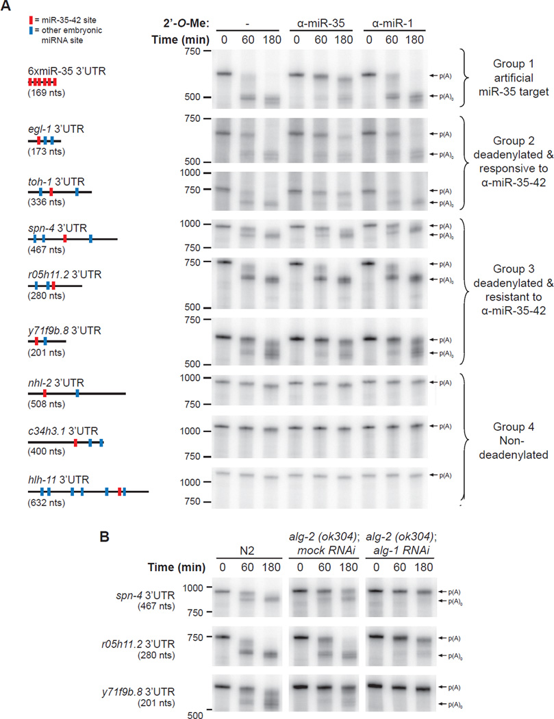
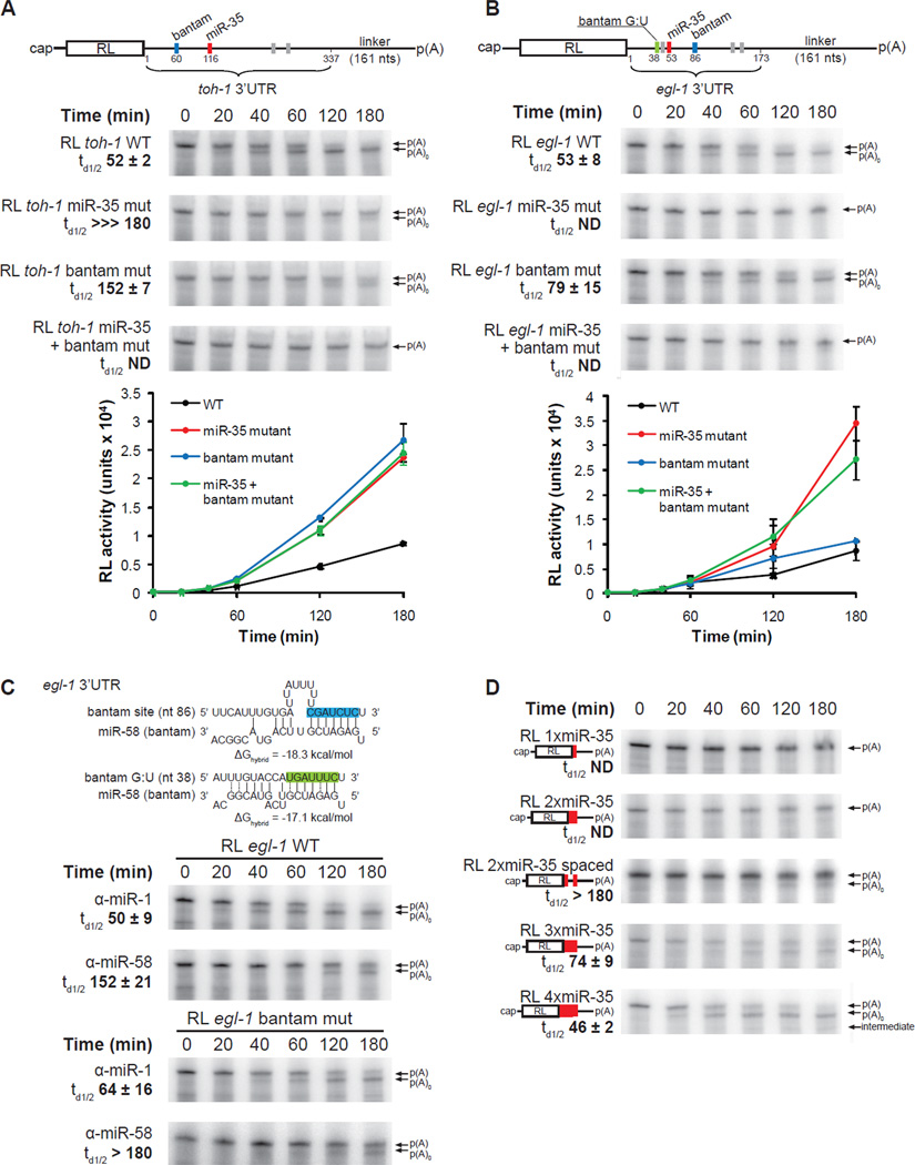
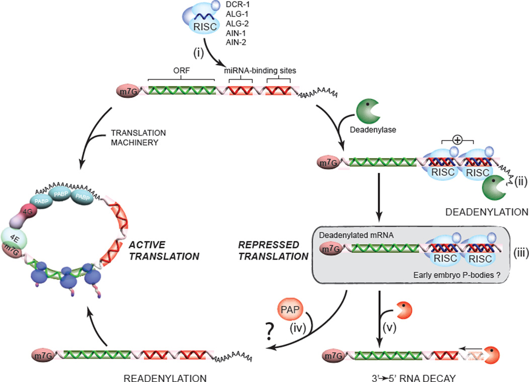
To understand how miRNA-mediated silencing impacts on embryonic mRNAs, we conducted a functional survey of abundant maternal and zygotic miRNA families in the C. elegans embryo. We show that the miR-35-42 and the miR-51-56 miRNA families define maternal and zygotic miRNA-induced silencing complexes (miRISCs), respectively, that share a large number of components. Using a cell-free C. elegans embryonic extract, we demonstrate that the miRISC directs the rapid deadenylation of reporter mRNAs with natural 3'UTRs. The deadenylated targets are translationally suppressed and remarkably stable. Sampling of the predicted miR-35-42 targets reveals that roughly half are deadenylated in a miRNA-dependent manner, but with each target displaying a distinct efficiency and pattern of deadenylation. Finally, we demonstrate that functional cooperation between distinct miRISCs within 3'UTRs is required to potentiate deadenylation. With this report, we reveal the extensive and direct impact of miRNA-mediated deadenylation on embryonic mRNAs.


Figures

Figure 1
Figure 1. miRISC programming by maternal and zygotic miRNA families in C. elegans embryos
(A) miRNAs and 2’-O-Me oligonucleotides used in this study. The seed region for each miRNA is highlighted in gray. (B) Expression profile of miR-35 by northern and real-time (qRT) PCR analysis. Total RNA from wild-type (N2) early-stage embryos (EE), middle-stage embryos (ME), late-stage embryos (LE), L1-, L4-, and adult-stage animals (Ad), or adult-stage (glp-4)bn2 (no germline) and fem-1(hc17) (no sperm) animals grown at 25°C. 5S ribosomal RNA (rRNA) is indicated as loading control. qRT-PCR results are presented as the mean from triplicate samples and error bars represent standard deviation. (C,D) Northern analysis of miR-52 and miR-58 (bantam) expression. (E) (Top) Schematic representation of the miRISC 2’-O-Me pulldown strategy. (Middle and bottom) Extracts prepared from wild-type (N2), alg-2(ok304), or alg-2(ok304); alg-1 RNAi embryos were incubated with the indicated 2’-O-Me matrices. Bound proteins were probed for ALG-1 and ALG-2, and average percentage pulled down of two independent experiments is indicated in bold. Related data in Figures S1.
Figure 2
Figure 2. Cell-free miRNA-mediated translational repression by maternal and zygotic miRNAs
(A) Cap and poly(A) tail synergy in C. elegans embryonic extracts. The translation of 10 nM RL reporters bearing a physiological 5’ m7GpppG-cap, a 5’ ApppG-cap, and/or 3’ poly(A) tail was monitored over a 3-hour time-course. (B) Schematic representation of the RL reporter mRNAs used. Sequences of the miR-35- and miR-52-binding sites (6xmiR-35 and 6xmiR-52) and mutated binding sites (6xmiR-35 mut and 6xmiR-52 mut) are shown. (C,E) Translation time-course of RL 6xmiR-35 (C) and 6xmiR-52 mRNAs (E) with or without 50 nM specific (α-miR-35 (C), α-miR-52 (E)) or non-specific α-miR-1 2’-O-Me. (D,F) Dose-response translation de-repression by α-miR-35 (D) and α-miR-52 (F) 2’-O-Me for a 3h reaction. Each bar represents the mean from triplicate independent experiments and error bars indicate standard deviation.
Figure 3
Figure 3. Embryonic miRISCs direct deadenylation but do not promote target decay in vitro
(A) Deadenylation time-course of RL and RL 6xmiR-35 with the indicated 50 nM 2’-O- Me, and (B) of RL 6xmiR-35 in wild-type (N2), alg-2(ok304); mock (gfp) RNAi, or alg-2(ok304); alg-1 RNAi embryonic extracts. (C) Time-course of RL 6xmiR-35 WT and mutant translation and deadenylation. The same samples from each time-points were examined in translation (upper panel) and PAGE-autoradiography (lower panels). (D) Schematic representation of 3’RACE products from RL 6xmiR-35 at the indicated time-points. The indicated number of reads terminated a. within the RL open reading frame, b. between the miR-35 binding sites, c. within the first 40 nts 3’ of the miR-35 binding sites, d. within the middle region of the 3’UTR, e. within less than 25 nts 5’ of the poly(A) tail, f. within the poly(A) tail. (E) Deadenylation time-course of RL 6xmiR-35 mRNA bearing a m7GpppG cap or a ApppG cap. (F) Decay time-course of unadenylated reporters. Panels B and E are representative of two independent experiment, panels A, C and F are representative of triplicate experiments conducted using the same extract preparation. Half-deadenylation (td1/2) and half-life (t1/2decay) were quantified using ImageJ. +/− indicates standard deviation. Related data in Figure S3.
Figure 4
Figure 4. miRNA-mediated deadenylation is prevalent in embryos
(A) Deadenylation of natural 3’UTR reporters in embryonic extracts. 3’UTRs were fused to a truncated RL fragment (nts 764-936 (172-nt long)), for all UTRs screened except c34h3.1 where nts 491–936 where included. Reporters also encoded a 161-nt linker and a poly(A) tail of 87 nts. Schematic representation of the each 3’UTRs is depicted on the left (size in parentheses). Red bars denote miR-35-42 sites, blue bars denote sites for miRNAs that are known to be expressed in embryos (Stoeckius et al., 2009). Courses were realized with or without 50 nM 2’-O-Me (either α-miR-35 or α-miR-1(C-)). 3’UTRs are divided into four groups: 1. deadenylated artificial miR-35 target (6xmiR-35, (control)), 2. deadenylated 3’UTR targets that are responsive to α-miR-35, 3. deadenylated 3’UTR targets that are resistant to α-miR-35, 4. 3’UTRs not subjected to detectable deadenylation. (B) Time-course of group 3 in N2, alg-2(ok304); mock (gfp) RNAi and alg-2(ok304); alg-1 RNAi embryonic extracts. Experiments were reproduced at least twice in independent extract preparations. Related data in Figure S4.
Figure 5
Figure 5. Target deadenylation requires miRISC cooperation
(A,B) Deadenylation and translation time-courses of RL toh-1 WT (A) and RL egl-1 WT (B) 3’UTR reporters in wild-type (N2) embryo extract. Detailed schematic representation of 3’UTR reporter mRNAs is shown. Red bars indicate miR-35-42 sites, blue and green bars indicate sites for CeBantam family members, and gray bars indicate sites for miRNAs that were not detected and/or had no detectable functional implications in our system (See also Figure S5). (C) (Top) Pairing of the the egl-1 3’UTR miR-58 (bantam) sites; the site with canonical base-pairing in blue, and the non-canonical site containing G:U wobble base-pairing in green. (Middle and bottom) Deadenylation time-course of the RL egl-1 WT, and the RL egl-1 bantam mut mRNA (encodes mutations within the canonical bantam site) in the presence of 50 nM α-miR-58, or the negative control α-miR-1. (D) Deadenylation time-course of RL reporter mRNAs encoding 1 to 4 copies of miR-35 binding sites. The 2xmiR-35 spaced reporter contains two miR-35 separated by 29 nts. Translation and deadenylation assays were conducted as triplicate of independent experiments. Quantifications of time of half-deadenylation (td1/2) were realized using ImageJ. Error bars, and +/− indicate standard deviation. Related data in Figure S5.
Figure 6
Figure 6. A model for the deadenylation and decay of early embryo miRNA targets
The miRISC complex (ALG-1/2, AIN-1/2, DCR-1 and other accessory proteins), programmed by the abundant maternal and zygotic miRNA families, scans and recognizes mRNA targets (i). Through functional cooperation (indicated by a + sign), embryonic miRISC recruit and/or activate the deadenylase complex (CCR4/NOT was previously identified in a number of studies, including our own), and direct the rapid deadenylation of the target (ii). The stability of deadenylated mRNAs, and the association with GW182 proteins AIN-1 and AIN-2 on our target site baits in proteomics suggest that deadenylated targets may be protected and/or stored within the miRISC, or possibly within P-body-like structures (iii). One consequence of this stability is the possibility that deadenylation may be reverted, or outcompeted by poly(A) polymerase activities (PAP) (iv). Although this last hypothesis remains to be tested, evidence for competing deadenylation and polyadenylation activities exists in paradigms such as the germline and in the early embryo (Goldstrohm and Wickens, 2008). Finally, a fraction of the deadenylated mRNA pool may be decayed through a slow 3’→5’ route (v). This destabilization could be accelerated by the recruitment of decapping machinery by the miRISC, for example (See Discussion).

References

    1. Alvarez-Saavedra E, Horvitz HR. Many families of C. elegans microRNAs are not essential for development or viability. Curr Biol. 2010;20:367–373. - PMC - PubMed
    1. Ambros V, Lee RC, Lavanway A, Williams PT, Jewell D. MicroRNAs and Other Tiny Endogenous RNAs in C. elegans. Curr Biol. 2003;13:807–818. - PubMed
    1. Baek D, Villen J, Shin C, Camargo FD, Gygi SP, Bartel DP. The impact of microRNAs on protein output. Nature. 2008;455:64–71. - PMC - PubMed
    1. Bagga S, Bracht J, Hunter S, Massirer K, Holtz J, Eachus R, Pasquinelli AE. Regulation by let-7 and lin-4 miRNAs results in target mRNA degradation. Cell. 2005;122:553–563. - PubMed
    1. Bartel DP. MicroRNAs: target recognition and regulatory functions. Cell. 2009;136:215–233. - PMC - PubMed

Publication types