

In-vivo clinical validation of cardiac deformation and strain measurements from 4D ultrasound
- PMID: 21095877
- PMCID: PMC3123825
- DOI: 10.1109/IEMBS.2010.5626332
In-vivo clinical validation of cardiac deformation and strain measurements from 4D ultrasound
Abstract
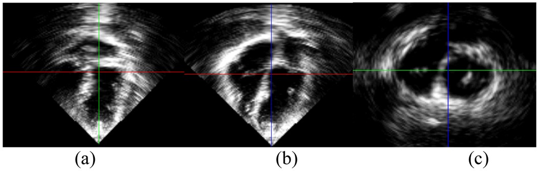
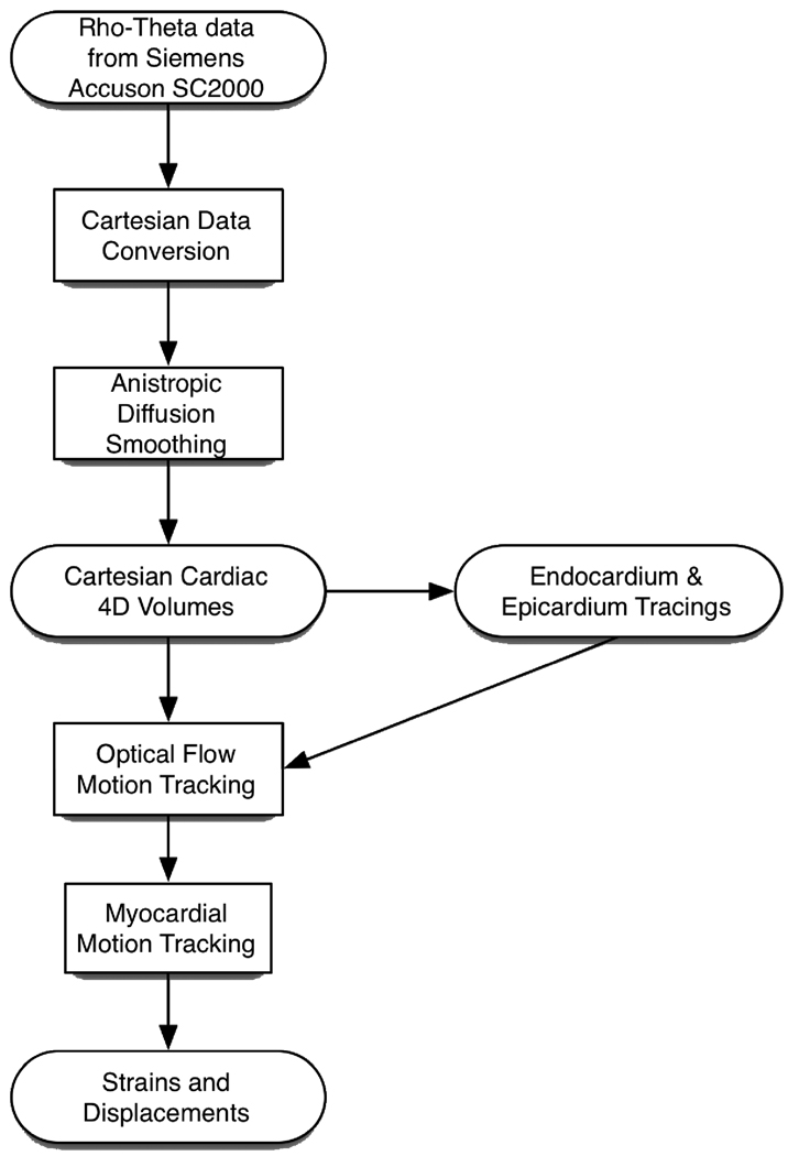
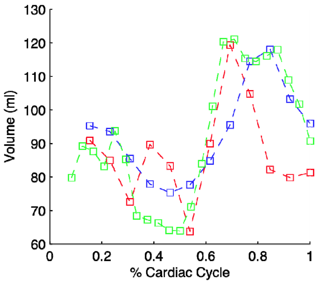
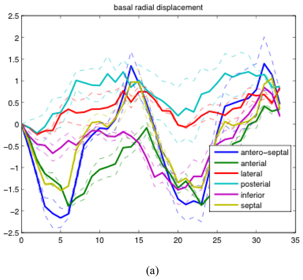
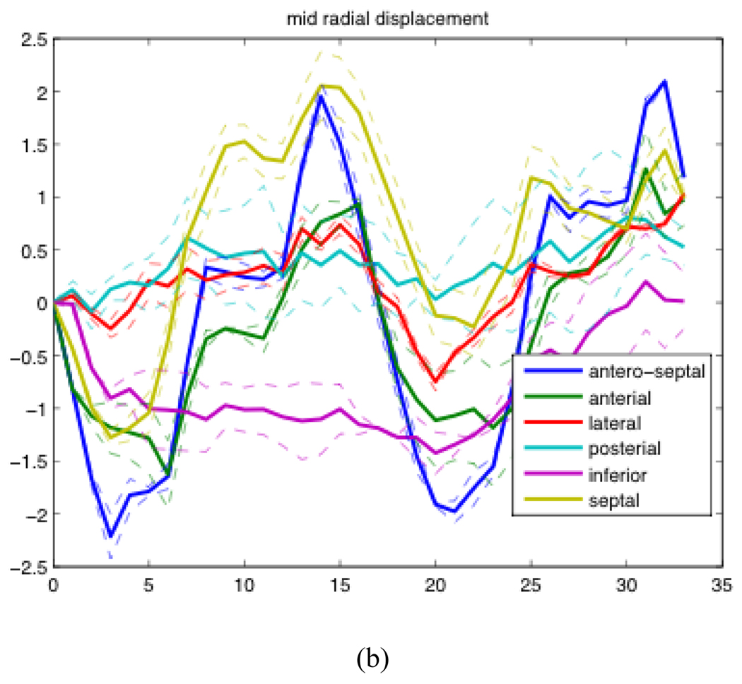
An important goal in clinical cardiology is the non-invasive quantification of regional cardiac deformation. While many methods have been proposed for the estimation of 3D left ventricular deformation and strains derived from 4D ultrasound, currently there is a lack of in vivo clinical validation of these algorithms on humans. In this paper, we describe the experiments used in validating cardiac deformation and strain estimates of 4D ultrasound using correlation-based optical flow tracking on two different COPD patients with normal left ventricular function. Validation of the algorithm was done by 1) validation of cardiac volume across multiple scans of the same patient and 2) validation of the repeatability of cardiac displacement and strain results from multiple scan acquisitions of the same patient. The preliminary results are encouraging with our algorithm producing consistent cardiac volume and strain results across multiple acquisitions. Furthermore, our derived 4D cardiac strains showed qualitatively correct results. We also observed particularly interesting results in the radial displacements of the posterior and lateral walls of our COPD patients.
Figures
Similar articles
-
Radial basis functions for combining shape and speckle tracking in 4D echocardiography.IEEE Trans Med Imaging. 2014 Jun;33(6):1275-89. doi: 10.1109/TMI.2014.2308894. IEEE Trans Med Imaging. 2014. PMID: 24893257 Free PMC article.
-
Clinical feasibility and validation of 3D principal strain analysis from cine MRI: comparison to 2D strain by MRI and 3D speckle tracking echocardiography.Int J Cardiovasc Imaging. 2017 Dec;33(12):1979-1992. doi: 10.1007/s10554-017-1199-7. Epub 2017 Jul 6. Int J Cardiovasc Imaging. 2017. PMID: 28685315 Free PMC article.
-
In-vivo validation of a new clinical tool to quantify three-dimensional myocardial strain using ultrasound.Int J Cardiovasc Imaging. 2016 Dec;32(12):1707-1714. doi: 10.1007/s10554-016-0962-5. Epub 2016 Aug 17. Int J Cardiovasc Imaging. 2016. PMID: 27535041
-
Current applications of fetal cardiac imaging technology.Curr Opin Obstet Gynecol. 2006 Apr;18(2):177-84. doi: 10.1097/01.gco.0000192987.99847.a4. Curr Opin Obstet Gynecol. 2006. PMID: 16601479 Review.
-
4D imaging of fetal right ventricle-feasibility study and a review of the literature.Int J Cardiovasc Imaging. 2022 Feb;38(2):319-329. doi: 10.1007/s10554-021-02407-9. Epub 2021 Sep 20. Int J Cardiovasc Imaging. 2022. PMID: 34545461 Free PMC article. Review.
Cited by
-
Evaluation of traditional and novel measures of cardiac function to detect anthracycline-induced cardiotoxicity in survivors of childhood cancer.J Cancer Surviv. 2014 Jun;8(2):183-9. doi: 10.1007/s11764-013-0326-2. Epub 2013 Dec 7. J Cancer Surviv. 2014. PMID: 24317971 Free PMC article.
-
Cardiac and respiratory-gated volumetric murine ultrasound.Int J Cardiovasc Imaging. 2018 May;34(5):713-724. doi: 10.1007/s10554-017-1283-z. Epub 2017 Dec 12. Int J Cardiovasc Imaging. 2018. PMID: 29234935 Free PMC article.
-
Modeling and incorporating cardiac-induced lung tissue motion in a breathing motion model.Med Phys. 2014 Apr;41(4):043501. doi: 10.1118/1.4866888. Med Phys. 2014. PMID: 24694158 Free PMC article.
References
-
- Canals R, et al. Volumetric ultrasound system for left ventricle motion imaging. Ultrasonics, Ferroelectrics and Frequency Control, IEEE Transactions on DOI - 10.1109/58.808877. 1999;vol. 46:1527–1538. - PubMed
-
- D'hooge J, et al. Regional strain and strain rate measurements by cardiac ultrasound: principles, implementation and limitations. Eur J Echocardiogr. 2000 Sep 1;vol. 1:154–170. - PubMed
-
- Ingrassia C, et al. Parameterization of left ventricular wall motion for detection of regional ischemia. Annals of Biomedical Imaging. 2005 Jan 1; - PubMed
-
- Park J, Park S. Strain analysis and visualization: left ventricle of a heart. Computers & Graphics. 2000;vol. 24:701–714.
Publication types
MeSH terms
Grants and funding
LinkOut - more resources
Full Text Sources
Medical