

Modeling the motor striatum under Deep Brain Stimulation in normal and MPTP conditions
- PMID: 21095944
- PMCID: PMC4105940
- DOI: 10.1109/IEMBS.2010.5626354
Modeling the motor striatum under Deep Brain Stimulation in normal and MPTP conditions
Abstract
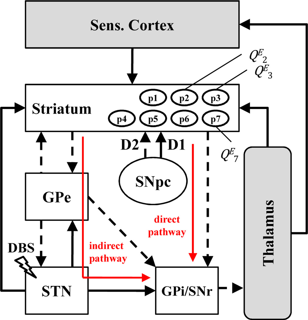
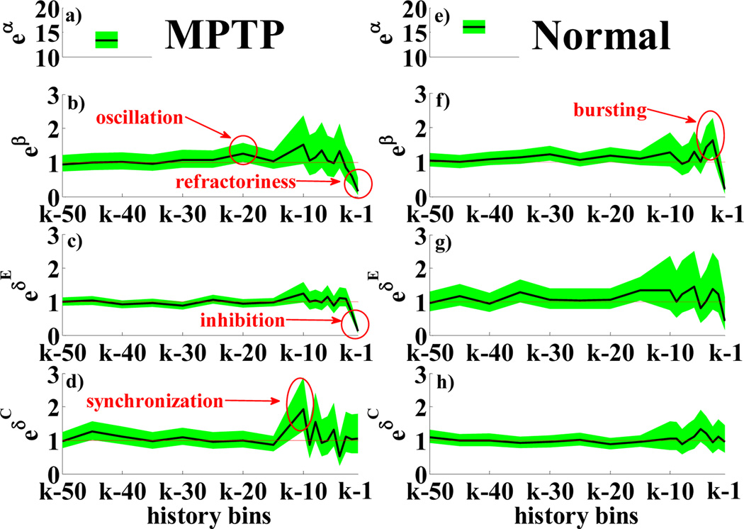
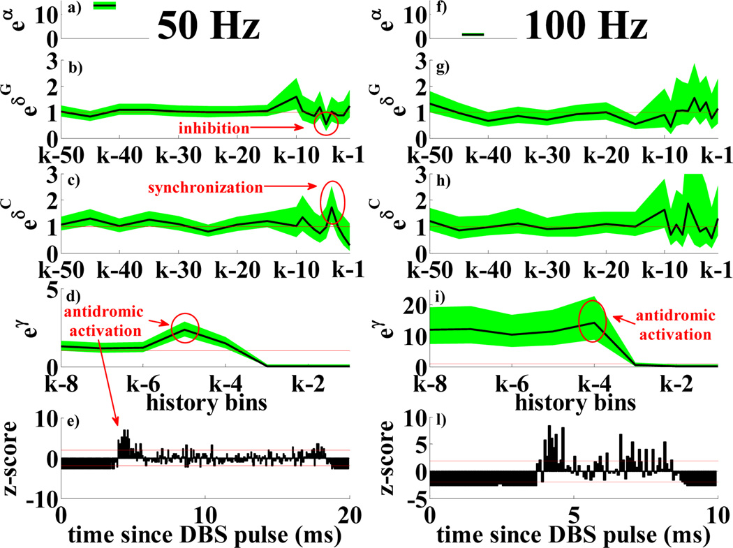
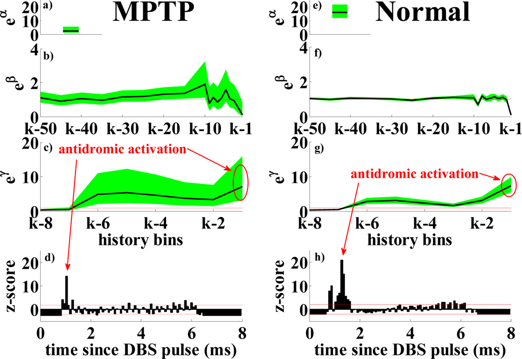
Striatum (STR) is the major input stage of the basal ganglia (BG). It combines information from cortex, subthalamic nucleus (STN) and external globus pallidus (GPe), and projects to the output stages of the BG, where selection between concurrent motor programs is performed. Parkinson's disease (PD) reduces the concentration of dopamine (DA, a neurotransmitter) in STR and changes in the level of DA correlate with the onset of PD motor disorders. Though STR plays a pivotal role in BG, its behavior under PD and Deep Brain Stimulation (DBS) is still unclear. We develop point-process models of the STR neurons as a function of the activity in GPe, cortex, and DBS. We use single unit recordings from a monkey under STN DBS at different frequencies before and after treatment with 1-methyl-4-phenyl-1,2,3,6-tetrahydro-pyridine (MPTP) to develop PD motor symptoms. The models suggest that STR neurons have prominent bursting activity in normal conditions, positive correlation with cortex (3-10 ms delay), and mild negative correlation with GPe (1-5 ms lag). DA depletion evokes 30-60 Hz oscillations, and increases the propensity of each neuron to be inhibited by surrounding neurons. DBS elicits antidromical activation, masks existent dynamics, reinforces dependencies between nuclei, and entrains at the stimulation frequency in both conditions.
Figures
References
-
- Gale JT, Amirnovin R, Williams ZM, Flaherty AW, Eskandar EN. From symphony to cacophony: Pathophysiology of the human basal ganglia in Parkinson disease. Neurosci. Biobehav. Rev. 2008 Mar;32:378–387. - PubMed
-
- DeLong MR, Wichmann T. Circuits and circuit disorders of the basal ganglia. Arch. Neurol. 2007 Jan;64:20–24. - PubMed
-
- Mink JW. The basal ganglia: focused selection and inhibition of competing motor programs. Prog. Neurobiol. 1996 Nov;50:381–425. - PubMed
-
- Brown P. Abnormal oscillatory synchronization in the motor system leads to impaired movement. Curr. Opin. Neurobiol. 2007 Dec;17:656–664. - PubMed
-
- Nakano K, Kayahara T, Tsutsumi T, Ushiro H. Neural circuits and functional organization of the striatum. J. Neurol. 2000 Sep;247(suppl. 5):V1–V15. - PubMed
MeSH terms
Substances
Grants and funding
LinkOut - more resources
Full Text Sources
Medical