. 2011 Feb;300(2):L204-15.
doi: 10.1152/ajplung.00292.2010. Epub 2010 Nov 19.

MCP-1/CCR2B-dependent loop upregulates MUC5AC and MUC5B in human airway epithelium

Affiliations

MCP-1/CCR2B-dependent loop upregulates MUC5AC and MUC5B in human airway epithelium

Maria E Monzon et al. Am J Physiol Lung Cell Mol Physiol. 2011 Feb.

Abstract

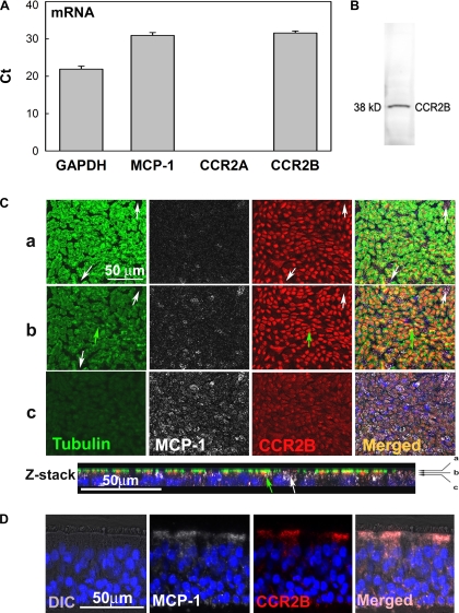
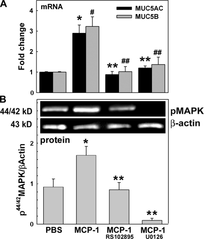
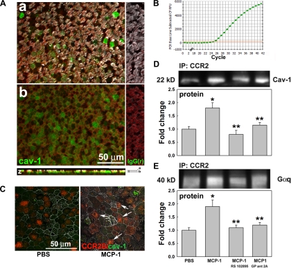
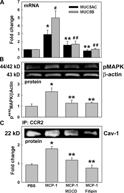
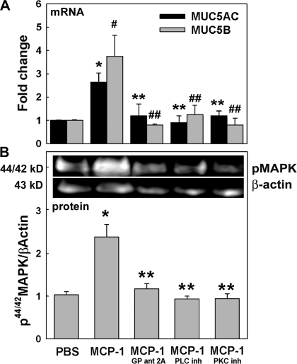
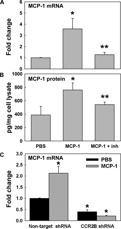
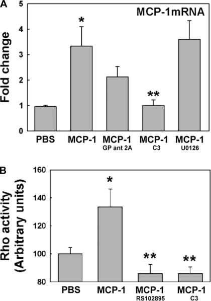
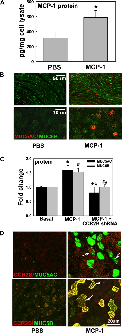
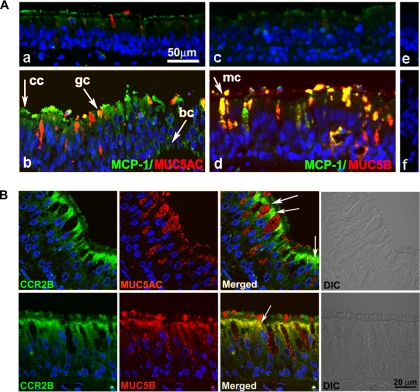
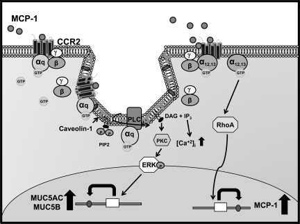
Cigarette smoke represents a major risk factor for the development of chronic obstructive pulmonary disease (COPD), a respiratory condition associated with airflow obstruction, mucus hypersecretion, chronic inflammation, and upregulation of inflammatory mediators such as the monocyte chemotactic protein-1 (MCP-1). MCP-1 through its receptor CCR2 induces chemotaxis and activates (44/42)MAPK, a kinase known to play a key role in mucin regulation in bronchial epithelium. In the present study we used differentiated primary cultures of normal human bronchial epithelial (NHBE) cells to test whether MCP-1 through its receptor CCR2 induces mucin upregulation. We have provided evidence that NHBE cells release MCP-1 to the epithelial surface and express the CCR2B isoform of the receptor mainly at the apical pole. In addition, we found that MCP-1 has a novel function in airway epithelium, increasing the two major airway mucins MUC5AC and MUC5B, an effect mediated, at least in part, by a cascade of events initiated by interaction of its receptor CCR2B with G(q) subunits in caveolae, followed by PLCβ, PKC, and (44/42)MAPK activation. We also have shown that MCP-1 is able to induce its own expression using the same receptor but through a different pathway that involves RhoA GTPase. Furthermore, we found that a single exposure to MCP-1 is enough to induce MCP-1 secretion and sustained mucin upregulation up to 7 days after initial exposure, an effect mediated by CCR2B as confirmed using short hairpin RNA. These results agree with our data in smoker's airway epithelium, where CCR2B is present in MUC5AC- and MUC5B-expressing cells and augmented MCP-1 expression is associated with increased MUC5AC and MUC5B immunolabeling, suggesting that the mechanisms described in primary cell cultures in the present study are operative in vivo. Therefore, therapeutic approaches targeting MCP-1/CCR2B may be useful in preventing not only influx of inflammatory cells to the airways but also mucus hypersecretion and goblet cell hyperplasia.


Figures

Fig. 1.
Fig. 1.
Monocyte chemotactic protein-1 (MCP-1) and its receptor, CC chemokine receptor 2 (CCR2B), are expressed in airway epithelial cells. A: GAPDH, MCP-1, CCR2A, and CCR2B mRNA expression in normal human bronchial epithelial (NHBE) cells was assessed by quantitative PCR (qPCR) and expressed as threshold cycle (Ct). Bar graphs show means ± SE (n = 10 different lung donors). B: CCR2B protein expression was confirmed by Western blotting. C: in addition, NHBE cells were labeled for MCP-1 (white), CCR2B (red), and acetylated tubulin (green; ciliated cell marker) by immunofluorescence (IF) and analyzed using confocal fluorescence microscopy (×63 objective lens). Three x-y sections (a–c) are shown (top) and indicated with arrows in the Z-stack reconstruction (bottom). Green arrows show ciliated cells expressing CCR2B, whereas white arrows indicate nonciliated cells expressing CCR2B. D: MCP-1 (white) and CCR2B (red) localization in human tracheobronchial sections scanned using fluorescence microscopy (×40 objective lens). Nuclei were labeled with 4,6-diamidino-2-phenylindole(DAPI). Differential interference contrast (DIC) images are also shown.
Fig. 2.
Fig. 2.
MCP-1 induces mucin upregulation and 44/42MAPK activation through CCR2B. NHBE cells were exposed to apical PBS (control), MCP-1, or MCP-1 + RS102895 (CCR2B inhibitor) or U0126 [phosphorylated 44/42MAPK (p44/42MAPK) inhibitor]. A: samples (n = 5 different lung donors) were analyzed for MUC5AC and MUC5B mRNA expression by qPCR. Results are expressed as fold change vs. control. *P < 0.05 vs. MUC5AC in PBS. #P < 0.05 vs. MUC5B in PBS. **P < 0.05 vs. MUC5AC in MCP-1-treated cultures. ##P < 0.05 vs. MUC5B in MCP-1-treated cultures. B: p44/42MAPK and β-actin immunoblotting (top) and p44/42MAPK/β-actin (bottom). Blot images are representative of the results obtained in 3 lung donors. Bar graphs show means ± SE. *P < 0.05 vs. PBS (nontreated control). **P < 0.05 vs. MCP-1.
Fig. 3.
Fig. 3.
MCP-1 induces caveolin-1 (Cav-1) and Gq subunit interaction with CCR2B. A: Cav-1 protein was labeled in green, cellular cytoskeletal shape (F-actin) with phalloidin (red), and cilia with acetylated tubulin (white). Cells were scanned using confocal fluorescence microscopy (×63 objective lens); 2 x-y sections (a and b) are shown and indicated by arrows in the Z-reconstruction. B: Cav-1 mRNA expression. C: CCR2B (red)/Cav-1 (green) interaction in NHBE cells treated with PBS or MCP-1 was assessed by confocal microscopy using a ×63 objective lens, and colocalization is indicated (white arrows). D: CCR2B/Cav-1 interaction was quantified by coimmunoprecipitation, immunoprecipitation (IP), and Western blotting. E: CCR2B/Gq subunit interaction was tested by Gq pull-down using anti-CCR2B antibodies. Blot images are representative of the results obtained in 3 lung donors. Bar graphs show means ± SE obtained from 3 different lung donors. *P < 0.05 vs. PBS (nontreated control). **P < 0.05 vs. MCP-1.
Fig. 4.
Fig. 4.
MCP-1-induced mucin upregulation depends on 44/42MAPK activation and requires caveolae integrity. Cells were exposed to apical PBS (control) or MCP-1 in the presence or the absence of the caveolae disruptors methy-β-cyclodextrin (MβCD) or filipin. A: samples (n = 4 different lung donors) were analyzed for MUC5AC and MUC5B mRNA expression by qPCR. Results are expressed as fold change vs. controls. *P < 0.05 vs. MUC5AC control. #P < 0.05 vs. MUC5B control. **P < 0.05, MUC5AC vs. MCP-1. ##P < 0.05, MUC5B vs. MCP-1. B: 44/42MAPK phosphorylation was assessed by Western blotting: p44/42MAPK and β-actin immunoblotting (top) and p44/42MAPK/β-actin (bottom). *P < 0.05 vs. PBS. **P < 0.05 vs. MCP-1. C: CCR2B/Cav-1 interaction was evaluated using coimmunoprecipitation and Cav-1 immunoblotting (top) and expressed as fold change of Cav-1 pulled down by anti-CCR2B antibody vs. control (bottom). Blot images are representative of the results obtained in 3 lung donors. Bar graphs show means ± SE obtained from 3 different lung donors. *P < 0.05 vs. PBS. **P < 0.05 vs. MCP-1.
Fig. 5.
Fig. 5.
MCP-1-induced mucin upregulation and 44/42MAPK activation is dependent on Gq subunits, PLCβ, and PKC. Cells were exposed to apical PBS (control) or MCP-1 in the presence or absence of Gq (GP ant 2A), PLCβ, or PKC inhibitors. A: samples (n = 4 different lung donors) were analyzed for MUC5AC and MUC5B mRNA expression by qPCR. Results are expressed as fold change vs. the corresponding basal control. *P < 0.05 vs. MUC5AC control (PBS). #P < 0.05 vs. MUC5B control (PBS). **P < 0.05 vs. MCP-1-induced MUC5AC. ##P < 0.05 vs. MCP-1-induced MUC5B. B: p44/42MAPK and β-actin immunoblotting (top) and p44/42MAPK/β-actin (bottom). Blot images are representative of the results obtained in 3 lung donors. Bar graphs show means ± SE obtained from 3 different lung donors. *P < 0.05 vs. PBS. **P < 0.05 vs. MCP-1.
Fig. 6.
Fig. 6.
MCP-1 induces its own mRNA and protein expression through CCR2B. MCP-1 mRNA and protein expression were assessed in NHBE cells treated with PBS or MCP-1 by qPCR and ELISA at day 1 after initial treatment. Effect of a CCR2B inhibitor (inh; RS102895) was assessed at the mRNA (A) and protein (B) levels. *P < 0.05 vs. PBS. #P < 0.05 vs. MCP-1. C: MCP-1 mRNA expression in NHBE cells infected with nontarget or CCR2B short hairpin (sh)RNA expressing lentivirus and exposed to PBS or MCP-1. Bar graphs show means ± SE obtained from 3 different lung donors. *P < 0.05 vs. PBS nontarget shRNA.
Fig. 7.
Fig. 7.
MCP-1 induces its own mRNA expression through RhoA GTPase activation. A: NHBE cultures exposed to PBS or MCP-1 in the presence or absence of inhibitors for Gq signaling (GP ant 2A), RhoA GTPase activation (C3), or 44/42MAPK activation (U0120) were analyzed for MCP-1 mRNA expression by qPCR. B: in addition, PBS- or MCP-1-treated NHBE cells in the presence or absence of CCR2B (RS102895) or RhoA inhibitors (C3) were assessed for RhoA activation using an ELISA-like assay. Bar graphs show means ± SE obtained from 4 different lung donors. *P < 0.01 vs. PBS. **P < 0.05 vs. MCP-1.
Fig. 8.
Fig. 8.
Sustained MUC5AC and MUC5B upregulation is dependent on MCP-1 and CCR2B. A: NHBE cultures exposed to PBS or MCP-1 were assessed for MCP-1 by ELISA. Bar graphs show means ± SE obtained from 4 different lung donors. *P < 0.05 vs. basal MCP-1. B: NHBE cells were also assessed for MUC5AC (red) and MUC5B (green) protein expression by IF at day 7 from the initial stimuli. Cultures were analyzed using fluorescence microscopy with ×10 (top images) or ×63 objective lenses (bottom images). C: anti-CCR2B shRNA blocked MCP-1-induced MUC5AC and MUC5B protein. Bar graphs show means ± SE from 3 different lung donors. *P < 0.05, MCP-1-induced MUC5AC vs. baseline. #P < 0.05, MCP-1-induced MUC5B vs. baseline. **P < 0.05, CCR2B-blocked MCP-1-induced MUC5AC vs. baseline. ##P < 0.05, CCR2B-blocked MCP-1-induced MUC5B vs. baseline. D: CCR2B immunolabeling (red) with MUC5AC (left, green) or MUC5B (right, green) in NHBE cells exposed to PBS or MCP-1.
Fig. 9.
Fig. 9.
Increased MUC5AC and MUC5B are associated to MCP-1 and CCR2B. A: human tracheobronchial sections from a nonsmoker donor (a, c, e) and 2 smoker donors (b, d, f) were double-labeled with anti-MUC5AC (a and b; red) or anti-MUC5B (c and d; red) and anti-MCP-1 (a–d; green). Nonimmune IgGs are depicted in e and f. Arrows indicate ciliated (cc), goblet (gc), MUC5B-positive (mc), and basal cells (bc). B: human tracheobronchial sections from smoker donors were also labeled with CCR2B (green) and MUC5AC (top; red) or MUC5B (bottom; red). Nuclei were labeled with DAPI. Slides were scanned using confocal microscopy (×63 objective lens). Images represent variability of 8 different lung donors.
Fig. 10.
Fig. 10.
Schematic of CCR2B signal transduction pathways that regulate MUC5AC, MUC5B, and MCP-1 expression. MCP-1 induces CCR2B/Gq interaction in caveolae. GTP-bound Gq-protein's effector interaction domain is exposed and activates PLCβ, which promotes phosphoinositol 4,5-bisphosphate (PIP2) hydrolysis into 1,2-diacylglycerol (DAG) and inositol 1,4,5-trisphosphate (IP3). DAG promotes PKC activation. Activated PKC phosphorylates 44/42MAPK, which upregulates MUC5AC and MUC5B. In addition, MCP-1 also induces CCR2B/G12/13 interaction associated with increased RhoA GTPase activity and upregulation of MCP-1.

Similar articles

Cited by

References

    1. Cigarette smoking among adults—United States, 1992, and changes in the definition of current cigarette smoking. MMWR Morb Mortal Wkly Rep 43: 342–346, 1994 - PubMed
    1. Ashida N, Arai H, Yamasaki M, Kita T. Distinct signaling pathways for MCP-1-dependent integrin activation and chemotaxis. J Biol Chem 276: 16555–16560, 2001 - PubMed
    1. Barnes PJ. New concepts in chronic obstructive pulmonary disease. Annu Rev Med 54: 113–129, 2003 - PubMed
    1. Belperio JA, Keane MP, Burdick MD, Lynch JP, 3rd, Xue YY, Berlin A, Ross DJ, Kunkel SL, Charo IF, Strieter RM. Critical role for the chemokine MCP-1/CCR2 in the pathogenesis of bronchiolitis obliterans syndrome. J Clin Invest 108: 547–556, 2001 - PMC - PubMed
    1. Budd DC, Willars GB, McDonald JE, Tobin AB. Phosphorylation of the Gq/11-coupled m3-muscarinic receptor is involved in receptor activation of the ERK-1/2 mitogen-activated protein kinase pathway. J Biol Chem 276: 4581–4587, 2001 - PubMed

Publication types

MeSH terms