. 2011 Jan;17(1):182-8.
doi: 10.1261/rna.2222811. Epub 2010 Nov 19.

Ligand-induced conformational capture of a synthetic tetracycline riboswitch revealed by pulse EPR

Affiliations

Ligand-induced conformational capture of a synthetic tetracycline riboswitch revealed by pulse EPR

Dorith Wunnicke et al. RNA. 2011 Jan.

Abstract

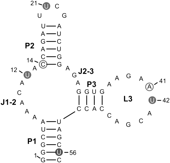
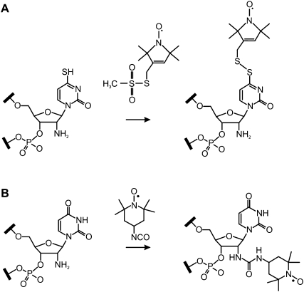
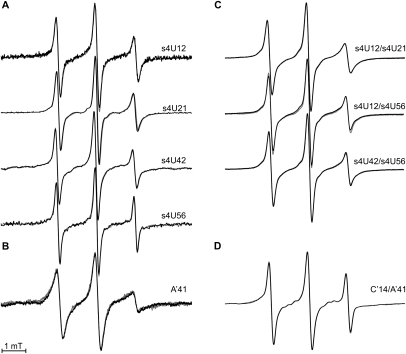
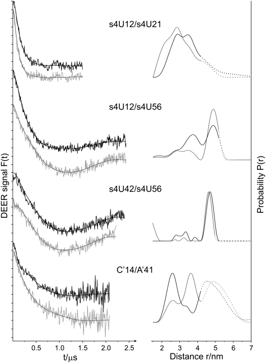
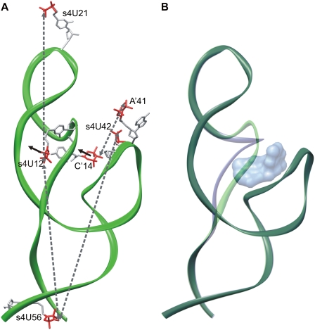
RNA aptamers are in vitro-selected binding domains that recognize their respective ligand with high affinity and specificity. They are characterized by complex three-dimensional conformations providing preformed binding pockets that undergo conformational changes upon ligand binding. Small molecule-binding aptamers have been exploited as synthetic riboswitches for conditional gene expression in various organisms. In the present study, double electron-electron resonance (DEER) spectroscopy combined with site-directed spin labeling was used to elucidate the conformational transition of a tetracycline aptamer upon ligand binding. Different sites were selected for post-synthetic introduction of either the (1-oxyl-2,2,5,5-tetramethylpyrroline-3-methyl) methanethiosulfonate by reaction with a 4-thiouridine modified RNA or of 4-isocyanato-2,6-tetramethylpiperidyl-N-oxid spin label by reaction with 2'-aminouridine modified RNA. The results of the DEER experiments indicate the presence of a thermodynamic equilibrium between two aptamer conformations in the free state and capture of one conformation upon tetracycline binding.


Figures

FIGURE 1.
FIGURE 1.
Secondary structure predicted for the Tc aptamer according to Hanson et al. (2005). Thiouridines (U) spin labeled with MTS are marked by full gray circles, 2′-aminouridine labeled with 4-isocyanato-TEMPO are depicted by open gray circles.
FIGURE 2.
FIGURE 2.
Strategy for the post-synthetic introduction of MTS using the selective reaction with 4-thiouridine (A) and of 4-isocyanato-TEMPO using the selective reaction with 2′-aminouridine (B).
FIGURE 3.
FIGURE 3.
Room temperature continuous wave EPR spectra measured at X-band in the absence (black lines) and in the presence of Tc (gray lines). (A,C) Single and double mutants labeled with MTS and (B,D) single and double mutants labeled with the 4-isocyanato-TEMPO. All plots are normalized by amplitude.
FIGURE 4.
FIGURE 4.
DEER analysis of the RNA double mutants s4U12/s4U21, s4U12/s4U56, s4U42/s4U56, and C′14/A′41 in the absence (black) and in the presence of Tc (gray). (Left) Background-corrected dipolar evolution data F(t). Tick marks are separated by 0.02. (Right) Distance distributions P(r) obtained using DeerAnalysis2006 (Jeschke et al. 2006). In the dotted range, intermolecular spin–spin interaction may contribute.
FIGURE 5.
FIGURE 5.
Schematic representations of the Tc aptamer conformations. The regions J1-2, J2-3, and L3 of the X-ray structure of the circularly permuted Tc-dependent aptamer (Xiao et al. 2008) was complemented with a schematic representation of the stems P1 and P2 based on the chemical and enzymatic probing experiments (Hanson et al. 2005). (A) Interspin distances between nitroxides at positions 12, 14, 21, 41, 42, and 56, as indicated by gray-dotted arrows were used as constraints during modeling. Black arrows indicate the displacement of the nitroxides at positions 12 and 14 upon addition of Tc. (B) Two aptamer conformations (green, blue) are in equilibrium in the free state. Upon addition of Tc (shown in surface representation) the aptamer conformation depicted in blue is trapped.

Similar articles

Cited by

References

    1. Beier C, Steinhoff HJ 2006. A structure-based simulation approach for electron paramagnetic resonance spectra using molecular and stochastic dynamics simulations. Biophys J 91: 2647–2664 - PMC - PubMed
    1. Berens C, Hillen W 2003. Gene regulation by tetracyclines. Eur J Biochem 270: 3109–3121 - PubMed
    1. Bordignon E, Steinhoff HJ 2007. Membrane protein structure and dynamics studied by site-directed spin labeling ESR. In ESR spectroscopy in membrane biophysics (ed. Hemminga MA, Berliner LJ), pp. 129–164 Springer Science and Business Media, New York
    1. Cai Q, Kusnetzow AK, Hubbell WL, Haworth IS, Gacho GPC, Van Eps N, Hideg K, Chambers EJ, Qin PZ 2006. Site-directed spin labeling measurements of nanometer distances in nucleic acids using a sequence-independent nitroxide probe. Nucleic Acids Res 34: 4722–4730 - PMC - PubMed
    1. Edwards TE, Okonogi TM, Robinson BH, Sigurdsson ST 2001. Site-specific incorporation of nitroxide spin-labels into internal sites of the TAR RNA; structure-dependent dynamics of RNA by EPR spectroscopy. J Am Chem Soc 123: 1527–1528 - PubMed

Publication types

LinkOut - more resources