. 2011 Jan;77(2):669-83.
doi: 10.1128/AEM.01952-10. Epub 2010 Nov 19.

Genomic and functional analyses of Rhodococcus equi phages ReqiPepy6, ReqiPoco6, ReqiPine5, and ReqiDocB7

Affiliations

Genomic and functional analyses of Rhodococcus equi phages ReqiPepy6, ReqiPoco6, ReqiPine5, and ReqiDocB7

E J Summer et al. Appl Environ Microbiol. 2011 Jan.

Abstract

The isolation and results of genomic and functional analyses of Rhodococcus equi phages ReqiPepy6, ReqiDocB7, ReqiPine5, and ReqiPoco6 (hereafter referred to as Pepy6, DocB7, Pine5, and Poco6, respectively) are reported. Two phages, Pepy6 and Poco6, more than 75% identical, exhibited genome organization and protein sequence likeness to Lactococcus lactis phage 1706 and clostridial prophage elements. An unusually high fraction, 27%, of Pepy6 and Poco6 proteins were predicted to possess at least one transmembrane domain, a value much higher than the average of 8.5% transmembrane domain-containing proteins determined from a data set of 36,324 phage protein entries. Genome organization and protein sequence comparisons place phage Pine5 as the first nonmycobacteriophage member of the large Rosebush cluster. DocB7, which had the broadest host range among the four isolates, was not closely related to any phage or prophage in the database, and only 23 of 105 predicted encoded proteins could be assigned a functional annotation. Because of the relationship of Rhodococcus to Mycobacterium, it was anticipated that these phages should exhibit some of the features characteristic of mycobacteriophages. Traits that were identified as shared by the Rhodococcus phages and mycobacteriophages include the prevalent long-tailed morphology and the presence of genes encoding LysB-like mycolate-hydrolyzing lysis proteins. Application of DocB7 lysates to soils amended with a host strain of R. equi reduced recoverable bacterial CFU, suggesting that phage may be useful in limiting R. equi load in the environment while foals are susceptible to infection.


Figures

FIG. 1.
FIG. 1.
Negative-stained images of new rhodococcal siphophages. ReqiDocB7 (A), ReqiPine5 (B), ReqiPoco6 (C), and ReqiPepy6 (D) negatively stained with 2% (wt/vol) aqueous uranyl acetate. Bars, 200 nm.
FIG. 2.
FIG. 2.
Dot plot alignment of the nucleotide sequences of Pepy6 and Poco6. Pepy6 and Poco6 are closely related, colinear phages with isolated blocks of nonidentity. The percentages of identity of the longer aligned blocks are indicated.
FIG. 3.
FIG. 3.
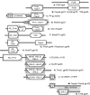
Genome maps of Pepy6 and Poco6 and related phages and prophages. (A) Genome maps of phages ReqiPepy6 and ReqiPoco6. Purple shading between the genome maps indicates segments of DNA that align with greater than 75% identity. The colors of the genes or proteins in the gene maps indicate the relationship of the encoded protein with other proteins as follows. Yellow indicates a homologue is encoded by phage 1706. Yellow genes with green outlines encode homologues of phage 1706 virion-associated proteins. The 1706 gene is indicated below the Rhodococcus phage gene. Turquoise indicates that there was no homologue in 1706, nor was a functional annotation possible. Dark blue indicates proteins for which a functional annotation could be made but lacked homologues in 1706. Green proteins possess 56 invariant, N-terminal amino acid residues, indicated by peach bars. Genes outlined in red encode proteins with predicted transmembrane helices. Regions shaded in peach indicate locations of gene insertions relative to phage 1706. (B) Genome maps of Lactococcus lactis phage 1706 and prophage elements BRYFOR and CLOSS21, present in the genomic sequences of Bryantella formatexigens DSM 14469 and Clostridium sp. strain SS2/1 genome, respectively. The phage 1706 genome map was derived from the GenBank entry with accession no. EU081845. The BRYFOR prophage encompasses the region encoding locus tags BRYFOR_08504 to BRYFOR_08569. The CLOSS21 prophage encompasses the region encoding locus tags CLOSS21_01492 to CLOSS21_01580. In order to align with phage 1706, the BRYFOR and CLOSS21 maps are drawn with the order of prophages changed. The locations of the prophage genomic termini, between the coding region of BRYFOR gene73 and gene1 and CLOSS21 gene89 and gene1, are indicated on the genome maps by turquoise shading. The colors of the genes or proteins indicate the relationship of the encoded protein with other proteins as follows: yellow, homologue encoded by Pepy6 and Poco6; gray, no homologue encoded by Pepy6 or Poco6. A green outline indicates that the protein encoded by the gene is a homologue of phage 1706 virion-associated protein. A red outline indicates that the protein encoded by the gene has predicted transmembrane helices. The gene number of the corresponding protein encoded by Pepy6 and 1706 is indicated below each map (-, no protein). Regions shaded in peach indicate locations of gene insertions in the map relative to phage 1706. Select functional annotations are shown above genes by the following abbreviations: e15, Salmonella phage epsilon15 tail fiber protein; lip, lipase; hol, holin; tmp, tape measure protein; Prim, primase; pol, DNA polymerase; HNH, homing endonuclease; Int, integrase; endo, endonuclease; ligA, ligase A domain; CMP deaminase, deoxycytidinylate deaminase. Homologous gene products in Pepy6 and 1706 (Pepy6yp and 1706yp, respectively) are indicated below each map.
FIG. 4.
FIG. 4.
Novel combinations and permutations of DNA metabolism-associated conserved domains in phages and proteins encoded by bacterial genes. (A) Lactococcus phage P087 (gp4); (B) Pepy6 (gp72), Poco6 (gp72), and Lactococcus phage 1706 (gp55); (C) satellite phage P4 (RPBPP4); (D) Xylella phage Xfas53 (gp23); (E) Vibrio phage Vp2 (VP2p2); (F) Burkholderia phage Bcep781 (gp53); (G) Pine5 (gp46) and Rosebush (gp54); (H) DocB7 (gp105); (I) Escherichia coli O157:H7 EDL933 (z1129); (J) DocB7 (gp48); (K) Pine5 (gp45) and mycobacteriophage Rosebush (gp50); (L) CLOS21 (CLOSS21_01549); (M) Pepy6 (gp102) and Poco6 (gp102); (N) Lactococcus phage 1706 (gp67). Related domains are shaped and shaded similarly. Domain abbreviations are based on the Conserved Domain Database entries as follows: PolB, COG0417; Prim_Cterm, COG3378; AE_Prim, cl01287; DEAD HELIC, CD00046; SSL2, COG1061; LigA, CL03295; PriCT-2D5N, pfam08707; D5_N superfamily, cl07360; Prim Z, cl07038; VirE, COG5545; RepA, COG3598; COG4643, COG4643.
FIG. 5.
FIG. 5.
Complex relationships between potential tail fibers encoded by Pepy6 and Poco6. (A) Alignment of the first 56 residues of Pepy6 gp1, gp2, gp4, gp5, and gp7 with Poco6 gp1, gp2, gp3, gp5, gp6, and gp8 and the amino-terminal residues of PSSM4_087 (encoded by Prochlorococcus phage P-SSM4 genes), XF_2114 (encoded by Xylella fastidiosa prophage Xfp6 genes), and 3396_48 (encoded by Streptococcus phage phi3396 genes). Invariant residues are shaded. (B) Schematic alignment of Pepy6 gp1, gp2, gp4, gp5, and gp7 with Poco6 gp1, gp2, gp3, gp5, gp6, and gp8 and also to e15 (tail fiber, encoded by Salmonella phage epsilon15 gene), PSSM4_087 (encoded by Prochlorococcus phage P-SSM4 gene), 3396_48 (encoded by Streptococcus phage phi3396 gene) and (FRAAL2677 encoded by Frankia alni ACN14a gene). Alignment scores are color coded as indicated. Conserved domains are indicated by shaded regions.
FIG. 6.
FIG. 6.
Genome maps of Pine5 and DocB7. Phage genome maps are drawn to scale with the coding strand indicated by the position of the genes above (rightwards) and below (leftwards) the central ruler. (A) Pine5 aligned with mycobacteriophage Rosebush. The Rosebush map was generated from the GenBank entry with accession no. AY129334.1. Purple lines between Pine5 and Rosebush indicate regions of greater than 60% DNA identity between the two phages. Genes are color coded to indicate similarity with other proteins in the public database as follows: yellow, homologues are found in the mycobacteriophages Rosebush, Qyrzula, Phlyer, Phaedrus, Pipefish, Nigel, Cooper, Chah, PG1, Orion, Colbert, Puhltonio, and UncleHowie; green, homologues in the public database but not in Rosebush; blue, no homologue in the public database; gray, Rosebush genes without homologues in Pine5. Genes with red outlines encode proteins with predicted transmembrane domains. Annotations of selected genes are abbreviated as follows: PAPSr, phosphoadenosine phosphosulfate reductase; sub, subunit; RuvC, Holliday junction resolvases. (B) DocB7.Genes are color coded to indicate similarity with other proteins in the public database as follows: yellow genes encode proteins with at least one homologue in the public database; blue genes, no homologue in the public database. Genes with red outlines encode proteins with predicted transmembrane domains. The 2,483-bp region highlighted in peach lacks recognizable protein-coding genes (no cds, no protein-coding sequences) and contains 13 perfect inverted repeats and one direct repeat.
FIG. 7.
FIG. 7.
Effect of phage application on recovery of R. equi from soil. Phage effect on R. equi was evaluated via 7 treatment groups. Each treatment group contained 3 parallel subsamples, where 3 sterile soil matrixes were inoculated independently with R. equi strain ATCC 33701 and were subjected to further treatment. R. equi was inoculated at an initial concentration of 1.0 × 10⁵ CFU/g soil. The R. equi levels were determined before (0-h) and after (48-h) incubation in the absence of phage treatment (No phage). The R. equi CFU levels were also determined following 48 h of incubation in the presence of phage DocB7. Phage DocB7 was applied at initial multiplicity of infections (MOIs) of 1,000, 100, 10, 1, and 0.1 PFU/CFU. After each subsample was analyzed in two replicate samples, the mean values± standard deviations of the subsamples (error bars) were plotted. Two independent experiments were carried out, and the results of one representative experiment are shown.

Similar articles

Cited by

  • Reconstructing viral genomes from the environment using fosmid clones: the case of haloviruses.
    Garcia-Heredia I, Martin-Cuadrado AB, Mojica FJ, Santos F, Mira A, Antón J, Rodriguez-Valera F. Garcia-Heredia I, et al. PLoS One. 2012;7(3):e33802. doi: 10.1371/journal.pone.0033802. Epub 2012 Mar 30. PLoS One. 2012. PMID: 22479446 Free PMC article.
  • Antimicrobial Resistance in Equines: A Growing Threat to Horse Health and Beyond-A Comprehensive Review.
    Kabir A, Lamichhane B, Habib T, Adams A, El-Sheikh Ali H, Slovis NM, Troedsson MHT, Helmy YA. Kabir A, et al. Antibiotics (Basel). 2024 Jul 29;13(8):713. doi: 10.3390/antibiotics13080713. Antibiotics (Basel). 2024. PMID: 39200013 Free PMC article. Review.
  • Tales of diversity: Genomic and morphological characteristics of forty-six Arthrobacter phages.
    Klyczek KK, Bonilla JA, Jacobs-Sera D, Adair TL, Afram P, Allen KG, Archambault ML, Aziz RM, Bagnasco FG, Ball SL, Barrett NA, Benjamin RC, Blasi CJ, Borst K, Braun MA, Broomell H, Brown CB, Brynell ZS, Bue AB, Burke SO, Casazza W, Cautela JA, Chen K, Chimalakonda NS, Chudoff D, Connor JA, Cross TS, Curtis KN, Dahlke JA, Deaton BM, Degroote SJ, DeNigris DM, DeRuff KC, Dolan M, Dunbar D, Egan MS, Evans DR, Fahnestock AK, Farooq A, Finn G, Fratus CR, Gaffney BL, Garlena RA, Garrigan KE, Gibbon BC, Goedde MA, Guerrero Bustamante CA, Harrison M, Hartwell MC, Heckman EL, Huang J, Hughes LE, Hyduchak KM, Jacob AE, Kaku M, Karstens AW, Kenna MA, Khetarpal S, King RA, Kobokovich AL, Kolev H, Konde SA, Kriese E, Lamey ME, Lantz CN, Lapin JS, Lawson TO, Lee IY, Lee SM, Lee-Soety JY, Lehmann EM, London SC, Lopez AJ, Lynch KC, Mageeney CM, Martynyuk T, Mathew KJ, Mavrich TN, McDaniel CM, McDonald H, McManus CJ, Medrano JE, Mele FE, Menninger JE, Miller SN, Minick JE, Nabua CT, Napoli CK, Nkangabwa M, Oates EA, Ott CT, Pellerino SK, Pinamont WJ, Pirnie RT, Pizzorno MC, Plautz EJ, Pope WH, Pruett KM, Rickstrew G, Rimple PA, Rinehart CA, Robinson KM, Rose VA, Russell DA, Schick AM, Schlossman J,… See abstract for full author list ➔ Klyczek KK, et al. PLoS One. 2017 Jul 17;12(7):e0180517. doi: 10.1371/journal.pone.0180517. eCollection 2017. PLoS One. 2017. PMID: 28715480 Free PMC article.
  • Lytic Spectra of Tailed Bacteriophages: A Systematic Review and Meta-Analysis.
    Pchelin IM, Smolensky AV, Azarov DV, Goncharov AE. Pchelin IM, et al. Viruses. 2024 Dec 4;16(12):1879. doi: 10.3390/v16121879. Viruses. 2024. PMID: 39772189 Free PMC article.
  • Complete Genome Sequence of Salmonella enterica Serovar Typhimurium Siphophage Skate.
    Rohren M, Xie Y, O'Leary C, Kongari R, Gill J, Liu M. Rohren M, et al. Microbiol Resour Announc. 2019 Jul 3;8(27):e00541-19. doi: 10.1128/MRA.00541-19. Microbiol Resour Announc. 2019. PMID: 31270198 Free PMC article.

References

    1. Ackermann, H. W. 2007. 5500 phages examined in the electron microscope. Arch. Virol. 152:227-243. - PubMed
    1. Adindla, S., and L. Guruprasad. 2003. Sequence analysis corresponding to the PPE and PE proteins in Mycobacterium tuberculosis and other genomes. J. Biosci. 28:169-179. - PubMed
    1. Akoh, C. C., G. C. Lee, Y. C. Liaw, T. H. Huang, and J. F. Shaw. 2004. GDSL family of serine esterases/lipases. Prog. Lipid Res. 43:534-552. - PubMed
    1. Alvarez, V. M., et al. 2008. Bioremediation potential of a tropical soil contaminated with a mixture of crude oil and production water. J. Microbiol. Biotechnol. 18:1966-1974. - PubMed
    1. Ashelford, K. E., M. J. Day, and J. C. Fry. 2003. Elevated abundance of bacteriophage infecting bacteria in soil. Appl. Environ. Microbiol. 69:285-289. - PMC - PubMed

Publication types

Associated data

LinkOut - more resources