. 2011 Feb;193(3):715-22.
doi: 10.1128/JB.00796-10. Epub 2010 Nov 19.

Isolation and characterization of P1 adhesin, a leg protein of the gliding bacterium Mycoplasma pneumoniae

Affiliations

Isolation and characterization of P1 adhesin, a leg protein of the gliding bacterium Mycoplasma pneumoniae

Daisuke Nakane et al. J Bacteriol. 2011 Feb.

Abstract

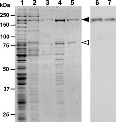
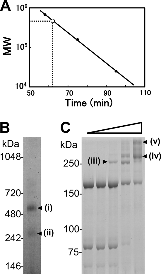
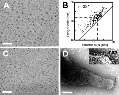
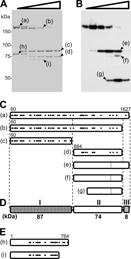
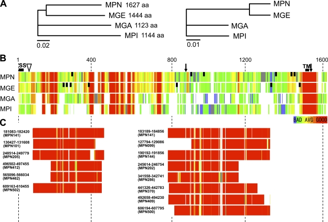
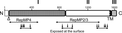
Mycoplasma pneumoniae, a pathogen causing human pneumonia, binds to solid surfaces at its membrane protrusion and glides by a unique mechanism. In this study, P1 adhesin, which functions as a "leg" in gliding, was isolated from mycoplasma culture and characterized. Using gel filtration, blue-native polyacrylamide gel electrophoresis (BN-PAGE), and chemical cross-linking, the isolated P1 adhesin was shown to form a complex with an accessory protein named P90. The complex included two molecules each of P1 adhesin and P90 (protein B), had a molecular mass of about 480 kDa, and was observed by electron microscopy to form 20-nm-diameter spheres. Partial digestion of isolated P1 adhesin by trypsin showed that the P1 adhesin molecule can be divided into three domains, consistent with the results from trypsin treatment of the cell surface. Sequence analysis of P1 adhesin and its orthologs showed that domain I is well conserved and that a transmembrane segment exists near the link between domains II and III.


Figures

FIG. 1.
FIG. 1.
Protein profiles of fractions in the P1-P90 complex isolation procedure. The protein fractions were applied to SDS-10% PAGE and subjected to CBB staining (lanes 1 to 5) or immunoblotting using an anti-P1 adhesion monoclonal antibody (lanes 6 and 7). Lanes 1 and 6, whole-cell lysate; lane 2, CHAPS-insoluble fraction; lane 3, octylglucoside-soluble fraction; lane 4, precipitate of 45 to 55% saturated ammonium sulfate; lanes 5 and 7, P1-P90 complex fraction eluted during gel filtration. Molecular masses are indicated on the left. The protein bands of P1 adhesin and P90 are marked by solid and open triangles, respectively.
FIG. 2.
FIG. 2.
Size and composition of isolated P1-P90 complex. (A) Size estimation by gel filtration. The retention time and apparent molecular mass during gel filtration are presented on the x and y axes, respectively. The solid circles show size standards: thyroglobulin, 670 kDa; aldolase, 158 kDa; and chymotrypsinogen, 25 kDa. The open circle shows the position of the P1-P90 complex. (B) Protein profiles of the P1-P90 complex analyzed by BN-PAGE. Molecular masses are indicated on the left. (C) Protein profiles of the P1-P90 complex cross-linked by various concentrations of BS3 as represented by the open triangle. The protein fraction was treated with 0, 0.002, 0.02, 0.2, and 2 mM BS3 and subjected to SDS-6.0% PAGE. Molecular masses are indicated on the left. The protein bands marked i to v were analyzed by PMF.
FIG. 3.
FIG. 3.
Molecular shape and dimensions of the P1-P90 complex. (A) Rotary-shadowed EM image. (B) Distribution of shorter and longer axes of the individual complex. The averages, shown by broken lines, are 18.8 ± 3.1 nm and 21.8 ± 3.1 nm for the shorter and longer axes, respectively. The diagonal solid line shows the plot of the same axes. (C) Rotary-shadowed EM image of Gli349, a leg protein of the fastest-gliding species, M. mobile (modified from reference 1). (D) Surface structure of M. pneumoniae. The area outlined by the dashed box is magnified in the inset to show the nap structures. Bars, 100 nm.
FIG. 4.
FIG. 4.
Partial digestion of the P1-P90 complex. (A) Protein profiles of the purified P1-P90 complex digested with various concentrations of trypsin. The protein fraction was treated with 0, 0.01, 0.03, 0.1, 0.3, and 1 mg/ml trypsin and subjected to SDS-8.75% PAGE. The resultant peptide fragments, marked a to i, were identified by PMF. (B) Immunoblot of M. pneumoniae cells treated with various concentrations of trypsin, using antibody against the C-terminal region of P1 adhesin for detection. The cells were treated with 0, 0.01, 0.03, 0.1, 0.3, and 1 mg/ml trypsin and subjected to SDS-PAGE and immunoblotting to detect the aa 1386 to 1394 sequence. The sizes of the resulting peptide fragments, marked e to g, were estimated to be 83, 77, and 48 kDa, respectively. The molecular masses are indicated on the left of panel A. (C) Mapping of products on the amino acid sequence of P1 adhesin. The 59 N-terminal residues predicted from the DNA sequence were processed in the mature form (46, 60). Protein bands a to d were identified by PMF. The positions of peptides detected in PMF are marked with solid dots. The N-terminal residues in bands a to d were determined by Edman analysis. The C terminus was estimated from the band size. The evident residue numbers of the ends are indicated. Peptide bands e to g were assigned from the band sizes, and the epitope positions are shown by gray lines. (D) Schematic of P1 adhesin, consisting of three domains, I, II, and III. The molecular mass is indicated below each domain. (E) Bands h and i mapped onto the amino acid sequence of P90, based on the results of PMF.
FIG. 5.
FIG. 5.
Sequence similarity of P1 adhesin homologs. (A) Phylogenetic tree of amino acid sequences of P1 adhesin orthologs (left) and 16S rRNA sequences (right) of M. pneumoniae (MPN) and its relatives, M. genitalium (MGE), M. gallisepticum (MGA), and M. pirum (MPI). The scale bars indicate the numbers of residue substitutions by position. (B) Multiple alignment of sequences of P1 adhesin orthologs performed by T-Coffee. The color indicates the sequence similarity score, as shown at the bottom right. The regions with no similarity to orthologs are colored gray. The N-terminal processing site is marked by a triangle between residues 59 and 60. The trypsin-sensitive sites are marked by arrows between residues 883 and 884 and ∼8 kDa from the C terminus. The predicted signal sequence (SS) and TM segment are labeled. The vertical black bars indicate the epitopes of adherence-inhibiting antibodies against P1 adhesin of M. pneumoniae and MgPa of M. genitalium (8, 43, 46). (C) Multiple alignment of amino acid sequences of P1 adhesin paralogs from M. pneumoniae performed with T-Coffee, showing the corresponding regions on the P1 adhesin sequence in panel B. The nucleotide numbers on the genome are indicated for the corresponding ORF. The paralogous sequences indicated on the left and right are classified as RepMP4 and RepMP2/3, respectively.
FIG. 6.
FIG. 6.
Schematic of P1 adhesin. The molecule is divided into domains I, II, and III, which are linked by predicted flexible hinges. Domain I is highly conserved. The TM segment is marked. The N-terminal 59 residues are removed during the maturation process at the position marked by an open triangle. The regions homologous to paralogs are indicated by lines marked RepMP4 and RepMP2/3. The binding sites of inhibitory antibodies, which should mark exposed regions, are indicated by filled triangles (8, 43, 46). Domain III is inside the cell and probably interacts with other proteins in the cell. Two molecules of P1 are predicted to fold into a globular complex with two molecules of P90.

Similar articles

Cited by

References

    1. Adan-Kubo, J., A. Uenoyama, T. Arata, and M. Miyata. 2006. Morphology of isolated Gli349, a leg protein responsible for glass binding of Mycoplasma mobile gliding revealed by rotary-shadowing electron microscopy. J. Bacteriol. 188:2821-2828. - PMC - PubMed
    1. Aluotto, B. B., R. G. Wittler, C. O. Williams, and J. E. Faber. 1970. Standardized bacteriologic techniques for the characterization of mycoplasma species. Int. J. Syst. Bacteriol. 20:35-58.
    1. Arata, T. 1998. Electron microscopic observation of monomeric actin attached to a myosin head. J. Struct. Biol. 123:8-16. - PubMed
    1. Burgos, R., et al. 2006. Mycoplasma genitalium P140 and P110 cytadhesins are reciprocally stabilized and required for cell adhesion and terminal-organelle development. J. Bacteriol. 188:8627-8637. - PMC - PubMed
    1. Catrein, I., R. Herrmann, A. Bosserhoff, and T. Ruppert. 2005. Experimental proof for a signal peptidase I like activity in Mycoplasma pneumoniae, but absence of a gene encoding a conserved bacterial type I SPase. FEBS J. 272:2892-2900. - PubMed

Publication types

MeSH terms

Substances

LinkOut - more resources