

doi: 10.1128/JB.01196-10.
Epub 2010 Nov 19.
Chemotaxis to the quorum-sensing signal AI-2 requires the Tsr chemoreceptor and the periplasmic LsrB AI-2-binding protein
Affiliations
- PMID: 21097621
- PMCID: PMC3021223
- DOI: 10.1128/JB.01196-10
Item in Clipboard
Chemotaxis to the quorum-sensing signal AI-2 requires the Tsr chemoreceptor and the periplasmic LsrB AI-2-binding protein
J Bacteriol.
2011 Feb.
Abstract
AI-2 is an autoinducer made by many bacteria. LsrB binds AI-2 in the periplasm, and Tsr is the l-serine chemoreceptor. We show that AI-2 strongly attracts Escherichia coli. Both LsrB and Tsr are necessary for sensing AI-2, but AI-2 uptake is not, suggesting that LsrB and Tsr interact directly in the periplasm.
Figures
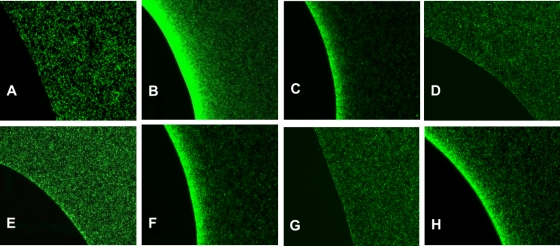
Chemotactic responses to l -serine and AI-2, demonstrated by the results of μPlug assays. The μPlug assay was carried out as described in the text and as previously reported (10, 19); the distribution of dead, RFP-labeled cells is not shown, but it was always uniform except when the dead cells were displaced around the plug by accumulating GFP-labeled motile cells. (A) Distribution of wild-type (CV1) cells in the absence of any attractant in the plug. (B) Distribution of wild-type (CV1) cells with 200 μM l -serine in the plug. (C) Distribution of wild-type (CV1) cells with 200 μM AI-2 in the plug. (D) Distribution of Δtsr (CV5) cells with 200 μM l -serine in the plug. (E) Distribution of Δtsr (CV5) cells with 200 μM AI-2 in the plug. (F) Distribution of lsrBΩKanr (MJ101) cells with 200 μM l -serine in the plug. (G) Distribution of lsrBΩKanr (MJ101) cells with 200 μM AI-2 in the plug. (H) Distribution of lsrCΩKanr (MJ102) cells with 200 μM AI-2 in the plug.
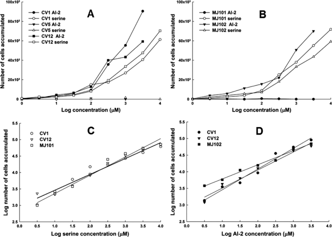
Responses of cells to l -serine and AI-2 in the capillary assay. Assays were performed at 32°C, essentially as described by Adler (1). Cells were resuspended in CB to a cell density of about 5 × 10⁷/ml. Assays were run twice, with three capillaries per run for each strain under each condition. Data shown are averages of six capillaries, and the standard deviations (not shown for purposes of clarity) ranged from 10 to 20%. The background accumulations in buffer-only capillaries were in the range of 500 to 1,000 cells. (A) Normalized values (with buffer-only control subtracted) of CV1 (wild-type) cells, CV5 (Δtsr) cells, and CV12 (Δtar-tap Δtrg) cells exposed to capillaries containing l -serine or AI-2. (B) Normalized values of MJ101 (lsrBΩKanr) cells and MJ102 (lsrCΩKanr) cells exposed to capillaries containing l -serine or AI-2. (C) Data for the responses of CV1, CV12, and MJ101 cells to l -serine, plotted on a log-log scale. The straight lines are linear regressions that can be extrapolated back to a threshold value. The extrapolated threshold concentrations, as predicted by Weber's law, are 1.7 × 10⁻¹² M for strain CV12 and 3.5 × 10⁻¹² M for strains CV1 and MJ101. The regression lines for CV12 and MJ101 were identical. (D) Data for the responses of CV1, CV12, and MJ102 cells to AI-2, plotted on a log-log scale. The linear regressions can be extrapolated back to a threshold value. The extrapolated threshold concentrations are 1.6 × 10⁻¹¹ for strain CV1, 4.6 × 10⁻¹² M for strain CV12, and 2.5 × 10⁻¹⁴ M for strain MJ102.
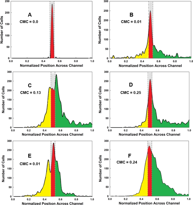
Assays of chemotactic behavior in the μFlow device. Cells were grown and prepared as for the μPlug assay, as described previously (9). The CMC was calculated according to the methods described in the text and elsewhere (10, 18). (A) Typical distribution of RFP-labeled dead cells, shown in red. The distribution of cells from one run is shown; it is typical for that found for RFP-labeled dead cells in all runs. The area occupied by dead cells is delineated by the gray bar enclosed in dashed lines. (B) Typical distribution of CV1 (wild-type) GFP-labeled cells in the absence of a chemoeffector gradient. The distribution of cells moving in the up-gradient direction beyond the “dead” zone is highlighted in green, and the distribution of cells moving in the down-gradient direction is highlighted in yellow. GFP-labeled cells remaining in the region occupied by dead cells (highlighted in red) were not included in the calculations of CMC values. (C) Typical distribution of CV1 cells in a 0-to-200 μM nonlinear gradient of l -serine. (D) Typical distribution of CV1 cells in a 0-to-200 μM nonlinear gradient of AI-2. (E) Typical distribution of CV1 cells in a 0-to-200 μM linear gradient of l -serine. (F) Typical distribution of CV1 cells in a 0-to-200 μM linear gradient of l -serine. All assays were run a minimum of three times. The CMC values obtained are indicated on each graph.
References
-
- Adler, J. 1973. A method for measuring chemotaxis and use of the method to determine optimum conditions for chemotaxis by Escherichia coli. J. Gen. Microbiol. 74:77-91. - PubMed
-
- Aksamit, R. R., and D. E. Koshland, Jr. 1974. Identification of the ribose binding protein as the receptor for ribose chemotaxis in Salmonella typhimurium. Biochemistry 13:4473-4478. - PubMed
-
- Bansal, T., P. Jesudhasan, S. Pillai, T. K. Wood, and A. Jayaraman. 2008. Temporal regulation of enterohemorrhagic Escherichia coli virulence mediated by autoinducer-2. Appl. Microbiol. Biotechnol. 78:811-819. - PubMed
-
- Bassler, B. L. 2002. Small talk: cell-to-cell communication in bacteria. Cell 109:421-424. - PubMed
Publication types
MeSH terms
Substances
Grants and funding
LinkOut - more resources
Full Text Sources
Molecular Biology Databases
