Review
. 2011 Mar;89(3):359-72.
doi: 10.1189/jlb.0910538. Epub 2010 Nov 19.

The paradox of the neutrophil's role in tissue injury

Affiliations
Review

The paradox of the neutrophil's role in tissue injury

George B Segel et al. J Leukoc Biol. 2011 Mar.

Abstract

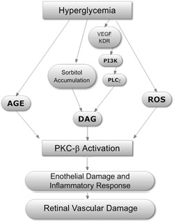
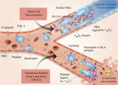
The neutrophil is an essential component of the innate immune system, and its function is vital to human life. Its production increases in response to virtually all forms of inflammation, and subsequently, it can accumulate in blood and tissue to varying degrees. Although its participation in the inflammatory response is often salutary by nature of its normal interaction with vascular endothelium and its capability to enter tissues and respond to chemotactic gradients and to phagocytize and kill microrganisms, it can contribute to processes that impair vascular integrity and blood flow. The mechanisms that the neutrophil uses to kill microorganisms also have the potential to injure normal tissue under special circumstances. Its paradoxical role in the pathophysiology of disease is particularly, but not exclusively, notable in seven circumstances: 1) diabetic retinopathy, 2) sickle cell disease, 3) TRALI, 4) ARDS, 5) renal microvasculopathy, 6) stroke, and 7) acute coronary artery syndrome. The activated neutrophil's capability to become adhesive to endothelium, to generate highly ROS, and to secrete proteases gives it the potential to induce local vascular and tissue injury. In this review, we summarize the evidence for its role as a mediator of tissue injury in these seven conditions, making it or its products potential therapeutic targets.


Figures

Figure 1
Figure 1
Metabolic alterations in diabetes mellitus, type II, leading to retinal vascular damage. The activation of isomers of PKC, particularly PKC‐β, appears central to the pathophysiological changes resulting from hyperglycemia, leading to the development of diabetic retinopathy [2, 4]. Although atypical PKCs also play a role in glucose metabolism, their role in the genesis of diabetes is unclear [3]. There are multiple pathways for the initiation of PKC‐β activity, examples of which are shown. Hyperglycemia results in the formation of AGE, which can lead to activation of PKC [2, 5]. Increased polyol flux and the accumulation of sorbitol, likewise, result in increased DAG production and PKC activation [2]. Heightened levels of growth factors, such as VEGF, are found in the eyes of patients with diabetic retinopathy, and VEGF binds to an endothelial membrane KDR, which activates PI3K [6]. The latter phosphorylates PLCγ‐generating DAG. Hyperglycemia‐induced generation of ROS, such as superoxide, also produces PKC activation. The activation of PKC‐β is key to the development of retinal vascular endothelial damage. Important in the pathological events and the development of microaneurisms is the concomitant induction of neutrophil adhesion, the release of cytokines and chemokines [4]. Inhibitors of PKC‐β, such as ruboxistaurin, prevent endothelial damage and loss of vision in diabetic patients. This figure is adapted from a figure in ref. [2].
Figure 2
Figure 2
A schematic representation of the major pathophysiological events in the development of sickle cell vaso‐occlusion and TRALI. In sickle cell vaso‐occlusion, there is adhesion of neutrophils to the endothelial cell surface, mediated by E‐selectin ligand‐1 (ESL‐1) on the neutrophil and E‐selectin on the endothelial cell surface. Sickle cells (SS) also adhere to the endothelium via ligands (e.g., CD36, CD47, α4β1) interacting with endothelial cell vascular cell adhesion molecule‐1, αVβ3, fibronectin, thrombospondin, and others, in some cases, mediated by von Willebrand factor. Adhesion of red cells and platelets to neutrophils occurs and may be mediated by αMβ2 neutrophil integrins. The red cell and platelet ligands are uncertain. This intercellular reaction involving red cells, neutrophils, and platelets (and possibly monocytes) results in vascular occlusion and tissue hypoxia [60]. In TRALI, antibodies present in transfusion products with specificity against HNAs (e.g., HNA‐1a, ‐2a, or ‐3a, HLA‐A2) react with their requisite antigen and initiate neutrophil activation. A two‐event model is proposed for the pathophysiology of TRALI, in which the first is the underlying inflammatory disease, inducing activation of the vascular endothelium of the lung and consequent neutrophil sequestration. The second event is the antibody antigen‐induced activation of neutrophils, which adhere to E‐selectin on the endothelial surface via their surface E‐selectin ligand‐1, similar to the process in sickle cell vaso‐occlusion. Platelets bind to neutrophil αMβ2 by an as‐yet unknown, complementary molecule. The resultant production of ROS in conjunction with inflammatory cytokines and chemokines leads to capillary permeability and fluid leak and the clinical syndrome. The inflammatory cytokines are released from the endothelial and inflammatory cells, and this contributes to the vascular effects. TRALI, induced by HNA‐3a, is particularly severe, and this antigen is encoded by the gene for the choline transporter‐like protein‐2 (SLC44A2), in which a single nucleotide polymorphism substitutes arginine for glutamine at position 154 [61, 62]. An animal model of TRALI has been used to identify a possible alternative mechanism of the disease involving the transfusion of an antibody to MHC I antigen on endothelial cells in the lung and activation of neutrophils via their FcγR [63]. Antineutrophil antibodies are, however, the principal pathogenetic mechanism in humans [64]. SS, homozygous sickle hemoglobin. This figure is adapted from a figure in ref. [63].

References

    1. Silliman, C. C. , McLaughlin, N. J. (2006) Transfusion‐related acute lung injury. Blood Rev. 20, 139–159. - PubMed
    1. Setter, S. M. , Campbell, R. K. , Cahoon, C. J. (2003) Biochemical pathways for microvascular complications of diabetes mellitus. Ann. Pharmacother. 37, 1858–1866. - PubMed
    1. Liu, X. J. , He, A. B. , Chang, Y. S. , Fang, F. D. (2006) Atypical protein kinase C in glucose metabolism. Cell. Signal. 18, 2071–2076. - PubMed
    1. Das Evcimen, N. , King, G. L. (2007) The role of protein kinase C activation and the vascular complications of diabetes. Pharmacol. Res. 55, 498–510. - PubMed
    1. Bloomgarden, Z. T. (2002) The epidemiology of complications. Diabetes Care 25, 924–932. - PubMed