. 2011 Jan 28;286(4):2843-52.
doi: 10.1074/jbc.M110.125526. Epub 2010 Nov 20.

Oxidative inactivation of the lipid phosphatase phosphatase and tensin homolog on chromosome ten (PTEN) as a novel mechanism of acquired long QT syndrome

Affiliations

Oxidative inactivation of the lipid phosphatase phosphatase and tensin homolog on chromosome ten (PTEN) as a novel mechanism of acquired long QT syndrome

Xiaoping Wan et al. J Biol Chem. .

Abstract

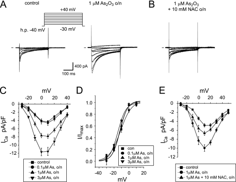
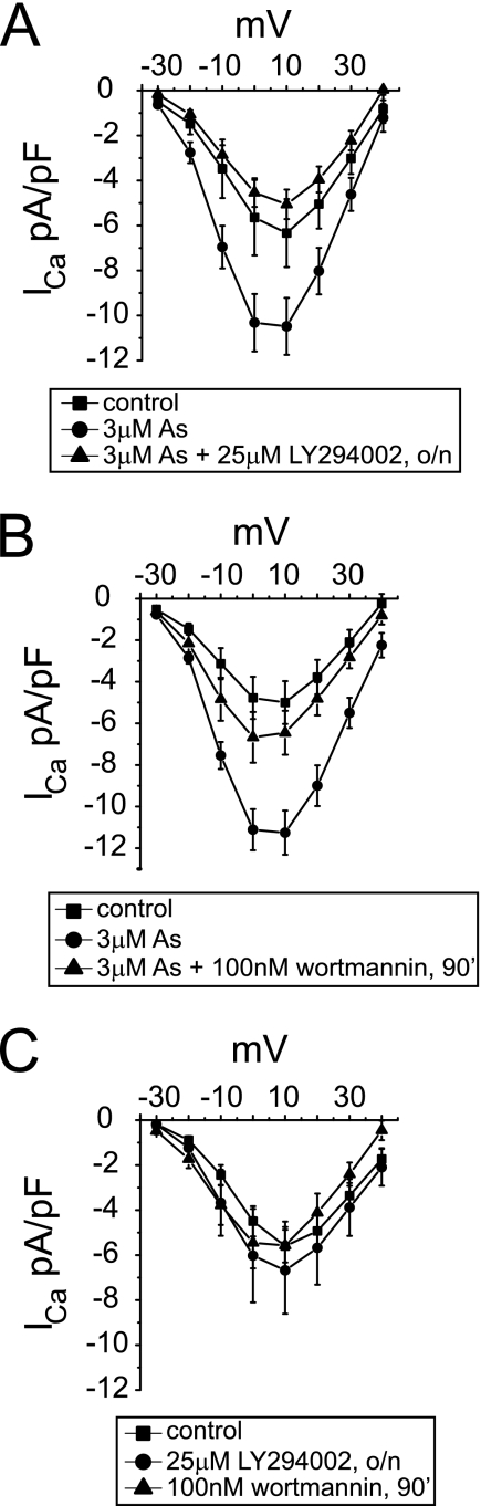
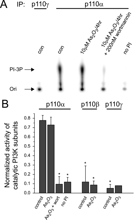
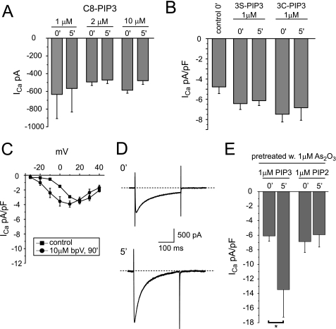
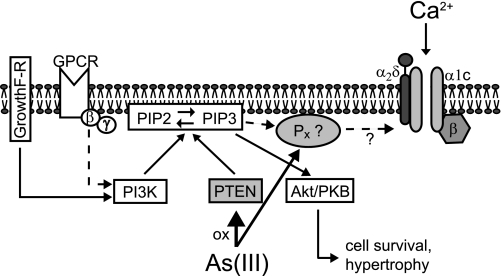
The most common cause of cardiac side effects of pharmaco-therapy is acquired long QT syndrome, which is characterized by abnormal cardiac repolarization and most often caused by direct blockade of the cardiac potassium channel human ether a-go-go-related gene (hERG). However, little is known about therapeutic compounds that target ion channels other than hERG. We have discovered that arsenic trioxide (As(2)O(3)), a very potent antineoplastic compound for the treatment of acute promyelocytic leukemia, is proarrhythmic via two separate mechanisms: a well characterized inhibition of hERG/I(Kr) trafficking and a poorly understood increase of cardiac calcium currents. We have analyzed the latter mechanism in the present study using biochemical and electrophysiological methods. We find that oxidative inactivation of the lipid phosphatase PTEN by As(2)O(3) enhances cardiac calcium currents in the therapeutic concentration range via a PI3Kα-dependent increase in phosphatidylinositol 3,4,5-triphosphate (PIP(3)) production. In guinea pig ventricular myocytes, even a modest reduction in PTEN activity is sufficient to increase cellular PIP(3) levels. Under control conditions, PIP(3) levels are kept low by PTEN and do not affect calcium current amplitudes. Based on pharmacological experiments and intracellular infusion of PIP(3), we propose that in guinea pig ventricular myocytes, PIP(3) regulates calcium currents independently of the protein kinase Akt along a pathway that includes a secondary oxidation-sensitive target. Overall, our report describes a novel form of acquired long QT syndrome where the target modified by As(2)O(3) is an intracellular signaling cascade.


Figures

FIGURE 1.
FIGURE 1.
As2O3 increases cardiac calcium currents. A, calcium current traces elicited using 300-ms step depolarizations in increments of 10 mV from −30 to +40 mV (h.p. = −40 mV) in guinea pig ventricular myocytes cultured overnight (24 h) under control conditions or in the presence of 1 μm As2O3. h.p., holding potential. B, typical calcium current traces elicited on co-incubation with 1 μm As2O3 and 10 mm NAC. C, averaged I-V relationships measured under control conditions and on overnight incubation with 0.1, 1, and 3 μm As2O3. At 0 mV, calcium current density was −4.6 ± 0.6 pA/picofarads under control condition, −7.9 ± 0.5 pA/picofarads on overnight incubation with 1 μm As2O3, and −11.6 ± 1 pA/picofarads on incubation with 3 μm As2O3 (n = 5–15). D, steady-state activation of calcium currents measured under control conditions and upon overnight incubation with 0.1, 1, and 3 μm As2O3. E, averaged I-V relationships measured under control conditions, in the presence of 1 μm As2O3, and upon co-incubation with 1 μm As2O3, 10 mm NAC (n = 5–8). o/n, overnight.
FIGURE 2.
FIGURE 2.
As2O3-induced increases in cardiac calcium currents are abolished by PI3K inhibitors. Calcium current traces were elicited as described in the legend to Fig. 1. A, averaged I-V relationships showing current densities measured under control conditions, upon overnight (o/n) incubation with 3 μm As2O3, and upon co-incubation with 3 μm As2O3 + 25 μm LY294002 overnight; B, averaged I-V relationships measured under control conditions, upon overnight incubation with 3 μm As2O3, and following a 90-min incubation with 100 nm wortmannin after overnight exposure to 3 μm As2O3; C, neither LY294002 (overnight) nor wortmannin (90 min) affects control calcium currents (n = 5–15). Data are expressed as mean ± S.E.
FIGURE 3.
FIGURE 3.
As2O3 does not increase cardiac PI3K activity. Freshly isolated guinea pig ventricular myocytes were incubated for 4 h under control conditions or in the presence of 10 μm As2O3. Catalytic PI3K subunits p110α, p110β, and p110γ were immunoprecipitated from whole cell lysates. PI3K activity of immunocomplexes was assayed as a function of PI-3P production. A, shown is a typical autoradiogram of 32P-labeled PI-3P produced on immunoprecipitation (IP) of p110γ or p100α and resolved by TLC. Ori, origin of TLC migration. Incubation with As2O3 does not alter PI-3P production, whereas co-incubation with 200 nm wortmannin inhibits PI-3P production. PI-3P production is not observed upon the omission of substrate from the assay reaction (no PI). Note that the basal activity of PI3K-γ/p110γ is very low under control conditions. B, quantitative analysis of PI3K/p110 activities. Shown is normalized PI-3P production under control conditions or in the presence of 10 μm As2O3 for PI-3K/p110α, -β, and -γ. PI-3K/p110α activity is significantly inhibited by 200 nm wortmannin. Assays without the addition of PI were used as negative controls (n = 1–5). *, significant difference from PI-3K/p100α activity measured under control conditions (Dunnett's test, p < 0.05). Error bars, S.E.
FIGURE 4.
FIGURE 4.
A, Western blot showing PTEN expressed under control conditions (con) in guinea pig ventricular myocytes (gp-m) and following incubation with 10 μm As2O3 for 4 h. Co-incubation of 10 μm As2O3 together with 10 mm NAC prevented oxidation of PTEN. PTEN was immunoprecipitated from whole cell lysates prior to detection on non-reducing SDS-PAGE. Arrowhead, position of oxidized PTEN. B, quantitative analysis of PTEN oxidation in cardiomyocytes on exposure to As2O3 (n = 3–4). As2O3 increases the oxidized form of PTEN significantly (Dunnett's test, p < 0.05). C, detection of oxidized PTEN by phosphatase assay. Cardiomyocytes were incubated for 4 h either under control conditions or in the presence of 10 μm As2O3 and lysed in the presence of iodoacetamide. PTEN was immunoprecipitated and assayed for phosphatase activity under reducing conditions. Measurements are expressed as the amount of phosphate released from PIP3 by PTEN. Note, that phosphate release is different between control- and As2O3-treated myocytes at the p < 0.05 level. D, detection of total PTEN activity by phosphatase assay. Cardiomyocytes were incubated for 4 h either under control conditions or in the presence of 10 μm As2O3 and lysed under reducing conditions. Importantly, total phosphate release was not different between control and As2O3-treated myocytes. IP, immunoprecipitation; WB, Western blot. Error bars, S.E.
FIGURE 5.
FIGURE 5.
As2O3-induced calcium current increases are not blocked by Akt inhibitors. A, Western blot (WB) of whole cell lysates showing total Akt and phospho-Akt Ser473 (p-Akt) in guinea pig ventricular myocytes under control conditions (con), following incubation with 10 μm As2O3 for 4 h, and following co-incubation with 10 μm As2O3 and 5 μm Akt inhibitor VIII for 4 h. Note that As2O3 exposure initiates Akt phosphorylation/activation, which can be completely blocked by Akt inhibitor VIII. B, cardiac calcium current traces in B–D were elicited in guinea pig ventricular myocytes as described in Fig. 1. Averaged I-V relationships show that 5 μm Akt inhibitor VIII does not inhibit As2O3-induced calcium current increases. AktVIII was applied for 90 min upon overnight incubation of cardiomyocytes with 3 μm As2O3. Note that control currents are not affected by AktVIII. C, averaged I-V relationships showing that 10 μm SH-6 does not inhibit As2O3-induced calcium current increases. Cardiomyocytes were co-incubated overnight with both 10 μm SH-6 and 10 μm As2O3. D, a 30 μm concentration of the inhibitory peptide TAT-AktVII does not affect As2O3-induced calcium current increases. The inhibitory peptide was applied with the extracellular perfusate for 90 min upon overnight incubation of cardiomyocytes with 3 μm As2O3 (n = 5–8). Error bars, S.E.
FIGURE 6.
FIGURE 6.
As2O3-induced increases in cardiac calcium currents are blocked by PKC inhibitors. Calcium current traces were elicited in guinea pig ventricular myocytes as described in the legend to Fig. 1. A, averaged I-V relationships showing partial inhibition of As2O3-induced calcium current increases by 30 nm Goe6976. Goe6976 was applied for 90 min following overnight incubation of cardiomyocytes with 3 μm As2O3. Note that control currents are not affected by 30 nm Goe6976. B, averaged I-V relationships showing inhibition of As2O3-induced calcium current increases by 300 nm Goe6976 applied for 90 min. C, averaged I-V relationships showing complete inhibition of As2O3-induced calcium current increases by 300 nm BisI applied for 90 min (n = 5–23). D, As2O3-induced calcium current increases are not attenuated by incubation with a 10 μm concentration of the PKA inhibitor H89 applied for 90 min; averaged I-V relationships (n = 5). Error bars, S.E.
FIGURE 7.
FIGURE 7.
Changes in calcium current density on internal perfusion with PIP3. A, maximal calcium current amplitudes measured in guinea pig ventricular myocytes on internal perfusion with 1, 2, or 10 μm C8-PIP3 immediately (0′) and 5 min (5′) after gaining whole cell access (n = 3–4). B, maximal calcium current densities measured in cardiomyocytes on internal perfusion with a 1 μm concentration of the metabolically stabilized PIP3 derivative C8-PIP3 (3S-PIP3) or C8-PIP3 3-methylenephosphonate (3C-PIP3) immediately and 5 min after gaining whole cell access (n = 5–6). C, averaged I-V relationships measured under control conditions or following a 90-min incubation with a 10 μm concentration of the phosphatase inhibitor bpV. D, calcium current traces elicited in a cardiomyocyte with a depolarizing test pulse to +20 mV from a holding potential of −40 mV immediately and 5 min after the start of internal perfusion with 1 μm C8-PIP3. Calcium currents were recorded following overnight incubation with 1 μm As2O3. E, maximal current densities measured in cardiomyocytes on internal perfusion with 1 μm either PIP3 or PIP2 following overnight incubation with 1 μm As2O3 immediately and 5 min after gaining whole cell access (n = 5). Note that internal application of C8-PIP3 increases calcium current density significantly at the p < 0.05 level. Error bars, S.E.
FIGURE 8.
FIGURE 8.
Diagram of the proposed As2O3-modified signaling pathways. For a detailed description, see “Discussion.”

Similar articles

Cited by

References

    1. Shen Z. X., Shi Z. Z., Fang J., Gu B. W., Li J. M., Zhu Y. M., Shi J. Y., Zheng P. Z., Yan H., Liu Y. F., Chen Y., Shen Y., Wu W., Tang W., Waxman S., De Thé H., Wang Z. Y., Chen S. J., Chen Z. (2004) Proc. Natl. Acad. Sci. U.S.A. 101, 5328–5335 - PMC - PubMed
    1. Nasr R., Guillemin M. C., Ferhi O., Soilihi H., Peres L., Berthier C., Rousselot P., Robledo-Sarmiento M., Lallemand-Breitenbach V., Gourmel B., Vitoux D., Pandolfi P. P., Rochette-Egly C., Zhu J., de Thé H. (2008) Nat. Med. 14, 1333–1342 - PubMed
    1. Wang Z. Y., Chen Z. (2008) Blood 111, 2505–2515 - PubMed
    1. Platanias L. C. (2009) J. Biol. Chem. 284, 18583–18587 - PMC - PubMed
    1. Ohnishi K., Yoshida H., Shigeno K., Nakamura S., Fujisawa S., Naito K., Shinjo K., Fujita Y., Matsui H., Takeshita A., Sugiyama S., Satoh H., Terada H., Ohno R. (2000) Ann. Intern. Med. 133, 881–885 - PubMed

Publication types

MeSH terms