

Tricellulin is expressed in autotypic tight junctions of peripheral myelinating Schwann cells
- PMID: 21097846
- PMCID: PMC2989243
- DOI: 10.1369/jhc.2010.956326
Tricellulin is expressed in autotypic tight junctions of peripheral myelinating Schwann cells
Abstract
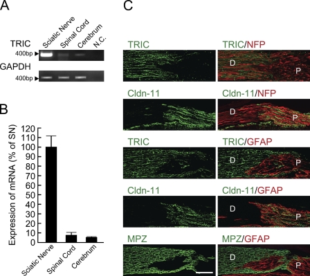
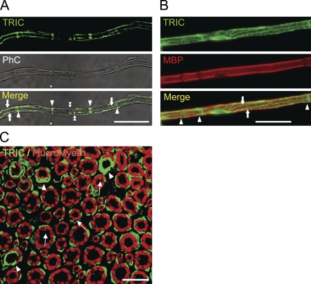
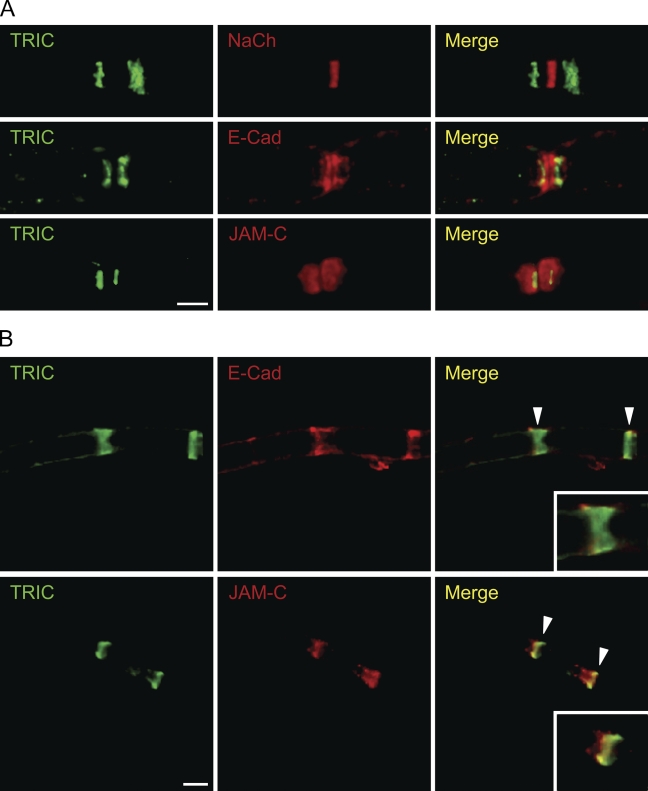
Autotypic tight junctions are formed by tight junction-like structures in three regions of myelinating Schwann cells, the paranodal loops, Schmidt-Lanterman incisures, and outer/inner mesaxons, and various tight junction molecules, including claudin-19 and junctional adhesion molecule (JAM)-C. Our findings demonstrate the identification and subcellular distribution of a novel tricellular tight junction protein, tricellulin (TRIC), in the autotypic tight junctions of mouse myelinating Schwann cells, compared with the autotypic adherens junction protein E-cadherin and the autotypic tight junction protein JAM-C, which are expressed in the paranodal loops, Schmidt-Lanterman incisures, and mesaxons. In real-time RT-PCR, the expression level of TRIC mRNA was about 10-fold higher in the sciatic nerve than in the spinal cord or cerebrum. In immunostaining, TRIC signals were completely restricted to the peripheral nervous system (PNS) and strongly concentrated at the paranodal loops, Schmidt-Lanterman incisures, and mesaxons of myelinating Schwann cells. In addition, TRIC was expressed in the thin region of the paranode and there was a gap between TRIC and the Na+ channel. Furthermore, TRIC was more distally located from the node than E-cadherin and was colocalized with JAM-C. It is possible that TRIC may be a component to maintain the integrity for PNS myelin function and morphology. This manuscript contains online supplemental material at http://www.jhc.org. Please visit this article online to view these materials.
Figures
Similar articles
-
Distinct claudins and associated PDZ proteins form different autotypic tight junctions in myelinating Schwann cells.J Cell Biol. 2002 Oct 28;159(2):361-72. doi: 10.1083/jcb.200207050. Epub 2002 Oct 28. J Cell Biol. 2002. PMID: 12403818 Free PMC article.
-
Tight junction proteins in human Schwann cell autotypic junctions.J Histochem Cytochem. 2009 Jun;57(6):523-9. doi: 10.1369/jhc.2009.951681. Epub 2009 Jan 19. J Histochem Cytochem. 2009. PMID: 19153196 Free PMC article.
-
Tight junction contact events and temporary gap junctions in the sciatic nerve fibres of the chicken during Wallerian degeneration and subsequent regeneration.J Neurocytol. 1982 Oct;11(5):839-58. doi: 10.1007/BF01153522. J Neurocytol. 1982. PMID: 7143029
-
[Cellular contacts in myelinated fibers of the peripheral nervous system].Med Sci (Paris). 2005 Feb;21(2):162-9. doi: 10.1051/medsci/2005212162. Med Sci (Paris). 2005. PMID: 15691487 Review. French.
-
Cellular junctions of myelinated nerves (Review).Mol Membr Biol. 2002 Apr-Jun;19(2):95-101. doi: 10.1080/09687680210130009. Mol Membr Biol. 2002. PMID: 12126235 Review.
Cited by
-
Barriers of the peripheral nerve.Tissue Barriers. 2013 Jul 1;1(3):e24956. doi: 10.4161/tisb.24956. Epub 2013 May 30. Tissue Barriers. 2013. PMID: 24665400 Free PMC article. Review.
-
Genetic deletion of Cadm4 results in myelin abnormalities resembling Charcot-Marie-Tooth neuropathy.J Neurosci. 2013 Jul 3;33(27):10950-61. doi: 10.1523/JNEUROSCI.0571-13.2013. J Neurosci. 2013. PMID: 23825401 Free PMC article.
-
Barrier function in the peripheral and central nervous system-a review.Pflugers Arch. 2017 Jan;469(1):123-134. doi: 10.1007/s00424-016-1920-8. Epub 2016 Dec 12. Pflugers Arch. 2017. PMID: 27957611 Review.
-
Tricellulin, α-Catenin and Microfibrillar-Associated Protein 5 Exhibit Concomitantly Altered Immunosignals along with Vascular, Extracellular and Cytoskeletal Elements after Experimental Focal Cerebral Ischemia.Int J Mol Sci. 2023 Jul 25;24(15):11893. doi: 10.3390/ijms241511893. Int J Mol Sci. 2023. PMID: 37569268 Free PMC article.
-
Molecular organization of tricellular tight junctions.Tissue Barriers. 2014 May 1;2:e28960. doi: 10.4161/tisb.28960. eCollection 2014. Tissue Barriers. 2014. PMID: 25097825 Free PMC article. Review.
References
-
- Ebnet K, Aurrand-Lions M, Kuhn A, Kiefer F, Butz S, Zander K, Meyer zu Brickwedde MK, et al. (2003) The junctional adhesion molecule (JAM) family members JAM-2 and JAM-3 associate with the cell polarity protein PAR-3: a possible role for JAMs in endothelial cell polarity. J Cell Sci 116:3879–3891 - PubMed
Publication types
MeSH terms
Substances
LinkOut - more resources
Full Text Sources
Molecular Biology Databases
Miscellaneous
