. 2011 Mar;39(6):2153-64.
doi: 10.1093/nar/gkq1139. Epub 2010 Nov 21.

Cooperation of RAD51 and RAD54 in regression of a model replication fork

Affiliations

Cooperation of RAD51 and RAD54 in regression of a model replication fork

Dmitry V Bugreev et al. Nucleic Acids Res. 2011 Mar.

Abstract

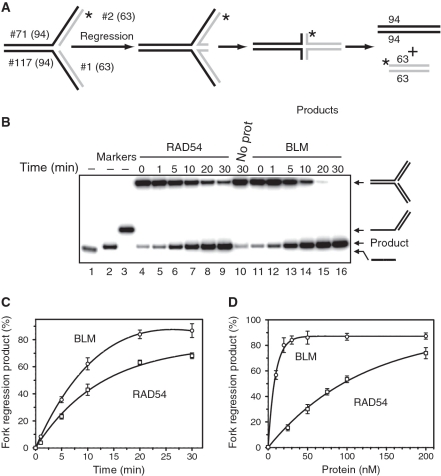
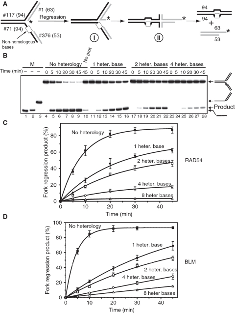
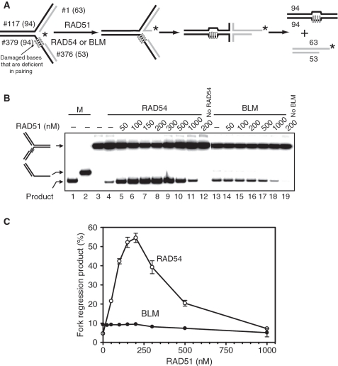
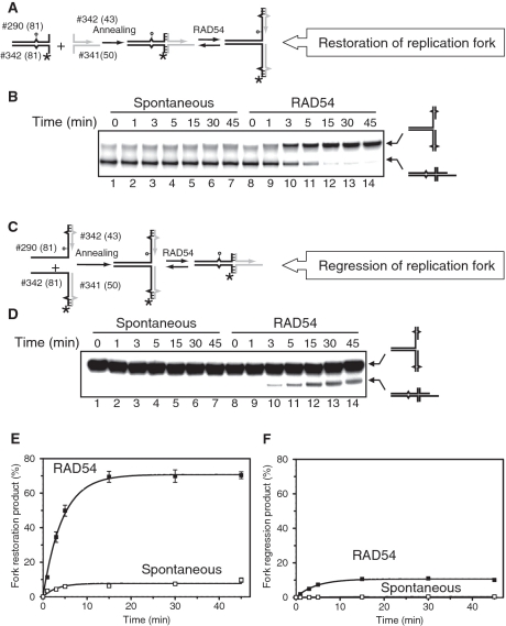
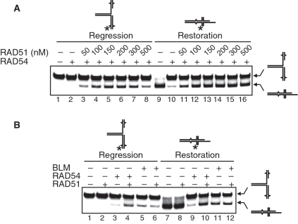
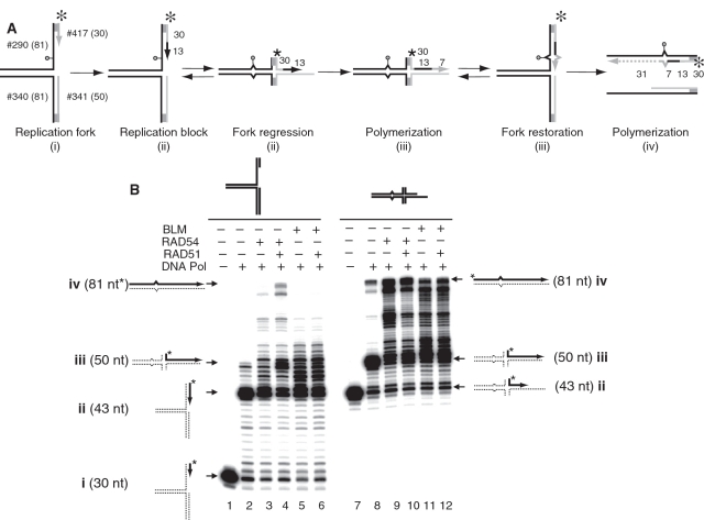
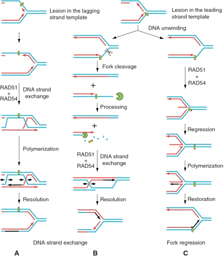
DNA lesions cause stalling of DNA replication forks, which can be lethal for the cell. Homologous recombination (HR) plays an important role in DNA lesion bypass. It is thought that Rad51, a key protein of HR, contributes to the DNA lesion bypass through its DNA strand invasion activity. Here, using model stalled replication forks we found that RAD51 and RAD54 by acting together can promote DNA lesion bypass in vitro through the 'template-strand switch' mechanism. This mechanism involves replication fork regression into a Holliday junction ('chicken foot structure'), DNA synthesis using the nascent lagging DNA strand as a template and fork restoration. Our results demonstrate that RAD54 can catalyze both regression and restoration of model replication forks through its branch migration activity, but shows strong bias toward fork restoration. We find that RAD51 modulates this reaction; by inhibiting fork restoration and stimulating fork regression it promotes accumulation of the chicken foot structure, which we show is essential for DNA lesion bypass by DNA polymerase in vitro. These results indicate that RAD51 in cooperation with RAD54 may have a new role in DNA lesion bypass that is distinct from DNA strand invasion.


Figures

Figure 1.
Figure 1.
Regression of model replication forks by RAD54 and BLM. (A) The experimental scheme. The asterisk marks the 32P-label. The numbers indicate the numbers and length (nt) of the individual strands in DNA substrates and products. (B) Analysis of fork regression by RAD54 and BLM by electrophoresis in an 8% polyacrylamide gel. Reactions were initiated by adding RAD54 (100 nM) (Lanes 4–9), BLM (10 nM) (Lanes 11–16), or storage buffer (Lane 10) to the DNA fork substrate (#71/2* + #117/1; see Supplementary Table S1). 32P-ssDNA (#2*) (Lane 1), 32P-dsDNA (#1/2*) and 32P-tailed DNA (#71/2*) (Lanes 1–3, respectively) were used as migration markers. (C) The kinetics of fork regression promoted by RAD54 or BLM presented as a graph. (D) The effect of RAD54 or BLM concentrations on the efficiency of fork regression. The reactions were initiated by addition of RAD54 or BLM (in the indicated concentrations) and were carried out for 15 min at 37°C. The spontaneous background was determined in identical reactions, except that RAD54 and BLM were replaced by their respective storage buffers, and was subtracted from the data. The experiments were repeated at least three times. The error bars indicate standard error of the mean (SEM).
Figure 2.
Figure 2.
RAD54 and BLM promote regression of replication forks containing heterologous bases. (A) The experimental scheme. Model replication forks contain regions of heterology of 1, 2, 4 or 8 bases (denoted by the white box) on the leading DNA strand template. The asterisk indicates the 32P-label. (B) Fork regression was initiated by addition of RAD54 (100 nM). The DNA products were analyzed by electrophoresis in an 8% polyacrylamide gel. Lanes 1–3 show DNA migration markers. Lane 10 shows the DNA products of spontaneous regression of a fully homologous replication fork in the absence of RAD54. Numbers of heterologous bases are indicated above the gel. (C) The kinetics of replication fork regression by RAD54 (100 nM) or (D) by BLM (50 nM) presented as a graph. The error bars indicate SEM.
Figure 3.
Figure 3.
RAD51 stimulates fork regression promoted by RAD54, but not by BLM. (A) The experimental scheme. The leading DNA strand template contains four heterologous bases. The asterisk indicates the 32P-label. (B) To promote fork regression RAD54 (100 nM) or BLM (50 nM) were incubated with the replication fork (#117/1* + #379/376) in the presence of RAD51 in indicated concentrations. Lanes 1 and 2 show DNA migration markers. In controls (Lanes 3, 4, 12, 13, 19), storage buffers were added instead of RAD51, RAD54, or BLM. The DNA products of the reaction were analyzed by electrophoresis in an 8% polyacrylamide gel. (C) The data from (B) are presented as graphs. The error bars indicate SEM.
Figure 4.
Figure 4.
RAD54 promotes restoration of replication forks more efficiently than fork regression. (A) The scheme of the chicken foot structure preparation and its regression to the replication fork. The circle indicates the position of iso-C that mimics a lesion on the leading DNA strand template. Unpaired single DNA bases are shown by carets. Hatched regions denote heterologous DNA terminal regions that prevent complete strand separation during fork regression. (B) RAD54 (100 nM) (Lanes 8–14) were incubated with the chicken foot structure (#341/342* + #340/290) (32 nM, molecules) for the indicated periods of time. In Lanes 1–7, RAD54 was replaced with storage buffer. The DNA products were analyzed by electrophoresis in an 8% polyacrylamide gel. (C) The scheme of the replication fork preparation and its regression to the chicken foot structure. (D) Spontaneous and Rad54-driven regression of replication forks. RAD54 (100 nM) (Lanes 8–14) were mixed with the replication fork structure (#290/342* + #340/341) (32 nM, molecules) and incubated for indicated periods of time. In Lane 1–7 RAD54 was replaced with storage buffer. Products of the reaction were analyzed by electrophoresis in an 8% polyacrylamide gel. (E and F) The data from (B) and (D) are presented as graphs. Background amounts of the DNA products (B and D, Lanes 1 and 8) were subtracted from the data before plotting. The error bars indicate SEM.
Figure 5.
Figure 5.
RAD51 stimulates RAD54-driven fork regression and inhibits fork restoration. (A) The effect of RAD51 concentration on a RAD54-dependent fork regression and restoration. RAD54 (100 nM) was mixed with the replication fork (#290/342* + #340/341) (32 nM, molecules) (Lanes 2–7) or the chicken foot structure (#341/342* + #340/290) (32 nM, molecules) (Lanes 10–15) in the presence of indicated concentrations of RAD51, or in the absence of RAD51 (Lanes 1 and 9). In the control reaction, the chicken fork structure was incubated in the absence of both RAD54 and RAD51 (Lane 8). (B) The effect of RAD54 (100 nM), RAD51 (200 nM) and BLM (50 nM) on the regression and restoration of the replication fork. Replication fork (#290/342* + #340/341) (32 nM, molecules) (Lanes 1–6) or chicken foot structure (#341/342* + #340/290) (32 nM, molecules) (Lanes 7–12) were mixed with indicated proteins. DNA products were analyzed by electrophoresis in an 8% polyacrylamide gel.
Figure 6.
Figure 6.
In vitro reconstitution of the DNA lesion bypass through the template switch mechanism. (A) The scheme of the lesion bypass via a mechanism that involves fork regression, DNA synthesis using the resulted chicken foot structure as a template, and fork restoration. The circle indicates the position of iso-C that mimics a DNA lesion at the stalled replication fork. The asterisk indicates the 32P-label. Shaded regions denote heterologous DNA terminal branches that prevent complete strand separation during fork regression. The numbers indicate the numbers and length (nt) of the DNA fragments in the substrates and the products of the reaction. (B) DNA polymerase reactions were carried out using DNA polymerase I Klenow Fragment (10 ng/ml) and the replication fork (#290/417* + #340/341) (32 nM, molecules) (left) or the chicken foot structure (#341/342* + #340/290) (32 nM, molecules) (right) as templates. RAD54 (100 nM), BLM (50 nM) and RAD51 (200 nM) were added to the reactions as indicated in ‘Materials and Methods’ section. The products of DNA synthesis were analyzed by electrophoresis in a 15% denaturing polyacrylamide gel.
Figure 7.
Figure 7.
Possible role of RAD54 and RAD51 proteins in DNA lesion bypass by replication forks. (A and B) Stalled replication forks can be rescued by HR via a mechanism that relies on DNA strand exchange activity of RAD51. RAD51 and auxiliary proteins including RAD54 promote pairing of either the daughter-strand gaps (A) or ssDNA tails produced as a result of fork cleavage (B) with undamaged chromatid. (C) Alternatively, stalled DNA replication can be restored through a mechanism that involves the DNA template strand switch. The important step of this mechanism involving fork regression and formation of the chicken foot structure may be carried out by the RAD51 and RAD54 proteins.

References

    1. Rothstein R, Michel B, Gangloff S. Replication fork pausing and recombination or ‘gimme a break’. Genes Dev. 2000;14:1–10. - PubMed
    1. Budzowska M, Kanaar R. Mechanisms of dealing with DNA damage-induced replication problems. Cell Biochem. Biophys. 2009;53:17–31. - PubMed
    1. Cox MM, Goodman MF, Kreuzer KN, Sherratt DJ, Sandler SJ, Marians KJ. The importance of repairing stalled replication forks. Nature. 2000;404:37–41. - PubMed
    1. Mirkin EV, Mirkin SM. Replication fork stalling at natural impediments. Microbiol. Mol. Biol. Rev. 2007;71:13–35. - PMC - PubMed
    1. Hoeijmakers JH. Genome maintenance mechanisms for preventing cancer. Nature. 2001;411:366–374. - PubMed

Publication types