. 2010 Nov 19;330(6007):1091-5.
doi: 10.1126/science.1197410.

Structure of the human dopamine D3 receptor in complex with a D2/D3 selective antagonist

Affiliations

Structure of the human dopamine D3 receptor in complex with a D2/D3 selective antagonist

Ellen Y T Chien et al. Science. .

Abstract

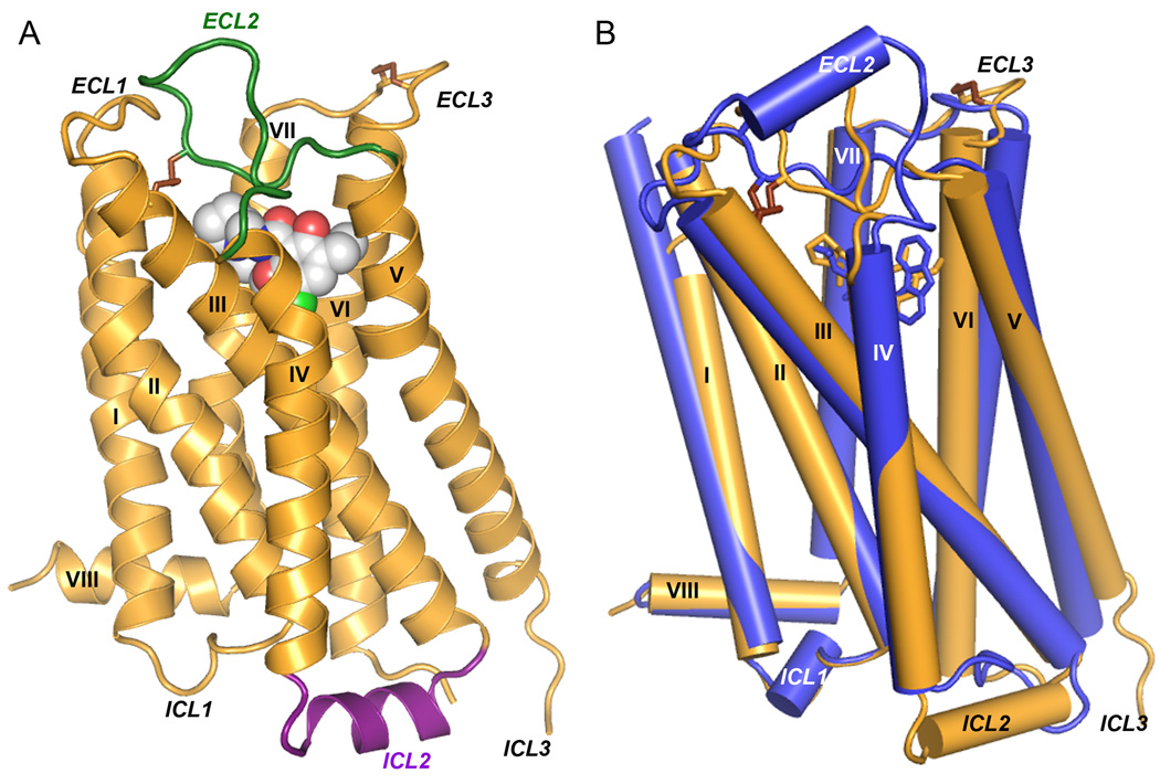
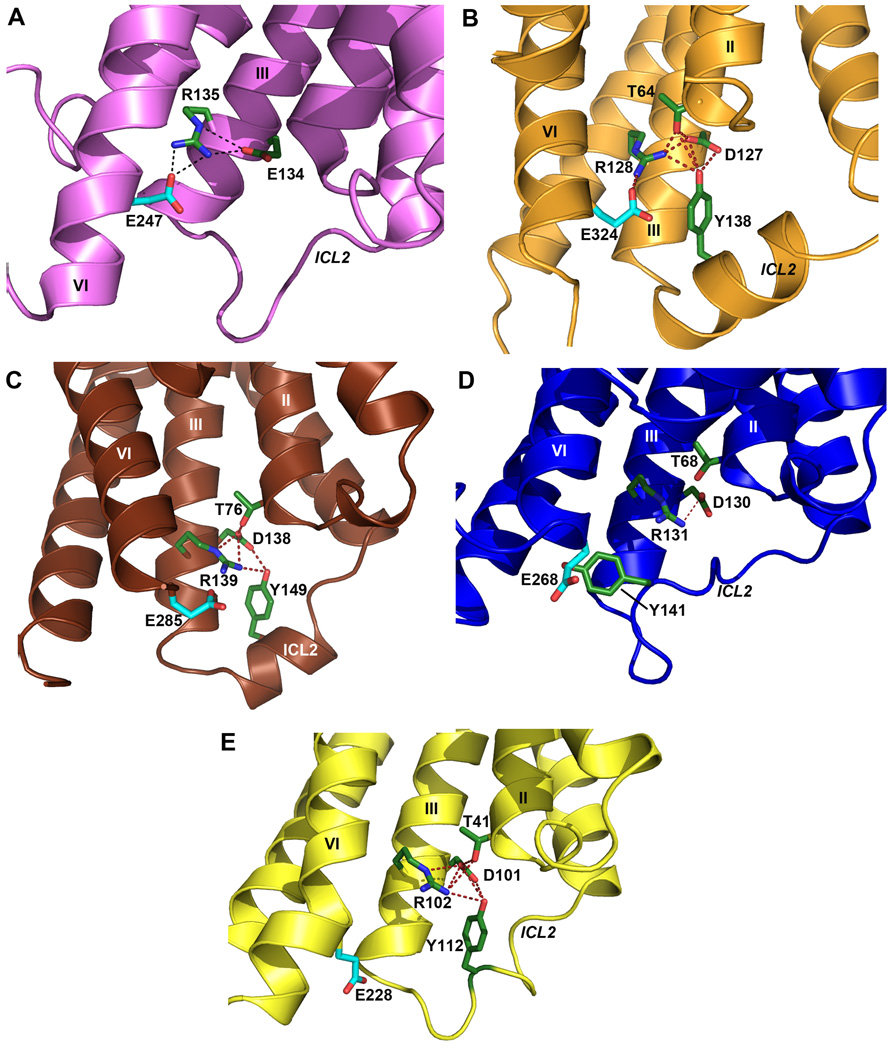
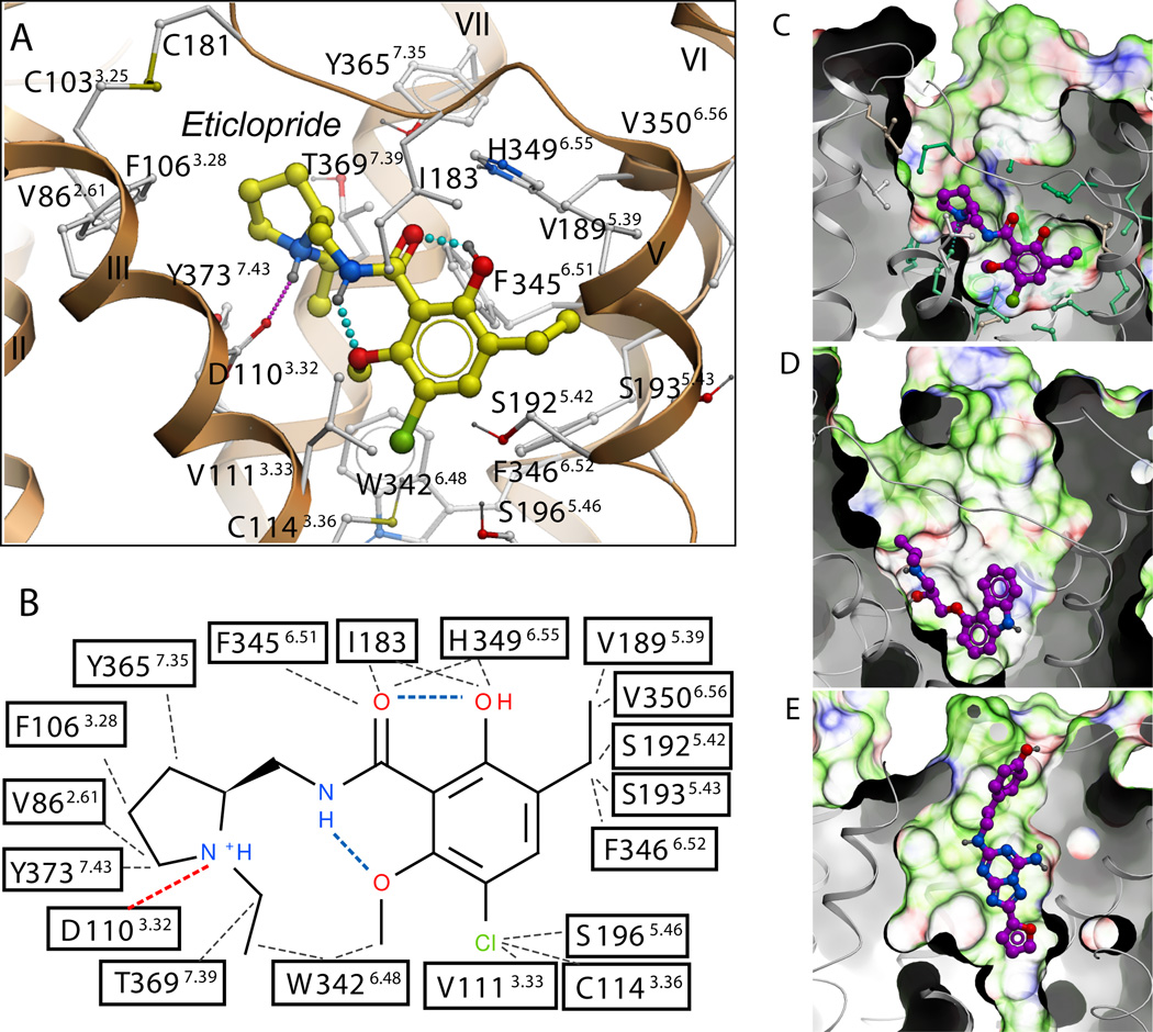
Dopamine modulates movement, cognition, and emotion through activation of dopamine G protein-coupled receptors in the brain. The crystal structure of the human dopamine D3 receptor (D3R) in complex with the small molecule D2R/D3R-specific antagonist eticlopride reveals important features of the ligand binding pocket and extracellular loops. On the intracellular side of the receptor, a locked conformation of the ionic lock and two distinctly different conformations of intracellular loop 2 are observed. Docking of R-22, a D3R-selective antagonist, reveals an extracellular extension of the eticlopride binding site that comprises a second binding pocket for the aryl amide of R-22, which differs between the highly homologous D2R and D3R. This difference provides direction to the design of D3R-selective agents for treating drug abuse and other neuropsychiatric indications.


Figures

Fig. 1
Fig. 1
Overall D3R structure with eticlopride and comparison with β2AR structure. (A) A model of the D3R with the bound ligand eticlopride in space-filling, ECL2 in green and ICL2 in purple (conformation of chain A shown). (B) Comparison of the transmembrane domains of D3R (brown) and β2AR (blue; PDB ID: 2RH1).
Fig. 2
Fig. 2
Conformation of ICL2 and ionic lock motif in D3R and other GPCR structures. As also seen in (A) the inactive Rhodopsin structure (PDB ID: 1U19), the conserved ionic lock motif D[E]RY is in a “locked” conformation in (B) the D3R structure, i.e. with a salt bridge formed between Arg1283.50 and Glu3246.30. In addition, the side chain of Tyr138 in the ICL2 α-helix of the D3R is inserted into the seven-TM bundle forming hydrogen bonds with Thr642.39, Arg1283.50 and Asp1273.49 (distances 3.0 Å, 3.2 Å and 3.2 Å, respectively), potentially stabilizing the ionic lock. There is no salt bridge between Arg3.50-Glu6.30 (and hence the “ionic lock” is “broken”) in other crystal structures of GPCR shown in panels (C) β1AR (PDB ID: 2VT4), (D) β2AR (PDB ID: 2RH1), (E) A2AAR (PDB ID: 3EML). In both the β1AR and A2AAR structures, however, the corresponding Tyr residue in ICL2 that aligns to Tyr138 in D3R forms two hydrogen bonds with the Asp3.49 and Arg3.50 side chains even in the absence of the closed ionic lock conformation. Salt bridges are shown as red dashed lines, and hydrogen bond interactions are shown as blue dashed lines.
Fig. 3
Fig. 3
Structural diversity of ligand binding sites in GPCR structures. (A) Close up of the eticlopride binding site showing the protein-ligand interaction. (B) Chemical structure of eticlopride and interactions with the D3R residues; hydrophobic contacts are colored in gray dots, hydrogen bonds in blue, and salt bridges in red. The ligand binding sites in (C) D3R, (D) β2AR (PDB ID: 2RH1), and (E) A2AAR (PDB ID: 3EML) crystal structures are shown in exactly the same orientation. A semi-transparent skin shows the molecular surface of the receptor, colored by the residue properties (green-hydrophobic, red-acidic, and blue-basic). Corresponding ligands, (C) eticlopride, (D) carazolol, and (E) ZM241385 are shown with carbon atoms colored magenta. For the D3R pocket, residues conserved between D3R and β2AR are colored turquoise and non-conserved are in gray.
Fig. 4
Fig. 4
The second binding pocket defined by R-22 is differentially modulated by non-conserved residues in D3R and D2R. (A) In addition to the core binding pocket, which essentially overlaps with that of eticlopride, the potential docking conformations of the core-constrained (see Supplementary information) D3R-selective compound R-22 position the extended aryl amide within a second binding pocket comprised by the junction of ECL1 and ECL2 and the interface of helices II, VII and I (dotted orange ellipse in A). (B) In the docking pose with the most extended conformation of R-22 (yellow), the ligand makes contact with several key conserved residues, including Asp1103.32, Tyr3737.43 and Glu902.65. The linker region of R-22 connecting the aryl amide and phenylpiperazine moieties (see fig. S1) is in a thinner representation. The 2,3-diCl-phenylpiperazine occupies essentially the same space as bound eticlopride (orange). (C–D) Close-up view of the interface of helices II, VII, and I of the D3R (C) and D2R (D) showing the results of molecular dynamics simulations indicating that the non-conserved regions of helix I and position 7.38 (orange) may orient key conserved contact residues differently and alter the shape of the second binding pocket, as reflected by the simulated distances between Glu902.65 and Tyr3737.43 in D3R (cyan) and D2R (magenta) (see fig. S7).

References

    1. Civelli O. In: Psychopharmacology : the fourth generation of progress. Bloom F, Kufper D, editors. NY: Raven press; 1995. pp. 155–161.
    1. Levant B. Pharmacol. Rev. 1997;49:231. - PubMed
    1. Sokoloff P, Giros B, Martres M, Bouthenet M, Schwartz J. Nature. 1990;347:146. - PubMed
    1. Shi L, Javitch JA. Annu. Rev. Pharmacol. Toxicol. 2002;42:437. - PubMed
    1. Sokoloff P, et al. Eur. J. Pharmacol. 1992;225:331. - PubMed

Publication types

Associated data