. 2011 Jan;107(1):89-99.
doi: 10.1093/aob/mcq221. Epub 2010 Nov 22.

Contrasting dynamics of radial O2-loss barrier induction and aerenchyma formation in rice roots of two lengths

Affiliations

Contrasting dynamics of radial O2-loss barrier induction and aerenchyma formation in rice roots of two lengths

Katsuhiro Shiono et al. Ann Bot. 2011 Jan.

Abstract

Background and aims: Many wetland species form aerenchyma and a barrier to radial O(2) loss (ROL) in roots. These features enhance internal O(2) diffusion to the root apex. Barrier formation in rice is induced by growth in stagnant solution, but knowledge of the dynamics of barrier induction and early anatomical changes was lacking.

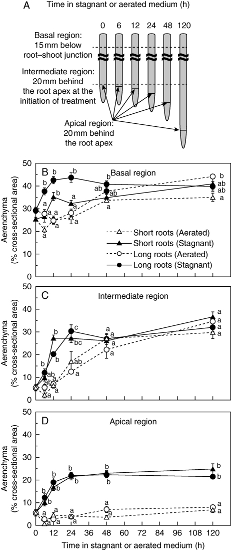
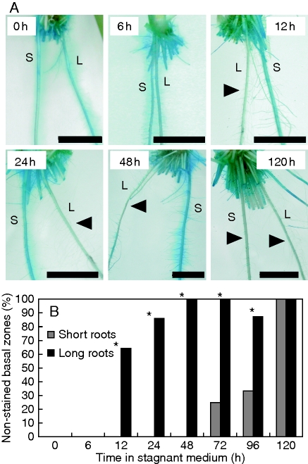
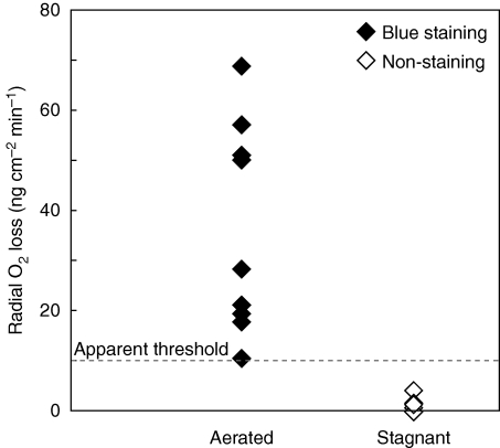
Methods: ROL barrier induction in short and long roots of rice (Oryza sativa L. 'Nipponbare') was assessed using cylindrical root-sleeving O(2) electrodes and methylene blue indicator dye for O(2) leakage. Aerenchyma formation was also monitored in root cross-sections. Microstructure of hypodermal/exodermal layers was observed by transmission electron microscopy (TEM).

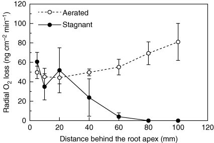
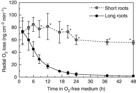
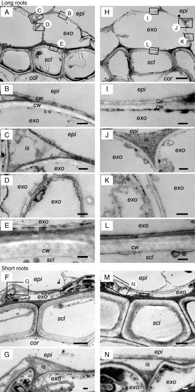
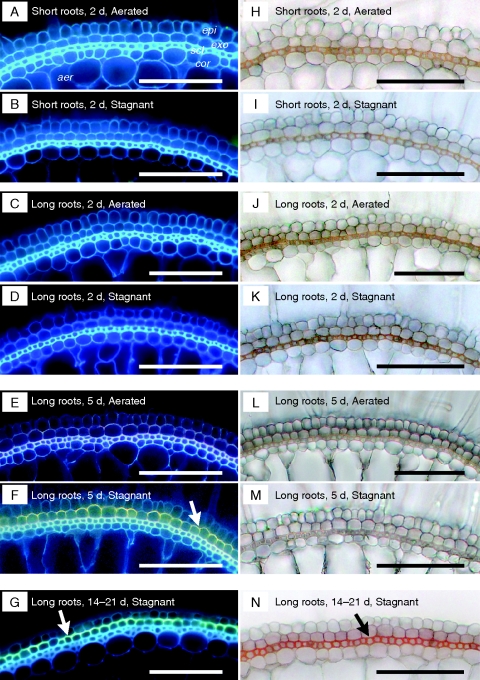
Key results: In stagnant medium, barrier to ROL formation commenced in long adventitious roots within a few hours and the barrier was well formed within 24 h. By contrast, barrier formation took longer than 48 h in short roots. The timing of enhancement of aerenchyma formation was the same in short and long roots. Comparison of ROL data and subsequent methylene blue staining determined the apparent ROL threshold for the dye method, and the dye method confirmed that barrier induction was faster for long roots than for short roots. Barrier formation might be related to deposition of new electron-dense materials in the cell walls at the peripheral side of the exodermis. Histochemical staining indicated suberin depositions were enhanced prior to increases in lignin.

Conclusions: As root length affected formation of the barrier to ROL, but not aerenchyma, these two acclimations are differentially regulated in roots of rice. Moreover, ROL barrier induction occurred before histochemically detectable changes in putative suberin and lignin deposits could be seen, whereas TEM showed deposition of new electron-dense materials in exodermal cell walls, so structural changes required for barrier functioning appear to be more subtle than previously described.


Figures

Fig. 1.
Fig. 1.
Rates of radial O2 loss along intact long adventitious roots of rice in O2-free medium with shoots in air, at 28 °C (mean ± s.e., n = 3). Plants were raised for 3–4 weeks in aerated nutrient solution, prior to transfer to stagnant deoxygenated agar nutrient solution for 5 d or aerated nutrient solution for 5 d. Lengths of adventitious roots (mean ± s.e., n = 3) were 154 ± 1 mm in aerated and 159 ± 3 mm in stagnant deoxygenated agar nutrient solution.
Fig. 2.
Fig. 2.
Time courses of rates of radial O2 loss at the basal part (15 mm below the root–shoot junction) of short and long adventitious roots following transfer into an O2-free medium with shoots in air, at 28 °C (short adventitious roots, n = 3; long adventitious roots, n = 4; mean ± s.e.). Plants were raised for 3–4 weeks in aerated nutrient solution, and then transferred for 48 h to O2-free stagnant 0·1 % agar solution that contained 5 mm KCl and 0·5 mm CaSO4. Short (65–90 mm) and long (105–130 mm) adventitious roots were used in experiments (lengths at commencement of treatment); after 48 h in the stagnant agar, root lengths were: short, 66–94 mm (growth rate 1·5 mm d−1); long, 110–131 mm (growth rate 3·4 mm d−1). *Significant difference between short and long adventitious roots (P < 0·05, two-sample t-test).
Fig. 3.
Fig. 3.
Comparison of the microstructure of the exodermis and the sclerenchyma in the basal part (15 mm below the root–shoot junction) of short or long adventitious roots grown continuously in aerated nutrient solution (short roots, F, G; long roots, A–E) or following transfer to stagnant deoxygenated agar nutrient solution for 48 h (short roots, M, N; long roots, H–L). Sections stained with uranyl acetate and lead citrate were observed by TEM. We observed the exodermis and sclerenchyma region (A, F, H, M; scale bar = 2 µm) and then specific cells at higher magnification (scale bar = 0·2 µm) for the epidermis side of the exodermis (B, C, G, I, J, N), between the exodermis cells (D, K), and also of the sclerenchyma side of the exodermis (E, L). Abbreviations: cor, cortex; cw, cell wall; epi, epidermis; exo, exodermis; is, intercellular space; scl, sclerenchyma. Plants were raised for 3–4 weeks in aerated nutrient solution, prior to transfer to stagnant deoxygenated agar nutrient solution for 48 h or aerated nutrient solution for 48 h. At the commencement of treatments the roots studied were short (65–90 mm) and long (105–130 mm) adventitious roots. Small boxes with letters in A and H and F and M indicate areas viewed at higher magnification and shown in the other panels of this figure.
Fig. 4.
Fig. 4.
Comparison of the development of suberin lamellae (A–G) and lignin deposition (H–N) of the exodermis and the sclerenchyma in the basal part (15 mm below the root–shoot junction) of short and long adventitious roots grown continuously in aerated nutrient solution [short roots, A, H; long roots: C, J (2 d), E, L (5 d)] or following transfer to stagnant deoxygenated agar nutrient solution for 2 d (short roots, B, I; long roots: D, K), 5 d (long roots: F, M) and 14–21 d (G, N). Suberin lamella is indicated by yellow–green fluorescence with Fluorol Yellow 088 (white arrow). Autofluorescence was detected as blue in the cell walls. Lignin with cinnamyl aldehyde groups stained orange/red with phloroglucinol-HCl (Black arrows). Abbreviations: aer, aerenchyma; cor, cortex; epi, epidermis; exo, exodermis; scl, sclerenchyma. Scale bars = 100 µm. Plants were raised for 3–4 weeks in aerated nutrient solution, prior to transfer to stagnant deoxygenated agar nutrient solution for 2 or 5 d, or aerated nutrient solution for 2 or 5 d. At the commencement of treatments the roots studied were short (65–90 mm) and long (105–130 mm) adventitious roots. For plants exposed to 14–21 d of treatments, seedlings were raised for 9 d in aerated nutrient solution, prior to transfer to stagnant deoxygenated agar nutrient solution for 14–21 d. Long roots in the continuous aerated controls (C, J and E, L) were of similar ages to those roots exposed to stagnant treatment for 14–21 d (G, N), so comparison of G, N can be made with C, J or E, L to see the influence of the 14–21 d stagnant treatment.
Fig. 5.
Fig. 5.
Aerenchyma at three positions along adventitious roots of rice, following transfer into aerated or stagnant deoxygenated agar nutrient solutions (A). Cross-sections at 15 mm below the root–shoot junction – termed ‘basal region’ (B), at 20 mm behind the root apex at the initiation of treatment – termed ‘intermediate region’ (C), and 20 mm behind the root apex at each time point – termed ‘apical region’ (D), were taken by hand with a razor blade and photographed under a microscope and the proportion of aerenchyma was determined. Plants were raised for 3–4 weeks in aerated nutrient solution, prior to transfer to stagnant deoxygenated agar nutrient solution for 0, 6, 12, 24, 48 or 120 h. Values are means ± s.e. Sample number and length of roots used in these measurements are given in Table 1. Different lower-case letters denote significant differences among root length classes and treatments (P < 0·05, one-way ANOVA and then Scheffé's test for multiple comparison).
Fig. 6.
Fig. 6.
Short (S) and long (L) adventitious roots of rice plants transferred into an O2-free agar solution with methylene blue. Methylene blue is colourless when reduced and blue when oxidized. Arrow denotes the ‘blue halos’ adjacent to adventitious roots. Plants were raised for 3–4 weeks in aerated nutrient solution, prior to transfer to stagnant deoxygenated agar nutrient solution for 0, 6, 12, 24, 48 or 120 h (A). Scale bar = 10 mm. Percentage of roots with ‘blue halos’ along short and long adventitious roots (B). Lengths at the commencement of treatment were: short (65–90 mm) and long (105–130 mm) adventitious roots. Sample numbers and length of roots used in these measurements are given in Supplementary Data Table S1, available online. *Significant difference between short and long adventitious roots (P < 0·05, Fisher's exact test).
Fig. 7.
Fig. 7.
Rates of radial O2 loss, and assessment of ‘blue halos’ formed with methylene blue treatment, for adventitious roots of rice grown in aerated or stagnant deoxygenated agar nutrient solution. The basal region of root, 11–50 mm below the root–shoot junction (i.e. 30–60 mm behind the root apex), was measured for ROL and then observed for staining with methylene blue. Before measurements, plants were raised for 8 d in aerated nutrient solution and then grown in either aerated or stagnant deoxygenated nutrient solution for 14–21 d. ROL was measured using cylindrical root-sleeving O2 electrodes in an O2-free agar solution with shoots in air, and subsequently the measured adventitious roots were transferred into O2-free agar solution containing methylene blue. Methylene blue is colourless when reduced and blue when oxidized. Measurements and observations were taken at 28 °C using intact adventitious roots 57–107 mm in length (three plants, n = 6) grown in aerated nutrient solution and 60–81 mm in length (four plants, n = 9) grown in stagnant deoxygenated agar nutrient solution. Dashed line: apparent threshold of methylene blue staining for 30–60 min.

References

    1. Armstrong J, Armstrong W. Phragmites australis – A preliminary study of soil-oxidizing sites and internal gas transport pathways. New Phytologist. 1988;108:373–382.
    1. Armstrong J, Armstrong W. Rice and Phragmites: effects of organic acids on growth, root permeability, and radial oxygen loss to the rhizosphere. American Journal of Botany. 2001;88:1359–1370. - PubMed
    1. Armstrong J, Armstrong W. Rice: sulfide-induced barriers to root radial oxygen loss, Fe2+ and water uptake, and lateral root emergence. Annals of Botany. 2005;96:625–638. - PMC - PubMed
    1. Armstrong J, Armstrong W, Beckett PM. Phragmites australis – Venturi- and humidity-induced pressure flows enhance rhizome aeration and rhizosphere oxidation. New Phytologist. 1992;120:197–207.
    1. Armstrong W. Radial oxygen losses from intact rice roots as affected by distance from the apex, respiration and waterlogging. Physiologia Plantarum. 1971;25:192–197.

Publication types