Review
. 2011 Feb 1;589(Pt 3):461-79.
doi: 10.1113/jphysiol.2010.191965. Epub 2010 Nov 22.

Mechanisms of closed-state inactivation in voltage-gated ion channels

Affiliations
Review

Mechanisms of closed-state inactivation in voltage-gated ion channels

Robert Bähring et al. J Physiol. .

Abstract

Inactivation of voltage-gated ion channels is an intrinsic auto-regulatory process necessary to govern the occurrence and shape of action potentials and establish firing patterns in excitable tissues. Inactivation may occur from the open state (open-state inactivation, OSI) at strongly depolarized membrane potentials, or from pre-open closed states (closed-state inactivation, CSI) at hyperpolarized and modestly depolarized membrane potentials. Voltage-gated Na(+), K(+), Ca(2+) and non-selective cationic channels utilize both OSI and CSI. Whereas there are detailed mechanistic descriptions of OSI, much less is known about the molecular basis of CSI. Here, we review evidence for CSI in voltage-gated cationic channels (VGCCs) and recent findings that shed light on the molecular mechanisms of CSI in voltage-gated K(+) (Kv) channels. Particularly, complementary observations suggest that the S4 voltage sensor, the S4S5 linker and the main S6 activation gate are instrumental in the installment of CSI in Kv4 channels. According to this hypothesis, the voltage sensor may adopt a distinct conformation to drive CSI and, depending on the stability of the interactions between the voltage sensor and the pore domain, a closed-inactivated state results from rearrangements in the selectivity filter or failure of the activation gate to open. Kv4 channel CSI may efficiently exploit the dynamics of the subthreshold membrane potential to regulate spiking properties in excitable tissues.


Figures

Figure 1
Figure 1. Kinetic schemes of activation and inactivation gating in VGCCs
C, O and I represent closed, open and inactivated states, respectively. C0 is the resting state, C1–Cn−1 represents a set of partially activated states, and Cn is the opening-permissive state. In three hypothetical situations, VGCCs may inactivate either from the O state only (Scheme I, open-state inactivation, OSI), from the closed state Cn only (Scheme II, closed-state inactivation, CSI) or from both open and closed states (Scheme III, OSI and CSI). IO and In are open-inactivated and closed-inactivated states, respectively; and I0–In−1 represents a set of closed-inactivated states directly associated with inactivation-permissive closed states. Transitions between C states are assumed to be strongly voltage dependent (f(Vm) indicates voltage-dependent transitions). Therefore, they involve conformational changes of the channel's voltage sensors. In specific kinetic models, n may range between 3 and 7 (text). VGCCs have four voltage sensors, which upon voltage-dependent activation move sequentially and independently in their tetrameric or pseudo-tetrameric assemblies. By contrast, the opening step and inactivation transitions may carry little or no voltage dependence. To open the pore, a single activation gate undergoes a concerted conformational change following activation of four voltage sensors and additional pre-open rearrangements. Whereas inactivation is tightly coupled to voltage-dependent activation in Schemes I and II, Scheme III assumes that activation and inactivation are independent or weakly coupled.
Figure 2
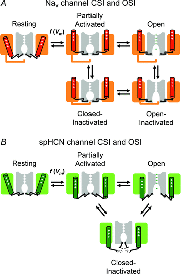
Figure 2. Cartoon representations of plausible mechanisms of inactivation gating in Nav channels (A) and spHCN channels (B)
Two opposing subunits (or pseudosubunits in Nav channels) of a fourfold symmetric channel are shown. Each subunit includes a voltage sensing domain (coloured) and a pore domain (grey). In the open conformation, ions are explicitly represented in the permeation pathway. As explained in Fig. 1, the activation steps are voltage dependent. Assuming a mechanical analogy, the voltage sensors and activation gate are shown ‘engaged’ in all conformations in the Nav channel. Inactivation of this channel occurs when an intracellular inactivation gate (‘hinged lid’) occludes the pore before or after opening of the activation gate. Note that the outward movement of the voltage sensor is necessary to allow interaction of the inactivation gate with its receptor site in the pore domain. By contrast, inactivation of spHCN channels may occur when the voltage sensors and activation gates ‘disengage’. Thus, the partially activated inactivation-permissive state may undergo inactivation, and ‘reclosure’ may cause inactivation from the open state.
Figure 6
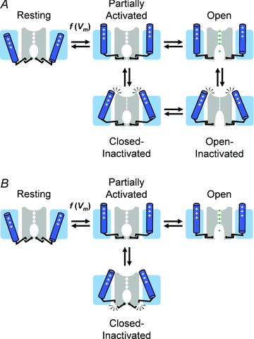
Figure 6. Conceptual cartoon representations of plausible general mechanisms of non-N-type inactivation gating in Kv channels
A, P/C-type mechanism of CSI and OSI. B, putative mechanism of CSI. Labelling and representations are as explained in Fig. 2. The voltage sensors play a central role in these mechanisms. Note that the P/C-type mechanism occurs when the channel's selectivity filter ‘collapses’ and this conformation is directly stabilized by the voltage sensors as they adopt a ‘relaxed’ conformation. Alternatively, the voltage sensors adopt a similar ‘relaxed’ conformation but the pore does not ‘collapse’ because there is no stable interaction between the voltage sensors and the selectivity filter. Instead, inactivation results when the voltage sensors and the activation gate ‘disengage’ as a result of a weak interaction (Fig. 2B). This mechanism can thus be seen as a ‘failure to open’ (Fig. 5).
Figure 5
Figure 5. Working structural model of slow inactivation in Kv4 channels
This hypothesis emerged from the studies by Dougherty et al. (2008) and Barghaan & Bähring (2009) (Figs 3 and 4). The simplified cartoons illustrating the significant states depict the relevant segments of two opposing channel subunits in the tetramer. Positively charged S4 voltage sensors are coloured grey; the S4S5 linkers are coloured green; and the S6 segments (bent at the PVPV motif) are coloured blue. Note that channel opening depends on maintaining physical contact between the S4S5 linkers and the distal portion of the S6 segments (post PVP). This contact is broken when the S4 segments slowly adopt an alternate ‘relaxed’ conformation. Consequently, the channel fails to open and becomes inactivated. Additional details can be found in the text.
Figure 3
Figure 3. A dynamic interaction between the S4S5 linker and the distal portion of the S6 segment underlies CSI in Kv4.2 channels (adapted fromBarghaan & Bähring, 2009)
Barghaan and Bähring used prepulse voltage protocols to asses CSI and thermodynamic double mutant cycle analysis to probe the combined role of the S4S5 linker and the S6 segment in Kv4.2 channel CSI. A, there is a significant energetic coupling (Ω= coupling coefficient) between residues S322 in the S4S5 linker and V404 in the S6 segment. B, there is also a similarly significant coupling between E323 and V404. Note that in such experiments the S6 residue S407 showed little or no coupling to the S4S5 linker residues examined. C, molecular model of the spatial relationships between the S4S5 linker and the distal portion of the S6 segment in Kv4.2 channels. Note a possible direct crosstalk between Glu 323 (E323) and Val 404 (V404). ©Barghaan & Bähring, 2009. Originally published in The Journal of General Physiology– doi:10.1085/jgp.200810073.
Figure 4
Figure 4. Slow gating charge immobilization precisely mirrors inactivation in Kv4.2 channels
A, simultaneous measurement of gating and ionic currents recorded under whole-cell patch-clamping conditions (adapted from Dougherty et al. 2008). The pulse protocol displayed below the current trace evoked the gating current (Ig) by stepping the voltage to the reversal potential (Er) of the ionic current, and the ionic Cs+ current (ICs) is subsequently observed by applying a large inward driving force (repolarization to −105 mV). In this experiment, VPP=−145 mV, a voltage insufficient to induce inactivation. Under these conditions, inactivation of gating and ionic currents is probed simultaneously by varying the duration and voltage of a conditioning prepulse (VPP) preceding the test pulse to the Er. B, the gating charge (Q=∫Ig) and ICs against the duration of VPP at −75 mV. Note, that the slow processes follow virtually identical trajectories; however, Q kinetics are biphasic (double exponential fit, continuous red line), indicating fast Q movement associated with voltage-dependent activation and a slow apparent Q-loss directly associated with inactivation. C, simulated gating current (top) evoked by a step depolarization from −150 to −75 mV and the corresponding Q kinetics (bottom). The shaded area under the gating current trace (i.e. integral, see above) is the corresponding Q. This simulation assumed a modified Zagotta–Hoshi–Aldrich model (Zagotta et al. 1994) as reported by Dougherty et al. (2008). The traces clearly show the fast observable component reflecting voltage-dependent activation (Q-on), and the slow undetectable component (Q-loss) reflecting apparent Q-immobilization precisely correlated with slow inactivation. Panels B and C are also adapted from Dougherty et al. (2008). ©Dougherty et al. 2008. Originally published in The Journal of General Physiology– doi:10.1085/jgp.200709938.

References

    1. Ahern CA, Eastwood AL, Dougherty DA, Horn R. An electrostatic interaction between TEA and an introduced pore aromatic drives spring-in-the-door inactivation in Shaker potassium channels. J Gen Physiol. 2009;134:461–469. - PMC - PubMed
    1. Aldrich RW. Inactivation of voltage-gated delayed potassium current in molluscan neurons. A kinetic model. Biophys J. 1981;36:519–532. - PMC - PubMed
    1. Aldrich RW. Fifty years of inactivation. Nature. 2001;411:643–644. - PubMed
    1. Aldrich RW, Stevens CF. Inactivation of open and closed sodium channels determined separately. Cold Spring Harb Symp Quant Biol. 1983;48:147–153. - PubMed
    1. Almers W, Armstrong CM. Survival of K+ permeability and gating currents in squid axons perfused with K+-free media. J Gen Physiol. 1980;75:61–78. - PMC - PubMed

Publication types

LinkOut - more resources