

Expression of a protein phosphatase 1 inhibitor, cdNIPP1, increases CDK9 threonine 186 phosphorylation and inhibits HIV-1 transcription
- PMID: 21098020
- PMCID: PMC3030381
- DOI: 10.1074/jbc.M110.196493
Expression of a protein phosphatase 1 inhibitor, cdNIPP1, increases CDK9 threonine 186 phosphorylation and inhibits HIV-1 transcription
Abstract
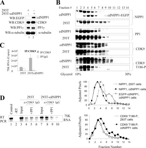
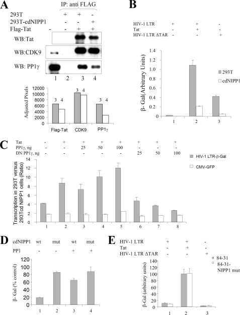
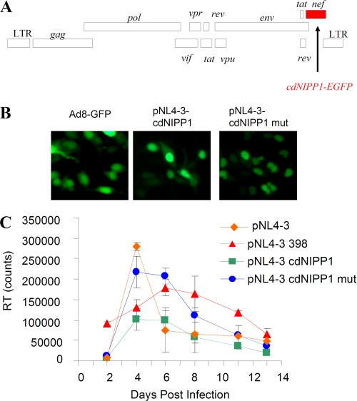
CDK9/cyclin T1, a key enzyme in HIV-1 transcription, is negatively regulated by 7SK RNA and the HEXIM1 protein. Dephosphorylation of CDK9 on Thr(186) by protein phosphatase 1 (PP1) in stress-induced cells or by protein phosphatase M1A in normally growing cells activates CDK9. Our previous studies showed that HIV-1 Tat protein binds to PP1 through the Tat Q(35)VCF(38) sequence, which is similar to the PP1-binding RVXF motif and that this interaction facilitates HIV-1 transcription. In the present study, we analyzed the effect of expression of the central domain of nuclear inhibitor of PP1 (cdNIPP1) in an engineered cell line and also when cdNIPP1 was expressed as part of HIV-1 pNL4-3 in place of nef. Stable expression of cdNIPP1 increased CDK9 phosphorylation on Thr(186) and the association of CDK9 with 7SK RNA. The stable expression of cdNIPP1 disrupted the interaction of Tat and PP1 and inhibited HIV-1 transcription. Expression of cdNIPP1 as a part of the HIV-1 genome inhibited HIV-1 replication. Our study provides a proof-of-concept for the future development of PP1-targeting compounds as inhibitors of HIV-1 replication.
Figures
Similar articles
-
Dephosphorylation of CDK9 by protein phosphatase 2A and protein phosphatase-1 in Tat-activated HIV-1 transcription.Retrovirology. 2005 Jul 27;2:47. doi: 10.1186/1742-4690-2-47. Retrovirology. 2005. PMID: 16048649 Free PMC article.
-
Small molecules targeted to a non-catalytic "RVxF" binding site of protein phosphatase-1 inhibit HIV-1.PLoS One. 2012;7(6):e39481. doi: 10.1371/journal.pone.0039481. Epub 2012 Jun 29. PLoS One. 2012. PMID: 22768081 Free PMC article.
-
Regulation of HIV-1 transcription by protein phosphatase 1.Curr HIV Res. 2007 Jan;5(1):3-9. doi: 10.2174/157016207779316279. Curr HIV Res. 2007. PMID: 17266553 Review.
-
Nuclear targeting of protein phosphatase-1 by HIV-1 Tat protein.J Biol Chem. 2005 Oct 28;280(43):36364-71. doi: 10.1074/jbc.M503673200. Epub 2005 Aug 29. J Biol Chem. 2005. PMID: 16131488
-
Regulation of CDK9 activity by phosphorylation and dephosphorylation.Biomed Res Int. 2014;2014:964964. doi: 10.1155/2014/964964. Epub 2014 Jan 12. Biomed Res Int. 2014. PMID: 24524087 Free PMC article. Review.
Cited by
-
Protein phosphatase-1 activates CDK9 by dephosphorylating Ser175.PLoS One. 2011 Apr 21;6(4):e18985. doi: 10.1371/journal.pone.0018985. PLoS One. 2011. PMID: 21533037 Free PMC article.
-
Cyclin-dependent kinase 7 (CDK7)-mediated phosphorylation of the CDK9 activation loop promotes P-TEFb assembly with Tat and proviral HIV reactivation.J Biol Chem. 2018 Jun 29;293(26):10009-10025. doi: 10.1074/jbc.RA117.001347. Epub 2018 May 9. J Biol Chem. 2018. PMID: 29743242 Free PMC article.
-
Increased iron export by ferroportin induces restriction of HIV-1 infection in sickle cell disease.Blood Adv. 2016 Dec 27;1(3):170-183. doi: 10.1182/bloodadvances.2016000745. Blood Adv. 2016. PMID: 28203649 Free PMC article.
-
Role of Divalent Cations in HIV-1 Replication and Pathogenicity.Viruses. 2020 Apr 21;12(4):471. doi: 10.3390/v12040471. Viruses. 2020. PMID: 32326317 Free PMC article. Review.
-
1E7-03, a low MW compound targeting host protein phosphatase-1, inhibits HIV-1 transcription.Br J Pharmacol. 2014 Nov;171(22):5059-75. doi: 10.1111/bph.12863. Br J Pharmacol. 2014. PMID: 25073485 Free PMC article.
References
-
- Nekhai S., Jeang K. T. (2006) Future Microbiol. 1, 417–426 - PubMed
-
- Yang Z., Zhu Q., Luo K., Zhou Q. (2001) Nature 414, 317–322 - PubMed
-
- Nguyen V. T., Kiss T., Michels A. A., Bensaude O. (2001) Nature 414, 322–325 - PubMed
-
- Yik J. H., Chen R., Nishimura R., Jennings J. L., Link A. J., Zhou Q. (2003) Mol. Cell 12, 971–982 - PubMed
Publication types
MeSH terms
Substances
Grants and funding
LinkOut - more resources
Full Text Sources
Other Literature Sources
Molecular Biology Databases
Miscellaneous
