. 2011 Jan 21;286(3):2143-54.
doi: 10.1074/jbc.M110.192245. Epub 2010 Nov 22.

Membrane-impermeable cross-linking provides evidence for homophilic, isoform-specific binding of desmosomal cadherins in epithelial cells

Affiliations

Membrane-impermeable cross-linking provides evidence for homophilic, isoform-specific binding of desmosomal cadherins in epithelial cells

Zhuxiang Nie et al. J Biol Chem. .

Abstract

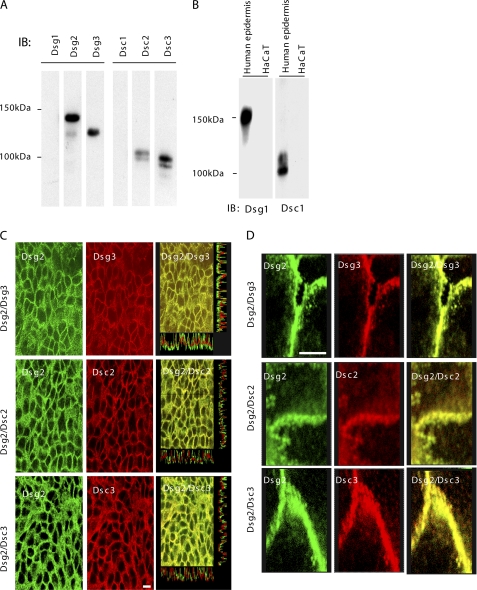
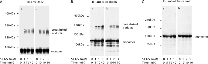
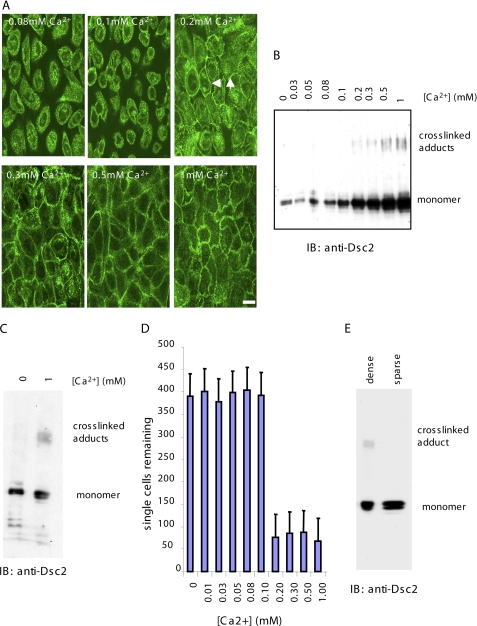
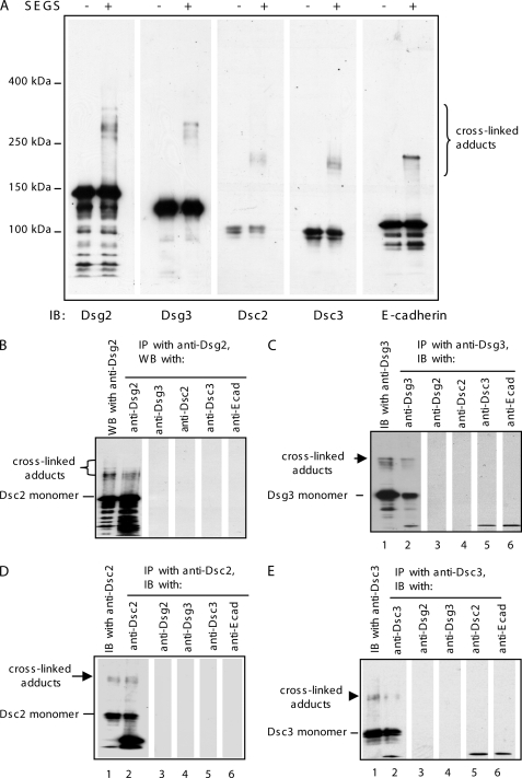
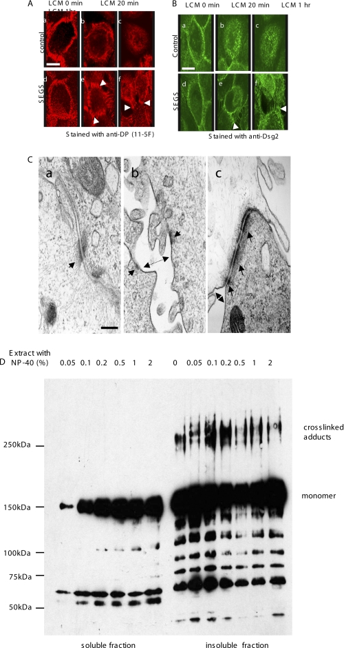
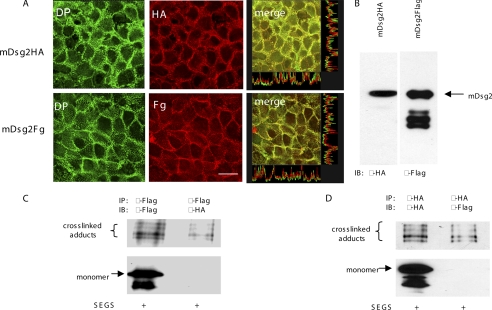
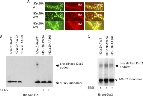
Desmosomes and adherens junctions are cadherin-based protein complexes responsible for cell-cell adhesion of epithelial cells. Type 1 cadherins of adherens junctions show specific homophilic adhesion that plays a major role in developmental tissue segregation. The desmosomal cadherins, desmocollin and desmoglein, occur as several different isoforms with overlapping expression in some tissues where different isoforms are located in the same desmosomes. Although adhesive binding of desmosomal cadherins has been investigated in a variety of ways, their interaction in desmosome-forming epithelial cells has not been studied. Here, using extracellular homobifunctional cross-linking, we provide evidence for homophilic and isoform-specific binding between the Dsc2, Dsc3, Dsg2, and Dsg3 isoforms in HaCaT keratinocytes and show that it represents trans interaction. Furthermore, the cross-linked adducts are present in the detergent-insoluble fraction, and electron microscopy shows that extracellular cross-linking probably occurs in desmosomes. We found no evidence for either heterophilic or cis interaction, but neither can be completely excluded by our data. Mutation of amino acid residues Trp-2 and Ala-80 that are important for trans interaction in classical cadherin adhesive binding abolished Dsc2 binding, indicating that these residues are also involved in desmosomal adhesion. These interactions of desmosomal cadherins may be of key importance for their ordered arrangement within desmosomes that we believe is essential for desmosomal adhesive strength and the maintenance of tissue integrity.


Figures

FIGURE 1.
FIGURE 1.
HaCaT cells express the Dsc2, Dsc3, Dsg2, and Dsg3 isoforms of desmosomal cadherins. A, extract of confluent HaCaT cells Western-blotted with isoform-specific desmosomal cadherin antibodies. The cells contain Dsg2, Dsg3, Dsc2, and Dsc3 but not Dsc1 and Dsg1. IB, immunoblot. B, positive controls for Dsc1 and Dsg1 antibodies on extract of human epidermis. C, desmosomal cadherins are co-localized on the cell surface. Confluent cells were double-stained with anti-Dsg2 and anti-Dsg3, anti-Dsg2 and anti-Dsc2, or anti-Dsg2 and anti-Dsc3 antibodies and examined by confocal microscopy. The merged images (right) demonstrate that Dsg2 is co-localized with each of the other isoforms. Scale bar, 10 μm. D, images are combined projections of a series of sequential slices and have been enlarged to show the precision of the co-localization. Scale bar, 5 μm.
FIGURE 2.
FIGURE 2.
Desmosomal cadherins of HaCaT cells can be cross-linked extracellularly with SEGS. A, confluent HaCaT cells were cross-linked with 1 mm SEGS for the times shown (panel a) or for 10 min at the SEGS concentrations shown (panel b), and whole cell extracts were then examined by Western blotting for Dsc2. The Dsc2a monomer had a mass of about 116 kDa. After cross-linking at all SEGS concentrations and all time points, cross-linked adducts of approximately double the size of the monomer were present. B, panels a and b, E-cadherin displayed a similar pattern under the same cross-linking conditions. C, cytoplasmic adherens junction protein α-catenin was not cross-linked under these conditions demonstrating that SEGS acts extracellularly. IB, immunoblot.
FIGURE 3.
FIGURE 3.
Desmosomal cadherin cross-linking occurs in a cell-cell adhesion-dependent manner. A, HaCaT cells were cultured for 24 h at the extracellular calcium concentrations indicated, and immunofluorescence was carried out for Dsc2. The threshold calcium concentration for desmosome assembly was 0.2 mm as indicated by the presence of Dsc2 staining at the cell periphery (white arrows). Scale bar, 15 μm. B, HaCaT cells were cultured at the same range of extracellular calcium concentrations as in A and cross-linked for 10 min with 1 mm SEGS, and cell extracts were examined by Western blotting for Dsc2. The threshold extracellular calcium concentration for appearance of cross-linked adducts was 0.2 mm, identical to that for desmosome assembly. C, no cross-linking was detected in cells cultured in calcium-free medium. Cells grown in the absence of calcium and in 1 mm calcium were cross-linked with 1 mm SEGS for 10 min. Western blotting for Dsc2 showed cross-linked adducts in the 1 mm extract only. D, dissociated HaCaT cells were aggregated in media containing the calcium concentrations shown, and the number of single cells remaining after 3 h was determined. As for desmosome assembly and cross-linking, the threshold calcium concentration for cells aggregation was 0.2 mm. E, no cross-linking was detectable in cells cultured in standard medium (1.8 mm calcium) at low density (>95% single cells) (sparse). IB, immunoblot.
FIGURE 4.
FIGURE 4.
Desmosomal cadherins bind homophilically and isoform-specifically. A, cross-linking produced slightly different adducts for each of the desmosomal cadherins and E-cadherin. Cell extracts from HaCaT cells with (+) or without (−) treatment with 1 mm SEGS for 10 min were blotted with desmosomal cadherin isoform-specific antibodies and E-cadherin. The cross-linked adducts were approximately double the size of the monomers and were slightly different in molecular weight in each case suggesting that they may correspond to homophilic dimers. B–E, cross-linked adducts contain only one desmosomal cadherin. Extracts of cross-linked HaCaT cells (1 mm SEGS for 10 min) were immunoprecipitated (IP) with each of the specific antibodies for the expressed desmosomal cadherins, and then equal amounts of extract were Western-blotted (WB) for one of the desmosomal cadherins and E-cadherin. Lane 1 in each panel shows a Western blot of the cross-linked cell extract with the antibody used for IP. Lanes 2–6 show Western blots of the immunoprecipitated material with the antibodies indicated at the top of the lanes. In each case only the antibody used for IP detected the cross-linked adduct. IB, immunoblot.
FIGURE 5.
FIGURE 5.
Desmosomes are cross-linked by SEGS. A and B, confluent HaCaT were either untreated (control) or treated with 1 mm SEGS for 10 min and exposed to calcium-free medium for 1 h, and then immunofluorescence was carried out for desmoplakin (A) or Dsg2 (B). (standard medium). In untreated cells, the majority of staining was internalized after LCM exposure (A, panels a–c, and B, panels a–c), but after SEGS treatment, a substantial amount of staining remained in intercellular processes at the cell surface (A, panels d–f, and B, panels d–f) (arrowheads). The cross-linking effect was seen in 100% of cells. Scale bars, 10 μm. C, electron microscopy confirms that desmosomes are cross-linked by SEGS. Panel a, standard medium control showing typical desmosomes and apposed plasma membranes. Panel b, after 1 h of LCM treatment, plasma membranes (double arrow) and desmosomal halves (arrows) had separated. Panel c, after cross-linking with 1 mm SEGS for 10 min followed by LCM treatment for 1 h, plasma membranes had separated (double arrow) but desmosomes (arrows) remained intact and became grouped at the cell surface. Scale bar, 200 nm. D, cross-linked adducts are in the Nonidet P-40-insoluble fraction. Confluent cells were cross-linked with 1 mm SEGS for 10 min and extracted with 1% Nonidet P-40 for 20 min, and the soluble and insoluble fractions were loaded onto a gel so that the amount of Dsg2 monomer in each lane was approximately equal and then Western-blotted for Dsg2 with monoclonal antibody 33-3D. Cross-linked adducts appeared in the insoluble fraction only.
FIGURE 6.
FIGURE 6.
Cross-linked desmosomal cadherins contain trans-interacting molecules. A, mDsg2-HA and mDsg2-FLAG were incorporated into desmosomes. Confluent transfected cells were double-stained with anti-desmoplakin antibody, 11-5F, and anti-HA or anti-FLAG. Co-localization of desmoplakin (DP) with HA-tagged or FLAG-tagged mDsg2 was observed under the confocal microscope. Scale bar, 5 μm. B, whole cell extracts of HaCaT cells with stable expression of pC-mDsg2HA or pC-mDsg2FLAG were Western-blotted (equal loading) and showed approximately equal expression of HA-tagged mDsg2 (mDsg2-HA) or FLAG-tagged mDsg2 (mDsg2-Fg) in the clones selected for the experiment. C and D, detection of trans-interacting cross-linked adducts. Transfected cells were mixed in a ratio of 1:2 of the type to be immunoprecipitated to the type to be Western-blotted after IP and cultured at confluent density. The cultures were cross-linked (1 mm SEGS for 10 min), extracted, and immunoprecipitated with the appropriate anti-tag antibody. The IPs were Western-blotted with the antibody used for IP and with the antibody against the other tag. In each case, the IP antibody recognized both the monomer and the cross-linked adduct, whereas the other antibody recognized only the cross-linked adducts.
FIGURE 7.
FIGURE 7.
Mutation in either Trp-2 or Ala-80 abolishes cross-linking of hDsc2. A, hDsc2 mutants were incorporated into desmosomes. Confluent cells expressing wild type hDsc2-HA or mutants hDsc2HAW2A or hDsc2HAA80I were double-stained with anti-HA and anti-DP, 11-5F. Co-localization of overexpressed wild type hDsc2 or its mutants with desmoplakin was observed by confocal microscopy. Scale bars, 5 μm. B, Western blot shows that wild type hDsc2 was cross-linked after SEGS treatment but that the mutants, which were expressed in comparable amounts, were not. C, as a control, an identical copy of the blot shown in B was stained with anti-hDsc2 antibody, showing that the endogenous Dsc2 was cross-linked in the presence of both mutants.

Similar articles

Cited by

References

    1. Huber O. (2003) Cell. Mol. Life Sci. 60, 1872–1890 - PMC - PubMed
    1. Garrod D. R., Merritt A. J., Nie Z. (2002) Curr. Opin. Cell Biol. 14, 537–545 - PubMed
    1. Garrod D. R., Merritt A. J., Nie Z. (2002) Mol. Membr. Biol. 19, 81–94 - PubMed
    1. Kimura T. E., Merritt A. J., Garrod D. R. (2007) J. Invest. Dermatol. 127, 775–781 - PubMed
    1. Dusek R. L., Godsel L. M., Green K. J. (2007) J. Dermatol. Sci. 45, 7–21 - PubMed

Publication types

LinkOut - more resources