. 2011 Feb 4;286(5):3323-31.
doi: 10.1074/jbc.M110.193854. Epub 2010 Nov 22.

Allosteric modulation of Ras-GTP is linked to signal transduction through RAF kinase

Affiliations

Allosteric modulation of Ras-GTP is linked to signal transduction through RAF kinase

Greg Buhrman et al. J Biol Chem. .

Abstract

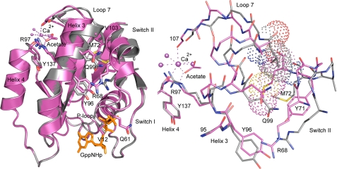
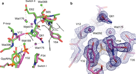
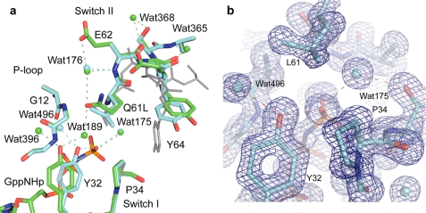
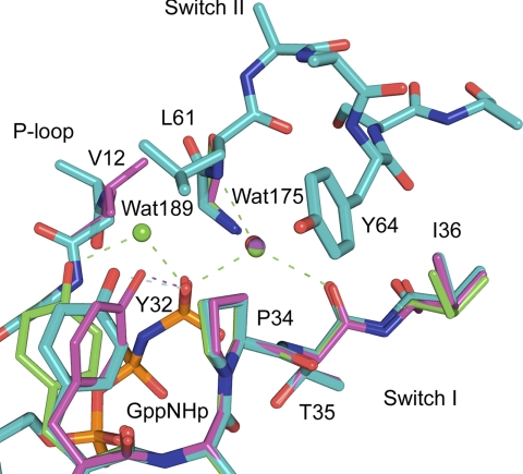
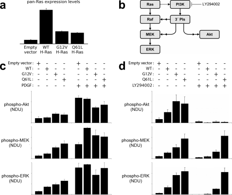
Ras is a key signal transduction protein in the cell. Mutants of Gly(12) and Gln(61) impair GTPase activity and are found prominently in cancers. In wild type Ras-GTP, an allosteric switch promotes disorder to order transition in switch II, placing Gln(61) in the active site. We show that the "on" and "off" conformations of the allosteric switch can also be attained in RasG12V and RasQ61L. Although both mutants have similarly impaired active sites in the on state, RasQ61L stabilizes an anti-catalytic conformation of switch II in the off state of the allosteric switch when bound to Raf. This translates into more potent activation of the MAPK pathway involving Ras, Raf kinase, MEK, and ERK (Ras/Raf/MEK/ERK) in cells transfected with RasQ61L relative to RasG12V. This differential is not observed in the Raf-independent pathway involving Ras, phosphoinositide 3-kinase (PI3K), and Akt (Ras/PI3K/Akt). Using a combination of structural analysis, hydrolysis rates, and experiments in NIH-3T3 cells, we link the allosteric switch to the control of signaling in the Ras/Raf/MEK/ERK pathway, supporting a GTPase-activating protein-independent model for duration of the Ras-Raf complex.


Figures

FIGURE 1.
FIGURE 1.
Superimposed structures of RasG12V with the allosteric switch in the on (magenta) and off (gray) conformations. Left panel, ribbon diagram of the structures showing the shift in Helix3/Loop7 and switch II. Arg68 and Arg97 are shown explicitly in stick form as is the calcium acetate bound in the allosteric site with coordinating water molecules and other key residues involved in activation of the allosteric switch. The GTP analog, GppNHp, is shown in orange. Right panel, interface between Helix3 and switch II with van der Waal's surfaces showing the clash that would occur between switch II residue Met72 in the on state (magenta) and the Loop7 residue Val103 in the off state (gray). The binding of calcium acetate shifts Helix3/Loop7, creating room for proper placement of switch II to complete the Ras active site. All figures in this article were created with PyMOL (Delano Scientific).
FIGURE 2.
FIGURE 2.
Active site of RasG12V in the on state. a, RasG12V (magenta) superimposed on the wild type protein (green) (PDB code 3K8Y). The off state in RasQ61L with the ordered switch II conformation (PDB code 2RGD) is shown in gray for comparison. Water molecules and dashed lines indicating H-bonds are colored as their respective protein models. b, 2FoFc electron density map contoured at 1.2 s shown for active site protein residues and water molecules within 6 Å of the O1γ atom of GppNHp.
FIGURE 3.
FIGURE 3.
Active site of RasQ61L in the on state. a, RasQ61L (cyan) superimposed on the wild type protein (green) (PDB code 3K8Y). The off state in RasQ61L with the ordered switch II conformation (PDB code 2RGD) is shown in gray for comparison. Water molecules and dashed lines indicating H-bonds are colored as their respective protein models. b, 2FoFc electron density map contoured at 1.2 s shown for active site protein residues and water molecules within 6 Å of the O1γ atom of GppNHp.
FIGURE 4.
FIGURE 4.
Allosteric switch in the off state in Ras. Superimposed active sites of RasG12V (magenta), RasQ61L (cyan) (PDB code 2RGD), and wild type Ras (green) (PDB code 2RGE) are shown. The nucleophilic and bridging water molecules are shown, WAT175 and WAT189, respectively. Water molecules and dashed lines indicating H-bonds are colored as their respective protein models.
FIGURE 5.
FIGURE 5.
Quantitative comparison of basal and growth factor-stimulated signal transduction in mouse fibroblasts expressing endogenous Ras or supplemented with expression of wild type, RasG12V, or RasQ61L variants. a, expression of wild type (WT) H-Ras or mutant H-Ras in NIH 3T3 fibroblasts was assessed by immunoblotting with pan-Ras antibodies, normalized by total ERK1/2 as a loading control (averages of two independent experiments). Control cells show endogenous Ras expression. b, ERK signaling network in mouse fibroblasts is composed of the canonical Ras-dependent pathway and a parallel PI3K-dependent pathway. The dotted arrow signifies that excessive levels of active Ras are required to mobilize PI3K signaling in these cells, and double arrows signify that additional signaling proteins, not depicted, are involved. c, phosphorylation of Akt1/2/3 (phospho-Akt), MEK1/2 (phospho-MEK), and ERK1/2 (phospho-ERK) in unstimulated and PDGF-BB-stimulated (1 nm, 15 min) NIH-3T3 fibroblasts were measured by quantitative immunoblotting alongside total Akt and total ERK as loading controls. The bar graphs show phosphorylation levels, expressed in normalized densitometry units (NDU) as described under “Experimental Procedures,” reported as mean ± S.E. (n = 4–5). d, same as c, except here the effect of PI3K inhibition (100 μm LY294002) on the unstimulated phosphorylation levels is assessed alongside controls with DMSO vehicle only (n = 3–6).

Similar articles

Cited by

References

    1. Cox A. D., Der C. J. (2003) Oncogene 22, 8999–9006 - PubMed
    1. McCubrey J. A., Steelman L. S., Chappell W. H., Abrams S. L., Wong E. W., Chang F., Lehmann B., Terrian D. M., Milella M., Tafuri A., Stivala F., Libra M., Basecke J., Evangelisti C., Martelli A. M., Franklin R. A. (2007) Biochim. Biophys. Acta 1773, 1263–1284 - PMC - PubMed
    1. Casey P. J., Solski P. A., Der C. J., Buss J. E. (1989) Proc. Natl. Acad. Sci. U.S.A. 86, 8323–8327 - PMC - PubMed
    1. Thapar R., Williams J. G., Campbell S. L. (2004) J. Mol. Biol. 343, 1391–1408 - PubMed
    1. Pacold M. E., Suire S., Perisic O., Lara-Gonzalez S., Davis C. T., Walker E. H., Hawkins P. T., Stephens L., Eccleston J. F., Williams R. L. (2000) Cell 103, 931–943 - PubMed

Publication types