. 2011 Feb 11;286(6):4107-16.
doi: 10.1074/jbc.M110.167502. Epub 2010 Nov 22.

Syk-dependent phosphorylation of CLEC-2: a novel mechanism of hem-immunoreceptor tyrosine-based activation motif signaling

Affiliations

Syk-dependent phosphorylation of CLEC-2: a novel mechanism of hem-immunoreceptor tyrosine-based activation motif signaling

Sonia Séverin et al. J Biol Chem. .

Abstract

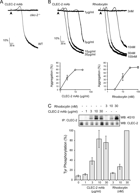
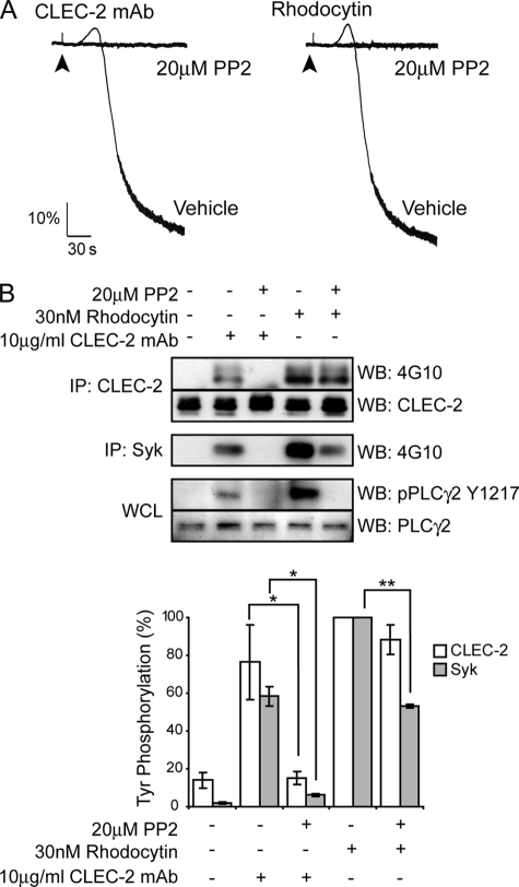
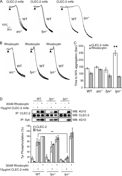
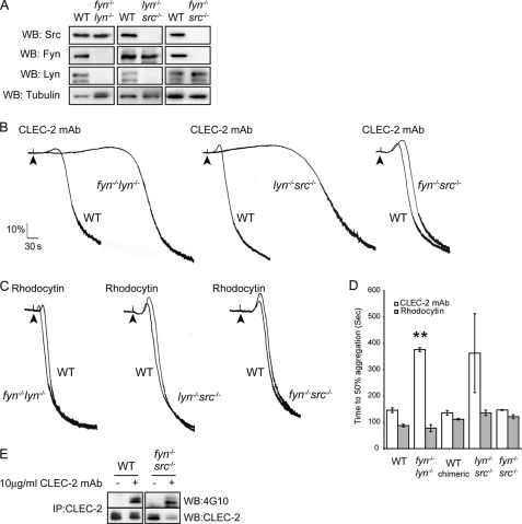
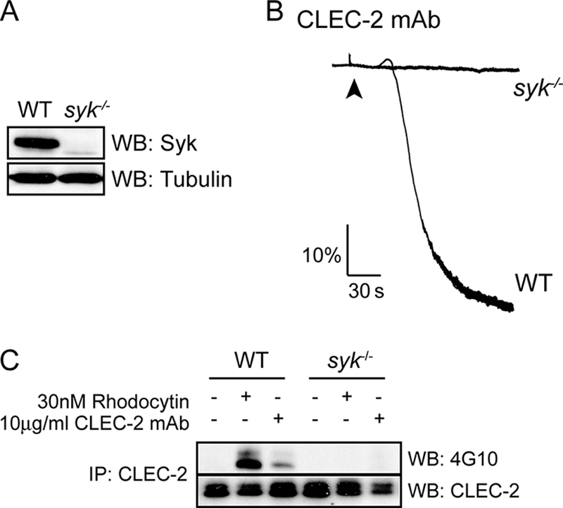
The C-type lectin-like receptor CLEC-2 signals via phosphorylation of a single cytoplasmic YXXL sequence known as a hem-immunoreceptor tyrosine-based activation motif (hemITAM). In this study, we show that phosphorylation of CLEC-2 by the snake toxin rhodocytin is abolished in the absence of the tyrosine kinase Syk but is not altered in the absence of the major platelet Src family kinases, Fyn, Lyn, and Src, or the tyrosine phosphatase CD148, which regulates the basal activity of Src family kinases. Further, phosphorylation of CLEC-2 by rhodocytin is not altered in the presence of the Src family kinase inhibitor PP2, even though PLCγ2 phosphorylation and platelet activation are abolished. A similar dependence of phosphorylation of CLEC-2 on Syk is also seen in response to stimulation by an IgG mAb to CLEC-2, although interestingly CLEC-2 phosphorylation is also reduced in the absence of Lyn. These results provide the first definitive evidence that Syk mediates phosphorylation of the CLEC-2 hemITAM receptor with Src family kinases playing a critical role further downstream through the regulation of Syk and other effector proteins, providing a new paradigm in signaling by YXXL-containing receptors.


Figures

FIGURE 1.
FIGURE 1.
Dose response curves in response to CLEC-2 mAb and rhodocytin. A, CLEC-2-deficient and litter-matched wild-type washed platelets (2 × 10⁸ platelets/ml) were stimulated with 10 μg/ml CLEC-2 mAb. B, murine washed platelets (2 × 10⁸ platelets/ml) were stimulated with increasing concentrations of CLEC-2 mAb or rhodocytin. Platelet aggregation was measured as a change in light transmission, using a lumi-aggregometer. Representative aggregation traces are shown. The addition of the agonist is indicated by an arrowhead. The data represent the means and standard error of at least eight independent experiments at 3 min of stimulation. C, CLEC-2 was immunoprecipitated (IP), and immunoprecipitates were immunoblotted with an anti-phosphotyrosine antibody and anti-CLEC-2 mAb as described under “Experimental Procedures.” The percentage of tyrosine phosphorylation was measured at 3 min of stimulation and is represented as the means and standard error of four independent experiments. WB, Western blotting.
FIGURE 2.
FIGURE 2.
Effect of Src family kinase inhibition on murine platelet in response to CLEC-2 mAb and rhodocytin. A, murine washed platelets (2 × 10⁸ platelets/ml) were incubated for 10 min with or without 20 μm PP2 and then stimulated with 10 μg/ml CLEC-2 mAb or 30 nm rhodocytin. Platelet aggregation was measured as a change in light transmission using a lumi-aggregometer. The addition of the agonist is indicated by an arrowhead. Representative aggregation traces from three to six independent experiments are shown. B, CLEC-2 and Syk were immunoprecipitated (IP), and immunoprecipitates were immunoblotted with an anti-phosphotyrosine antibody. CLEC-2 immunoprecipitates were also immunoblotted with anti-CLEC-2 mAb as described under “Experimental Procedures.” Whole cell lysates (WCL) were probed with pY1217 PLCγ2 antibody and reprobed with PLCγ2 antibodies. The percentage of tyrosine phosphorylation was measured at 3 min of stimulation and is represented as the means and standard error of three to six independent experiments. *, p < 0.05; **, p < 0.005 (significant difference according to two-tailed Student's t test). WB, Western blotting.
FIGURE 3.
FIGURE 3.
Lyn plays a critical role in CLEC-2-mediated signaling by antibody ligation but is not involved in response to rhodocytin. Fyn-, Lyn-, or Src-deficient washed platelets and their litter-matched wild-type platelets (2 × 10⁸ platelets/ml) were stimulated with 10 μg/ml CLEC-2 mAb (A) or 30 nm rhodocytin (B). Platelet aggregation was measured as a change in light transmission, using a lumi-aggregometer. Representative aggregation traces are shown. The addition of the agonist is indicated by an arrowhead. C, data represent the means of the time to get 50% of aggregation and standard error of three to six independent experiments. **, p < 0.005 (significant difference versus wild type, according to two-tailed Student's t test). D, Fyn- or Lyn-deficient washed platelets and their litter-matched wild-type platelets (2 × 10⁸ platelets/ml) were stimulated with 10 μg/ml CLEC-2 mAb or 30 nm rhodocytin for 3 min. CLEC-2 and Syk were immunoprecipitated (IP), and immunoprecipitates were immunoblotted with an anti-phosphotyrosine antibody. CLEC-2 immunoprecipitates were also immunoblotted with anti-CLEC-2 antibody as described under “Experimental Procedures.” The percentage of tyrosine phosphorylation was measured at 3 min of stimulation and represented as the means and standard error of three to six independent experiments. **, p < 0.005 (significant difference versus wild type, according to two-tailed Student's t test). WB, Western blotting.
FIGURE 4.
FIGURE 4.
A, whole cell lysates prepared from deficient platelets and their litter-matched wild-type platelets were immunoblotted with anti-Lyn, anti-Fyn, anti-Src, and anti-tubulin antibodies. B and C, washed platelets deficient in Fyn/Lyn, Fyn/Src, and Lyn/Src (2 × 10⁸ platelets/ml) were stimulated with 10 μg/ml CLEC-2 mAb (B) or 30 nm rhodocytin (C). Platelet aggregation was measured as a change in light transmission, using a lumi-aggregometer. Representative aggregation traces from three independent experiments are shown. The addition of the agonist is indicated by an arrowhead. D, data represent the means of the time to get 50% of aggregation and standard error of three independent experiments. **, p < 0.005 (significant difference versus wild type, according to two-tailed Student's t test). E, Fyn/Src-deficient washed platelets and their litter-matched wild-type platelets (2 × 10⁸ platelets/ml) were stimulated with 10 μg/ml CLEC-2 mAb for 3 min. CLEC-2 was immunoprecipitated (IP), and immunoprecipitates were immunoblotted with an anti-phosphotyrosine antibody. CLEC-2 immunoprecipitates were also immunoblotted with anti-CLEC-2 antibody as described under “Experimental Procedures.” Representative data from two independent experiments are shown. WB, Western blotting.
FIGURE 5.
FIGURE 5.
CD148 deficiency does not impair CLEC-2 signaling. A, CD148-deficient washed platelets and their litter-matched wild-type platelets (2 × 10⁸ platelets/ml) were stimulated with 10 μg/ml CLEC-2 mAb or 30 nm rhodocytin. Platelet aggregation was measured as a change in light transmission, using a lumi-aggregometer. Representative aggregation traces of four independent experiments are shown. The addition of the agonist is indicated by an arrowhead. B, CD148-deficient washed platelets and their litter-matched wild-type platelets (2 × 10⁸ platelets/ml) were stimulated with 10 μg/ml CLEC-2 mAb or 30 nm rhodocytin for 3 min. CLEC-2 and Syk were immunoprecipitated (IP), and immunoprecipitates were immunoblotted with an anti-phosphotyrosine antibody. CLEC-2 immunoprecipitates were also immunoblotted with anti-CLEC-2 mAb as described under “Experimental Procedures.” The percentage of tyrosine phosphorylation was measured at 3 min of stimulation and represented as means and standard error of four independent experiments. WB, Western blotting.
FIGURE 6.
FIGURE 6.
Syk is a key regulator of CLEC-2 signaling. A, whole cell lysates prepared from wild-type and Syk-deficient platelets were immunoblotted with anti-Syk and anti-tubulin antibodies. B, Syk-deficient washed platelets and their litter-matched control platelets (2 × 10⁸ platelets/ml) were stimulated with 10 μg/ml CLEC-2 mAb. Platelet aggregation was measured as a change in light transmission, using a lumi-aggregometer. Representative aggregation traces of three independent experiments are shown. The addition of the agonist is indicated by an arrowhead. C, after 3 min of stimulation with 10 μg/ml CLEC-2 mAb, CLEC-2 was immunoprecipitated (IP) from Syk-deficient platelets and their litter-matched control platelets (2 × 10⁸ platelets/ml) lysates and immunoblotted with an anti-phosphotyrosine antibody and anti-CLEC-2 mAb as described under “Experimental Procedures.” A representative blot of three independent experiments is shown. WB, Western blotting.
FIGURE 7.
FIGURE 7.
Role of Src family kinases in CLEC-2-mediated aggregation following multivalent ligands and podoplanin ligation. A, washed platelets (2 × 10⁸/ml) under basal, CLEC-2 mAb (10 μg/ml) or rhodocytin-stimulated (30 nm) conditions had their surface proteins cross-linked with the addition of 1.5 mm Sulfo-EGS cross-linking reagent, with a linker length of 1.6 nm (16 Å) as described under “Experimental Procedures.” Monomeric CLEC-2 was quantified, and the data represent the means and standard error of three independent experiments. **, p < 0.005 (significant difference versus wild type, according to two-tailed Student's t test). mAb, CLEC-2 mAb; Rh, rhodocytin. B, Lyn-deficient washed platelets and their litter-matched wild-type platelets (2 × 10⁸ platelets/ml) were stimulated with 2 μg/ml CLEC-2 mAb for 2 min and then with 10 μg/ml anti-rat Fc antibody for 5 min. Representative aggregation traces of three independent experiments are shown. The addition of the agonists is indicated by an arrowhead. C, Lyn-deficient washed platelets and their litter-matched wild-type platelets (2 × 10⁸ platelets/ml) incubated with or without 20 μm PP2 were stimulated with IgM CLEC-2 antibody. The data represent the means of the time to get 50% of aggregation and standard error of three to six independent experiments. *, p < 0.05 (significant difference versus wild type, according to two-tailed Student's t test). D, Lyn-deficient platelets and their litter-matched control platelets (2 × 10⁸ platelets/ml) incubated with or without 20 μm PP2 were stimulated in plasma with 1.5 × 10⁵ lymphatic endothelial cells (hLEC) or mouse podoplanin expressing CHO cells (mPodoCHO). Platelet aggregation was measured as a change in light transmission, using a lumi-aggregometer. Representative aggregation traces of three independent experiments are shown. The addition of the agonist is indicated by an arrowhead.

Similar articles

Cited by

References

    1. Senis Y. A., Tomlinson M. G., García A., Dumon S., Heath V. L., Herbert J., Cobbold S. P., Spalton J. C., Ayman S., Antrobus R., Zitzmann N., Bicknell R., Frampton J., Authi K. S., Martin A., Wakelam M. J., Watson S. P. (2007) Mol. Cell Proteomics 6, 548–564 - PMC - PubMed
    1. Suzuki-Inoue K., Fuller G. L., García A., Eble J. A., Pöhlmann S., Inoue O., Gartner T. K., Hughan S. C., Pearce A. C., Laing G. D., Theakston R. D., Schweighoffer E., Zitzmann N., Morita T., Tybulewicz V. L., Ozaki Y., Watson S. P. (2006) Blood 107, 542–549 - PubMed
    1. Colonna M., Samaridis J., Angman L. (2000) Eur. J. Immunol. 30, 697–704 - PubMed
    1. Kerrigan A. M., Dennehy K. M., Mourão-Sá D., Faro-Trindade I., Willment J. A., Taylor P. R., Eble J. A., Reis e Sousa C., Brown G. D. (2009) J. Immunol. 182, 4150–4157 - PMC - PubMed
    1. Sobanov Y., Bernreiter A., Derdak S., Mechtcheriakova D., Schweighofer B., Düchler M., Kalthoff F., Hofer E. (2001) Eur. J. Immunol. 31, 3493–3503 - PubMed

Publication types

MeSH terms