. 2011 Jan 28;286(4):2469-76.
doi: 10.1074/jbc.M110.148171. Epub 2010 Nov 22.

Binding of albumin promotes bacterial survival at the epithelial surface

Affiliations

Binding of albumin promotes bacterial survival at the epithelial surface

Arne Egesten et al. J Biol Chem. .

Abstract

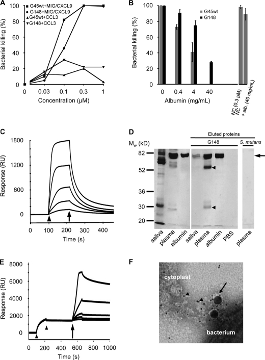
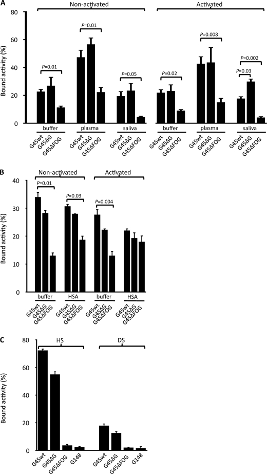
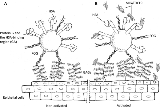
Human serum albumin (HSA) is the dominating protein in human plasma. Many bacterial species, especially streptococci, express surface proteins that bind HSA with high specificity and affinity, but the biological consequences of these protein-protein interactions are poorly understood. Group G streptococci (GGS), carrying the HSA-binding protein G, colonize the skin and the mucosa of the upper respiratory tract, mostly without causing disease. In the case of bacterial invasion, pro-inflammatory cytokines are released that activate the epithelium to produce antibacterial peptides, in particular the chemokine MIG/CXCL9. In addition, the inflammation causes capillary leakage and extravasation of HSA and other plasma proteins, environmental changes at the epithelial surface to which the bacteria need to respond. In this study, we found that GGS adsorbed HSA from both saliva and plasma via binding to protein G and that HSA bound to protein G bound and inactivated the antibacterial MIG/CXCL9 peptide. Another surface protein of GGS, FOG, was found to mediate adherence of the bacteria to pharyngeal epithelial cells through interaction with glycosaminoglycans. This adherence was not affected by activation of the epithelium with a combination of IFN-γ and TNF-α, leading to the production of MIG/CXCL9. However, at the activated epithelial surface, adherent GGS were protected against killing by MIG/CXCL9 through protein G-dependent HSA coating. The findings identify a previously unknown bacterial survival strategy that helps to explain the evolution of HSA-binding proteins among bacterial species of the normal human microbiota.


Figures

FIGURE 1.
FIGURE 1.
Characterization of the interactions between the antibacterial chemokine MIG/CXCL9, GGS, and HSA. A, bactericidal activity of MIG/CXCL9 against GGS. Bacteria (strains G45wt and G148) were incubated with MIG/CXCL9 or CCL3 at the indicated concentrations or with buffer alone. To calculate percent killing, the cfu present after exposure to the polypeptides were compared with the cfu obtained after incubation in buffer alone. The data shown represent mean ± S.E. from three separate experiments. B, HSA attenuates the bactericidal activity of MIG/CXCL9. Bacteria were incubated with recombinant MIG/CXCL9 (0.3 μm) for 1 h at 37 °C or with MIG/CXCL9 that had been preincubated with HSA at 0.4, 4, or 40 mg/ml (corresponding to the HSA content of 1, 10, and 100% plasma, respectively) for 20 min prior to exposure to GGS (strains G45wt and G148) for 1 h at 37 °C. The number of cfu after incubation in buffer alone was set to 100%. The antibacterial activity of the peptide novicidin (NC; 0.3 μm) was not significantly affected (p = 0.25) by the presence of albumin (alb.; 40 mg/ml). The data shown represent mean ± S.E. from three separate experiments. C, HSA binds MIG/CXCL9 in surface plasmon resonance experiments. HSA was immobilized on a sensor chip, and increasing concentrations of MIG/CXCL9 were injected over the surface. The start and stop of injection are denoted by an arrowhead and arrow, respectively. RU, response units. D, GGS adsorb HSA from saliva and plasma. Bacteria were incubated with undiluted saliva, 10% plasma, or 4 mg/ml HSA for 20 min. After washing with PBS, bound proteins were eluted from bacteria with glycine HCl. These eluted proteins were separated by SDS-PAGE and visualized by Coomassie Brilliant Blue staining. Bands corresponding to albumin (arrow) are seen in all lanes, except the PBS control. The bands of ∼25 and ∼56 kDa in the lane with material eluted from bacteria incubated with plasma (arrowheads) correspond to light and heavy chains of IgG, respectively. S. mutans strain α3201 was incubated with plasma, but no bound proteins were visible after elution. E, binding of albumin to immobilized GA modules, followed by a dose-dependent increase in binding of MIG/CXCL9 as determined by surface plasmon resonance. GA modules were immobilized on a sensor chip, followed by injection (between the two arrowheads) and binding of HSA to the chip. Thereafter, increasing concentrations of MIG/CXCL9 were injected over the surface (indicated by the arrow), displaying a stable binding to the surface-bound GA·HSA complexes. F, electron micrograph showing the interaction between cytoplasts derived from pharyngeal epithelial cells, MIG/CXCL9 (labeled with 4-nm colloidal gold particles; arrowheads), HSA (labeled with 15-nm colloidal gold particles; arrow), and bacteria.
FIGURE 2.
FIGURE 2.
Binding of GGS to pharyngeal epithelial cells. A and B, GGS strain G45. Wild-type G45 and isogenic mutants G45ΔFOG and G45ΔG, lacking protein FOG and protein G, respectively, were killed by heat and labeled with 125I. Pharyngeal epithelial cells were cultured in the absence (non-activated) or presence (activated) of a combination of IFN-γ (100 units/ml) and TNF-α (10 ng/ml). 125I-Labeled G45wt, G45ΔFOG, and G45ΔG bacteria were incubated for 15 min in PBS, 10% human plasma, and whole saliva (A) or 0.4 mg/ml HSA (B); washed; and incubated with epithelial cells for 3 h at 37 °C. After washing, the radioactivity of the remaining pellets was calculated, and the radioactivity in percent of total added radioactivity was determined. Values are mean ± S.E. of at least three separate experiments, and Student's t test for paired observations was used to calculate p values. C, GGS bind highly sulfated GAGs. G45wt, G45ΔFOG, G45ΔG, and G148 bacteria were incubated with 125I-labeled HS or DS for 1 h at room temperature. After washing, the radioactivity of the bacterial pellets was determined and compared with the total radioactivity added. Data represent mean ± S.E. of four separate experiments. p values were calculated using Student's t test for paired observations.
FIGURE 3.
FIGURE 3.
Bacterial survival on the surface of non-activated and activated pharyngeal epithelial cells. A, GGS (strain G45wt) were incubated with HSA (4 mg/ml) or PBS, washed, and added to non-activated or IFN-γ- and TNF-α-activated epithelial cells. Thereafter, bacteria and epithelial cells were co-incubated for 3 h. Following washing, the epithelial cells were detached and lysed, and the resulting debris was plated on agar plates. The number of cfu was calculated and related to the control (non-activated epithelial cells and strain G45wt pretreated with HSA). Values are mean ± S.E. from three separate experiments, and p values were calculated using Student's t test for paired observations. B, the same experiment as in A was performed with the isogenic mutant G45ΔG, devoid of protein G. Values represent mean ± S.E. of three separate experiments. C, electron microscopy of G45wt bacteria (GGS) incubated with non-activated pharyngeal epithelial cells (Ep). Ultrathin sections of cells and bacteria were incubated with anti-MIG/CXCL9 antibodies, followed by visualization of bound antibodies using secondary antibodies conjugated with 10-nm colloidal gold particles. The bacteria have a preserved integrity with a visible cell wall/plasma membrane. A few scattered gold particles are seen, indicating low or no presence of MIG/CXCL9. D, G45wt bacteria visualized at the surface of pharyngeal epithelial cells activated with a combination of IFN-γ and TNF-α. The integrity of bacteria is lost, and the cell wall/plasma membrane is no longer apparent. MIG/CXCL9 is seen both associated with the bacterial surface (arrows) and intracellularly (arrowheads). E, G45wt bacteria coated with HSA prior to exposure to activated pharyngeal epithelial cells. Bacterial integrity is preserved, and large amounts of MIG/CXCL9 are detected at the bacterial surface but not intracellularly (arrows). Scale bar = 1 μm. F, higher magnification (×10) of C showing the bacterial cell wall (W; indicated with a zigzag line) and the lipid bilayer of the plasma membrane (M) of a streptococcus in contact with a non-activated epithelial cell. G, disintegrated plasma membrane and cell wall with dispersed presence of MIG/CXCL9 in a streptococcus in contact with the cytokine-activated epithelium. H, higher magnification (×10) of A of a streptococcus coated with HSA. MIG/CXCL9, visualized as colloidal gold particles, accumulated at the cell wall, and the plasma membrane is intact.
FIGURE 4.
FIGURE 4.
Schematic and hypothetical representation of GGS at the epithelium of the oropharynx. A, under normal, non-activated conditions, GGS adhere to the epithelium via binding of FOG to highly sulfated GAGs. It is likely that the GA modules of protein G adsorb HSA from saliva, coating the bacteria also in the absence of an inflammatory stimulus causing vascular leakage and extravasation of HSA. B, in response to pro-inflammatory stimuli, epithelial cells produce antibacterial peptides, including MIG/CXCL9. This chemokine is associated with GAGs at the epithelial surface but is also present in a soluble form. HSA bound to protein G binds and attenuates the bactericidal activity of MIG/CXCL9. Also at the activated epithelial surface, the FOG protein remains an important mediator of bacterial adhesion.

Similar articles

Cited by

References

    1. Hashikawa S., Iinuma Y., Furushita M., Ohkura T., Nada T., Torii K., Hasegawa T., Ohta M. (2004) J. Clin. Microbiol. 42, 186–192 - PMC - PubMed
    1. Sylvetsky N., Raveh D., Schlesinger Y., Rudensky B., Yinnon A. M. (2002) Am. J. Med. 112, 622–626 - PubMed
    1. Sansonetti P. J. (2004) Nat. Rev. Immunol. 4, 953–964 - PubMed
    1. Hyland K. A., Brennan R., Olmsted S. B., Rojas E., Murphy E., Wang B., Cleary P. P. (2009) FEMS Immunol. Med. Microbiol. 55, 422–431 - PubMed
    1. Raeder R. H., Barker-Merrill L., Lester T., Boyle M. D., Metzger D. W. (2000) J. Infect. Dis. 181, 639–645 - PubMed

Publication types

MeSH terms