. 2011 Jan 15;71(2):528-37.
doi: 10.1158/0008-5472.CAN-10-2581. Epub 2010 Nov 22.

CaM kinase kinase beta-mediated activation of the growth regulatory kinase AMPK is required for androgen-dependent migration of prostate cancer cells

Affiliations

CaM kinase kinase beta-mediated activation of the growth regulatory kinase AMPK is required for androgen-dependent migration of prostate cancer cells

Daniel E Frigo et al. Cancer Res. .

Abstract

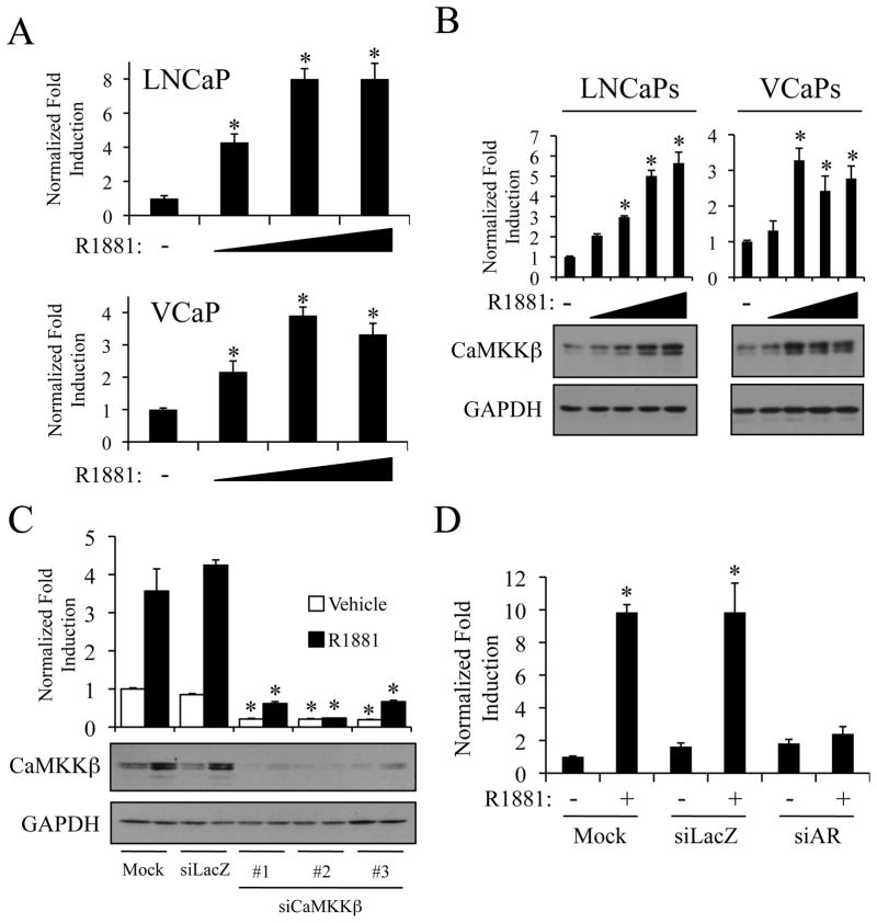
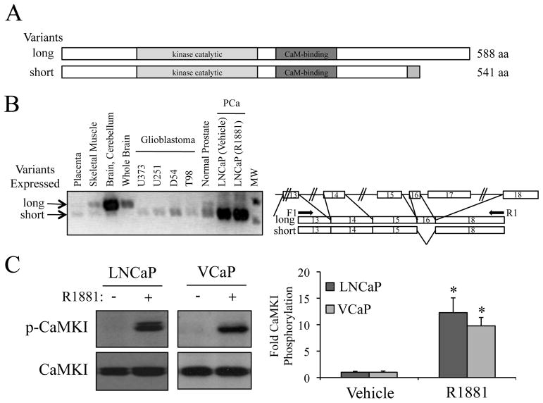
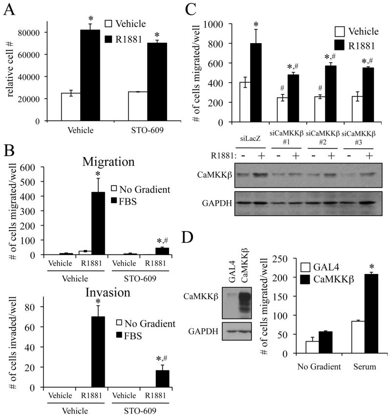
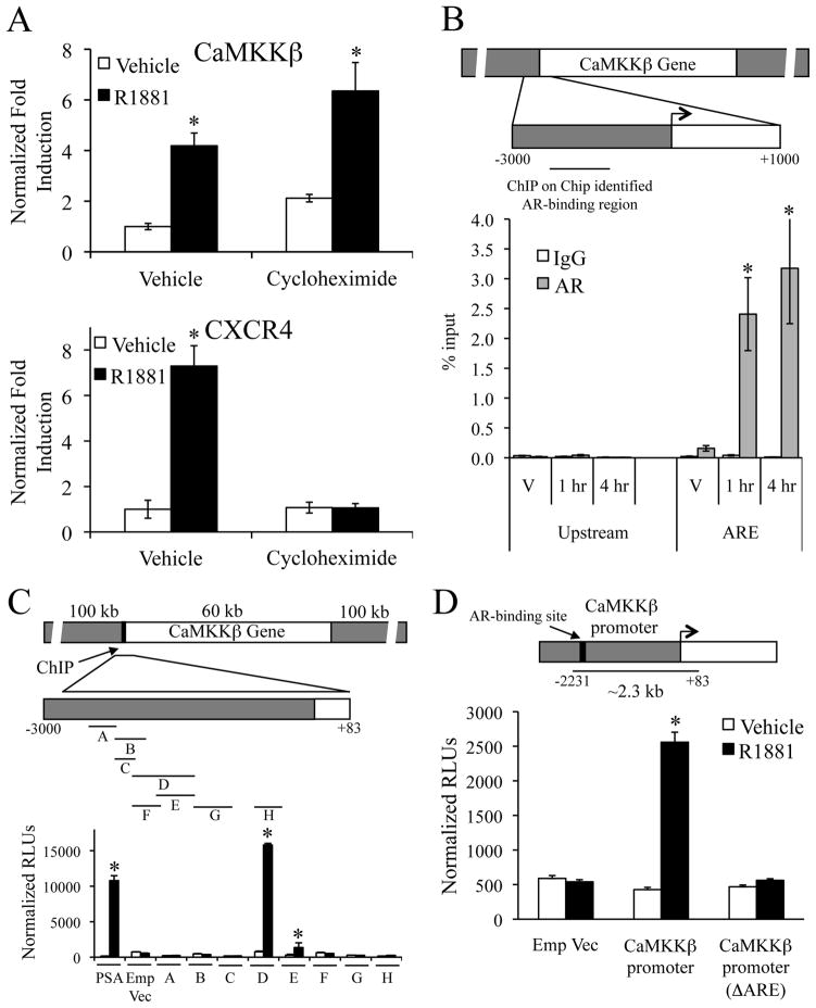
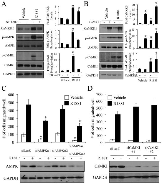
While patients with advanced prostate cancer initially respond favorably to androgen ablation therapy, most experience a relapse of the disease within 1-2 years. Although hormone-refractory disease is unresponsive to androgen-deprivation, androgen receptor (AR)-regulated signaling pathways remain active and are necessary for cancer progression. Thus, both AR itself and the processes downstream of the receptor remain viable targets for therapeutic intervention. Microarray analysis of multiple clinical cohorts showed that the serine/threonine kinase Ca2+/calmodulin-dependent protein kinase kinase β (CaMKKβ) is both highly expressed in the prostate and further elevated in prostate cancers. Using cellular models of prostate cancer, we have determined that androgens (a) directly increase the expression of a CaMKKβ splice variant and (b) increase functional CaMKKβ protein levels as determined by the phosphorylation of both CaMKI and AMP-activated protein kinase (AMPK), two of CaMKKβ's primary substrates. Importantly, inhibition of the CaMKKβ-AMPK, but not CaMKI, signaling axis in prostate cancer cells by pharmacological inhibitors or siRNA-mediated knockdown blocks androgen-mediated migration and invasion. Conversely, overexpression of CaMKKβ alone leads to both increased AMPK phosphorylation and cell migration. Given the key roles of CaMKKβ and AMPK in the biology of prostate cancer cells, we propose that these enzymes are potential therapeutic targets in prostate cancer.


Conflict of interest statement

Disclosure of potential conflicts of interest: None

Figures

Figure 1
Figure 1
Androgens increase CaMKKβ levels in an AR-dependent manner. LNCaP or VCaP cells were treated for 24 h with vehicle or increasing concentrations of the synthetic androgen R1881 (A-0.1, 1, and 10 nM; B-0.01, 0.1, 1, and 10 nM). A, after treatment, cells were lysed, and RNA was isolated and reversed transcribed. The expression of CaMKKβ was assessed using qPCR. B, after treatment, cells were subjected to western blot analysis and subsequent densitometry (top). CaMKKβ protein levels were normalized to GAPDH loading control. A and B, results are expressed as fold induction over vehicle-treated cells + SE (n = 3). *, significant changes from vehicle-treated cells. C, LNCaP cells were transiently transfected with mock or Stealth siRNAs targeting a negative control (siLacZ) or CaMKKβ(#1–3). Two days later, cells were treated for 24 h +/− 10 nM R1881. Whole-cell extracts were subjected to western blot analysis and densitometry (top) as described in B. *, significant changes from mock-transfected cells. D, LNCaP cells were transfected as described in C with mock or Stealth siRNAs targeting LacZ or AR and treated for 24 h. The expression of CaMKKβ was assessed as in A using qPCR.
Figure 2
Figure 2
The prostate expresses a different functional splice variant of CaMKKβ compared to brain A, schematic of CaMKKβ splice variants. B, RT-PCR using primers spanning specific exons (indicated in right schematic) was performed on cDNA generated from various tissues and cell lines. C, LNCaP or VCaP cells were treated for 24 h +/− 10 nM R1881. Cell lysates were then subjected to western blot analysis and subsequent densitometry (right). Phospho-CaMKI (p-CaMKI) protein levels were normalized to total CaMKI. Results are expressed as fold CaMKI phosphorylation over vehicle-treated cells + SE (n = 3). *, significant changes from vehicle-treated cells.
Figure 3
Figure 3
CaMKKβ is required and sufficient for the androgen-mediated migration and invasion of prostate cancer cells. A, LNCaP cells were plated in 96-well plates and grown for 3 d. Cells were treated +/− 1 nM R1881 and +/− 30 μM STO-609 on d 3, d 5, and d 7. On d 10, cells were lysed and the relative number of cells was measured with the fluorescent DNA binding dye FluoReporter Blue. Each sample was performed in triplicate, and results from a representative experiment are shown. Results are expressed as relative cell number ± SE (n = 2). *, significant changes from vehicle (no R1881)-treated cells. B, LNCaP cells were pretreated for 1 h +/− 30 μM STO-609 prior to overnight treatment +/− 10 nM R1881. Cells were then dissociated and reseeded into the top chamber for a Boyden migration or Matrigel extracellular matrix invasion assay. Fresh medium with the corresponding treatments was added to the top and bottom chambers while either no chemoattractant or 5% FBS (serum) was added to the bottom chamber. After 16 h, migrated cells were fixed, stained and counted in three different microscopic fields and added together. The results are expressed as mean ± SE (n = 3). *, significant changes from vehicle (no R1881)-treated cells. #, significant changes from vehicle (no STO-609)-treated cells. C top, LNCaP cells were transfected with indicated siRNAs. Two days after transfection, cells were treated +/− 10 nM R1881 and subjected to a Boyden migration assay as described in B. *, significant changes from vehicle-treated cells. #, significant changes from control (siLacZ)-transfected cells. C bottom, western blot to demonstrate CaMKKβ knockdown Quantification of these blots is presented in Supplementary Fig. S4D. D right, LNCaP cells stably expressing either GAL4 (control) or CaMKKβ were subjected to a migration assay as described in B using +/− 5% FBS as chemoattractant. The results are expressed as mean + SE (n = 3). *, significant changes from LNCaP-GAL4 cells. D left, western blot confirming CaMKKβ expression. Quantification of these blots is presented in Supplementary Fig. S4E.
Figure 4
Figure 4
Identification of the ARE that regulates CaMKKβ expression. A, LNCaP cells were pretreated for 1 h with vehicle or 1 μg/ml cycloheximide followed by vehicle or 10 nM R1881 for 24 h. CaMKKβ or CXCR4 mRNA levels were quantitated using qPCR. Results are expressed as fold induction over vehicle (no R1881)-treated cells ± SE (n = 3). *, significant changes from vehicle-treated cells. B, LNCaP cells were treated with vehicle (V) or 10 nM R1881 for 1 or 4 h. Cross-linked chromatin was immunoprecipitated with indicated antibodies. The precipitated DNA was amplified using primers spanning a region identified using ChIP on Chip data as a potential AR-binding site (indicated in top schematic) or a distal upstream region (negative control). The results are presented as percent input ± SE (n = 3). *, significant changes from IgG controls. C, various enhancer luciferase reporter constructs (depicted in top model) were transfected into LNCaP cells and treated overnight +/−10 nM R1881. After treatment, cells were harvested and assayed for luciferase activity. Luciferase values were normalized to β-galactosidase control. Data are the mean relative light units (RLUs) + SEM for one representative experiment performed in triplicate (n = 3). *, significant changes from vehicle-treated cells. D, CaMKKβ promoter constructs (depicted in top model) were transfected into LNCaP cells and then treated overnight with vehicle or 10 nM R1881. After treatment, cells were harvested and assayed for luciferase activity as in C. Emp Vec, empty vector.
Figure 5
Figure 5
Androgen-mediated migration occurs through a CaMKKβ-AMPK-dependent pathway. A, LNCaP cells were pretreated for 1 h +/− 30 μM STO-609 prior to overnight treatment +/− 10 nM R1881. Cell lysates were then subjected to western blot analysis and subsequent densitometry (right). CaMKKβ levels were normalized to GAPDH. Phospho-CaMKI (p-CaMKI) levels were normalized to total CaMKI. Phospho-AMPK (p-AMPK) levels were normalized to total AMPK. Results are expressed as fold induction/phosphorylation over double vehicle-treated cells + SE (n = 3). *, significant changes from vehicle-treated cells. B, LNCaP cells stably expressing either GAL4 or CaMKKβ were treated overnight +/− 10 nM R1881. Cell lysates were then subjected as in A to western blot analysis and densitometry (right). Results are expressed as fold induction/phosphorylation over LNCaP-GAL4 vehicle-treated cells + SE (n = 3). *, significant changes from LNCaP-GAL4 vehicle-treated cells. C and D, LNCaP cells were transfected with indicated siRNAs, treated and subjected to a migration assay (top) or western blot analysis (bottom) as in Fig. 3C. *, significant changes from control (siLacZ)-transfected cells. Quantification of the blots is presented in Supplementary Fig. S7.

References

    1. Cancer Facts and Figures. American Cancer Society; 2007.
    1. Isaacs JT, Isaacs WB. Androgen receptor outwits prostate cancer drugs. Nat Med. 2004;10:26–7. - PubMed
    1. Chen CD, Welsbie DS, Tran C, et al. Molecular determinants of resistance to antiandrogen therapy. Nature Med. 2004;10:33–9. - PubMed
    1. Frigo DE, Sherk AB, Wittmann BM, et al. Induction of Kruppel-like factor 5 expression by androgens results in increased CXCR4-dependent migration of prostate cancer cells in vitro. Mol Endocrinol. 2009 - PMC - PubMed
    1. Sherk AB, Frigo DE, Schnackenberg CG, et al. Development of a small molecule serum and glucocorticoid-regulated kinase 1 antagonist and its evaluation as a prostate cancer therapeutic. Cancer Res. 2008;68:1–9. - PMC - PubMed

Publication types

MeSH terms