. 2010 Dec 20;207(13):2817-30.
doi: 10.1084/jem.20100844. Epub 2010 Nov 22.

Transient activation of c-MYC expression is critical for efficient platelet generation from human induced pluripotent stem cells

Affiliations

Transient activation of c-MYC expression is critical for efficient platelet generation from human induced pluripotent stem cells

Naoya Takayama et al. J Exp Med. .

Abstract

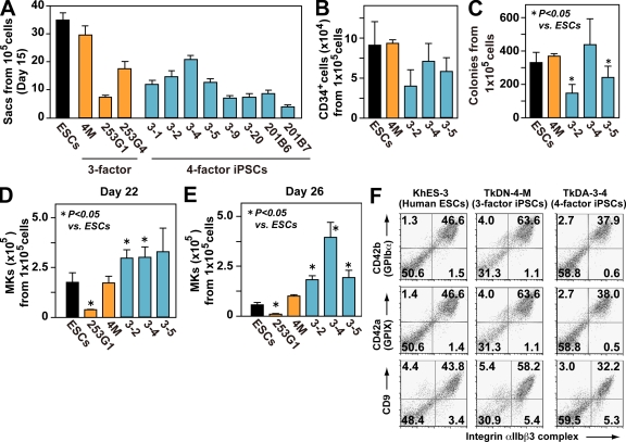
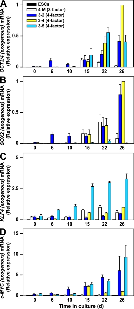
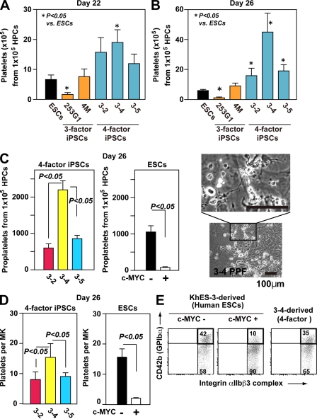
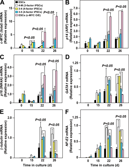
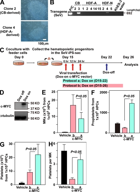
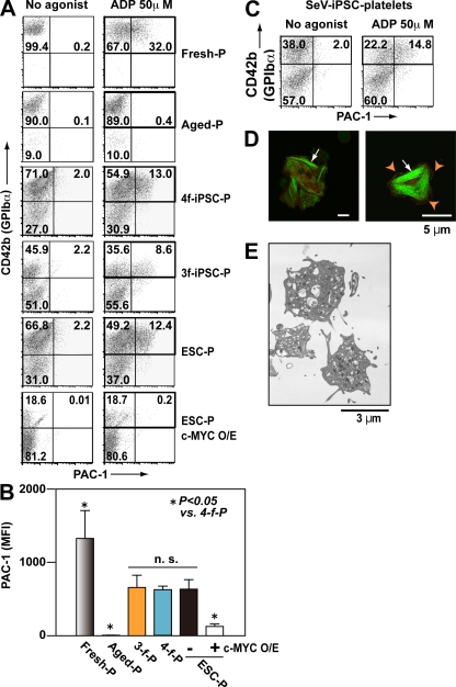
Human (h) induced pluripotent stem cells (iPSCs) are a potentially abundant source of blood cells, but how best to select iPSC clones suitable for this purpose from among the many clones that can be simultaneously established from an identical source is not clear. Using an in vitro culture system yielding a hematopoietic niche that concentrates hematopoietic progenitors, we show that the pattern of c-MYC reactivation after reprogramming influences platelet generation from hiPSCs. During differentiation, reduction of c-MYC expression after initial reactivation of c-MYC expression in selected hiPSC clones was associated with more efficient in vitro generation of CD41a(+)CD42b(+) platelets. This effect was recapitulated in virus integration-free hiPSCs using a doxycycline-controlled c-MYC expression vector. In vivo imaging revealed that these CD42b(+) platelets were present in thrombi after laser-induced vessel wall injury. In contrast, sustained and excessive c-MYC expression in megakaryocytes was accompanied by increased p14 (ARF) and p16 (INK4A) expression, decreased GATA1 expression, and impaired production of functional platelets. These findings suggest that the pattern of c-MYC expression, particularly its later decline, is key to producing functional platelets from selected iPSC clones.


Figures

Figure 1.
Figure 1.
Four-factor human iPSCs are better than three-factor iPSCs for megakaryopoiesis, which is independent of hematopoietic colony potential. (A) Numbers of ESC- and iPSC-sac–like structures generated from 105 cells (n = 3, means ± SEM from three independent experiments). (B) Numbers of CD34+ hematopoietic progenitors within ESC- or iPSC-sacs yielded from 105 human ESCs or iPSCs (n = 3, means ± SEM). (C) Numbers of hematopoietic colonies derived from 105 human ESCs or iPSCs within sacs (n = 3, means ± SEM). (D and E) Numbers of CD42b (GPIbα)+ MKs derived from 105 hematopoietic progenitors within sacs on day 22 (D) and day 26 (E; n = 5, means ± SEM). (F) Representative flow cytometry dot plots for khES-3–, TkDN-4-M–, and TkDA3-4–derived MKs examined on day 24.
Figure 2.
Figure 2.
Time-dependent changes in qPCR induced by exogenous reprogramming genes in three-factor or four-factor iPSCs. mRNA encoding exogenous OCT3/4 (A), SOX2 (B), KLF4 (C), and c-MYC (D) in human ES cells (ESCs), TkDN4-M (three-factor iPSCs), TkDA3-2, TkDA3-4, and TkDA3-5 (four-factor iPSCs) on day 0 or their derivatives (on days 6, 10, 15, 22, and 26 after initiation of MK-lineage culture) were examined by qPCR as described in the Materials and methods section. TkDA3-4–derived mature MKs (day 26) was assigned a value of 1.0 (n = 4, means ± SEM from two independent experiments).
Figure 3.
Figure 3.
Effects of reprogramming factors on megakaryopoiesis. (A–C) Each reprogramming factor was transduced, together with EGFP or KO markers, on day 15 of MK-lineage culture. Numbers of total and marker genes EGFP or KO-expressing cells in floating cells on day 17 (A) and CD42b+ MKs on days 22 (B) and 26 (C) were measured (n = 3, means ± SEM). (D) Representative flow cytometry dot plots of hESC-derived hematopoietic cells transduced with vehicle (EGFP), OCT3/4-KO, SOX2-EGFP, KLF4-EGFP, or c-MYC-EGFP on day 22. (E and F) On day 22, May-Giemsa staining (E) or ploidy analysis (F) of the cells transduced with vehicle or c-MYC was examined.
Figure 4.
Figure 4.
Level of c-MYC reactivation in individual iPSC-derived MKs may determine the efficiency of platelet generation in vitro. (A and B) Numbers of CD41a+CD42b+ platelets generated from hESCs or hiPSCs on days 22 (A) and 26 (B; peak of platelet generation; n = 5, means ± SEM). (C and D) Numbers of proplatelets (C) and platelets (D) derived from four-factor iPSCs and from ESC hematopoietic progenitors, with or without c-MYC transduction (ii; n = 5, means ± SEM). Representative photomicrographs of proplatelets are derived from four-factor iPSCs. (D) Numbers of platelets per MK was calculated as the total number of platelets divided by the total number of MKs on day 26 (n = 5, means ± SEM). (E) Representative flow cytometry dot plots show MKs derived from TkDA3-4 and KhES-3, with or without c-MYC transduction, on day 26.
Figure 5.
Figure 5.
Level of c-MYC expression affects INK4A/ARF locus genes and genes related to MK maturation during megakaryopoiesis from pluripotent stem cells. qRT-PCR analysis of total c-MYC (A, endogenous plus exogenous), p14ARF (B), p16INK4A (C), GATA1 (D), β1-tubulin (E), and NF-E2 p45 (F) expression in hESCs, with and without overexpression (O/E) of exogenous c-MYC, on days 22 and 26 (7 and 11 d after transduction) in three-factor hiPSCs (TkDN4-M) or in four-factor hiPSCs (TkDA3-4 and TkDA3-5) on days 0, 15, 22, and 26. All levels were normalized to the level of GAPDH expression (n = 4 of two independent samples). The levels of c-MYC (A), p14ARF (B), and p16INK4A (C) expression in an undifferentiated TkDA3-4 iPSC clone (day 0) or expression of the other genes (D–F) in TkDA3-4–derived mature MKs (day 26) was assigned a value of 1.0 (n = 4, means ± SEM).
Figure 6.
Figure 6.
Inducible c-MYC expression system enabling Sendai viral vector–based iPSCs without reactivation to recapitulate enhanced MK maturation with increased platelet generation. (A) Representative photomicrographs of SeV-iPSCs derived from CB CD34+CD45+ cells or HDFs. Original magnification, 100×. (B) RT-PCR analyses of Sendai virus Tg (harboring reprogramming factors) expression in SeV-iPSC clones (passage number 4) derived from CB, HDF-A (adult), and HDF-N (neonate). A sample of HDFs transduced with SeV is used as a positive control for the SeV Tg. (C) Scheme of c-MYC induction in SeV-iPSC–derived hematopoietic cells. Hematopoietic progenitors derived from SeV-iPSCs were transfected with DOX-inducible c-MYC O/E vector on day 15 and analyzed on day 26. In Protocol a, DOX was added only from days 15 to 22. In protocol b, DOX was added from days 15 through 26. (D) Representative Western blots of cell lysates with c-MYC O/E (DOX-on; protocol b) or without c-MYC O/E (DOX-off; Protocol a) on day 26. The α-tubulin levels indicate same protein value. (E–G) Numbers of CD42b (GPIbα)+ MKs (E), proplatelets (F), and platelets (G) on day 26 derived from 105 hematopoietic progenitors transfected with vehicle or DOX-inducible c-MYC O/E vector in protocol a or protocol b (n = 4, means ± SEM). (H) Numbers of platelets per MK generated on day 26 of culture (peak of platelet generation; n = 4, means ± SEM). Numbers of platelets per MK were calculated as the total number of platelets divided by the total number of MKs on day 26.
Figure 7.
Figure 7.
Integrin activation and the structure of human iPSC platelets are comparable to those in human PB-derived platelets. (A–C) Integrin activation in fresh human platelets (Fresh-P), aged human platelets (48-h incubation at 37°C; Aged-P), TkDN4-M (three-factor iPSC) platelets (3f-iPSC-P), TkDA3-4 (four-factor iPSC) platelets (4f-iPSC-P), and ESC platelets (ESC-P), with or without c-MYC O/E. The binding of PAC-1 (indicative of platelet activation) to individual platelets was quantified in the absence and presence of 50 µM ADP using flow cytometry. (A) Representative flow cytometry dot plots. Square indicates CD42b+ platelets. (B) Mean fluorescence intensity (MFI) of bound PAC-1, obtained from square gate in A. Error bars depict means ± SEM for four independent experiments (duplicate). (C) Representative flow cytometry analysis of PAC-1–bound platelets generated from integration-free SeV-iPSCs subjected to biphasic activation and, thereafter, decline of c-MYC expression as protocol b shown in Fig. 6 C. Square indicates CD42b+ platelets. (D) Spreading of iPSC platelets on fibrinogen. Human CD41a (red) and phalloidin (green) were used to identify F-actin fibers. Arrowheads indicate lamellipodia. Arrows indicate actin stress fibers. Bars, 5 µm. (E) Transmission electron micrographs of hiPSC (TkDA3-4) platelets on day 26. Bar, 3 µm.
Figure 8.
Figure 8.
Human iPSC platelets circulate in NOG mice and adhere to vessel during thrombus formation in vivo. (A) NOG (nod-scid/IL-2 γc-null) mice were irradiated (2.0 Gy) to induce thrombocytopenia. 9 d later, human iPSC platelets, human PB-derived platelets (107), or PBS alone was injected via the tail vein. Platelet chimerism was quantified by flow cytometry. Representative dot plots for the same experiment are shown. Circulation of injected platelets was evaluated after 2 and 24 h (orange squares). Experiments were independently performed three times. (B) Sequential images of circulating iPSC platelets. A combination of FITC-dextran (green) and 107 TMRE-stained iPSC platelets (red) in PBS was injected via the tail vein into NOG mice. Mesenteric capillaries were visualized using a confocal laser-scanning microscope. Red arrows indicate circulating iPSC platelets in vivo. Original videos are available as Video 2. (C) Representative sequential images of thrombus formation by iPSC platelets in a blood vessel. Hematoporphyrin was administrated to induce thrombus formation after laser-induced injury, as described previously (Nishimura et al., 2008; Takizawa et al., 2010). Red arrows indicate iPSC platelets in a developing thrombus. White arrows indicate host (mouse) platelets. Original videos are available as Videos 3–5. Experiments were independently performed three times. Bars, 10 µm.

Comment in

References

    1. Askew D.S., Ashmun R.A., Simmons B.C., Cleveland J.L. 1991. Constitutive c-myc expression in an IL-3-dependent myeloid cell line suppresses cell cycle arrest and accelerates apoptosis. Oncogene. 6:1915–1922 - PubMed
    1. Bergmeier W., Burger P.C., Piffath C.L., Hoffmeister K.M., Hartwig J.H., Nieswandt B., Wagner D.D. 2003. Metalloproteinase inhibitors improve the recovery and hemostatic function of in vitro-aged or -injured mouse platelets. Blood. 102:4229–4235 10.1182/blood-2003-04-1305 - DOI - PubMed
    1. Chanprasert S., Geddis A.E., Barroga C., Fox N.E., Kaushansky K. 2006. Thrombopoietin (TPO) induces c-myc expression through a PI3K- and MAPK-dependent pathway that is not mediated by Akt, PKCzeta or mTOR in TPO-dependent cell lines and primary megakaryocytes. Cell. Signal. 18:1212–1218 10.1016/j.cellsig.2005.09.010 - DOI - PubMed
    1. Choi K.D., Yu J., Smuga-Otto K., Salvagiotto G., Rehrauer W., Vodyanik M., Thomson J., Slukvin I. 2009. Hematopoietic and endothelial differentiation of human induced pluripotent stem cells. Stem Cells. 27:559–567 - PMC - PubMed
    1. Eilers M., Schirm S., Bishop J.M. 1991. The MYC protein activates transcription of the alpha-prothymosin gene. EMBO J. 10:133–141 - PMC - PubMed

Publication types

MeSH terms

Substances