. 2010 Nov 29;191(5):1013-27.
doi: 10.1083/jcb.201006006. Epub 2010 Nov 22.

An invasive podosome-like structure promotes fusion pore formation during myoblast fusion

Affiliations

An invasive podosome-like structure promotes fusion pore formation during myoblast fusion

Kristin L Sens et al. J Cell Biol. .

Abstract

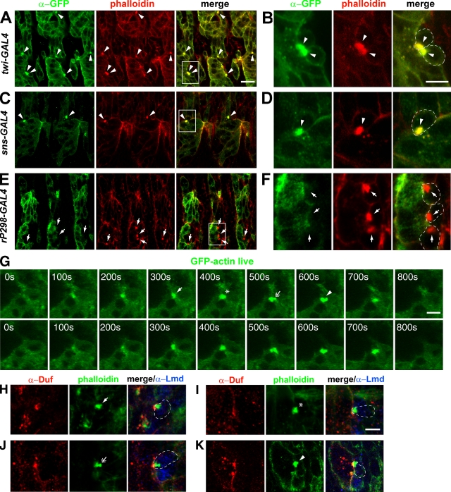
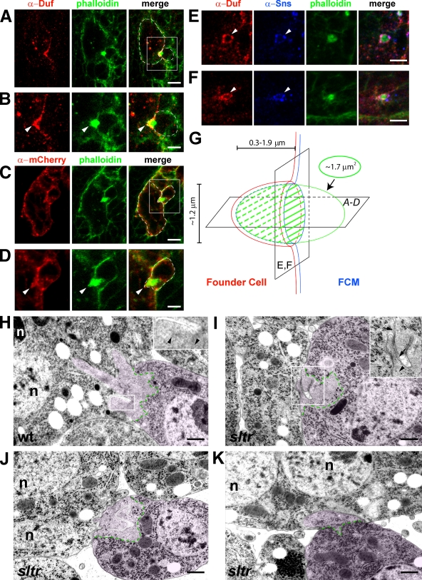
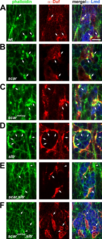
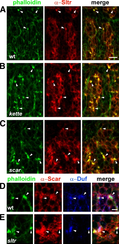
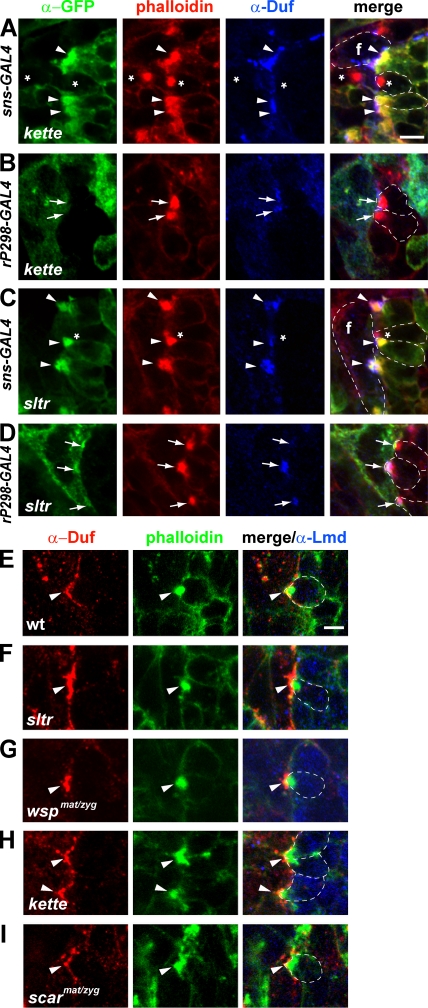
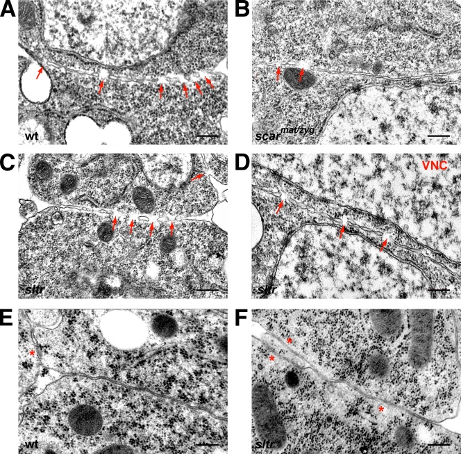
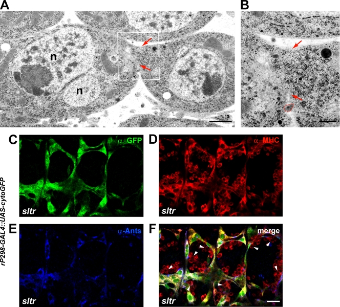
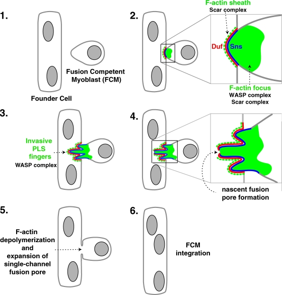
Recent studies in Drosophila have implicated actin cytoskeletal remodeling in myoblast fusion, but the cellular mechanisms underlying this process remain poorly understood. Here we show that actin polymerization occurs in an asymmetric and cell type-specific manner between a muscle founder cell and a fusion-competent myoblast (FCM). In the FCM, a dense F-actin-enriched focus forms at the site of fusion, whereas a thin sheath of F-actin is induced along the apposing founder cell membrane. The FCM-specific actin focus invades the apposing founder cell with multiple finger-like protrusions, leading to the formation of a single-channel macro fusion pore between the two muscle cells. Two actin nucleation-promoting factors of the Arp2/3 complex, WASP and Scar, are required for the formation of the F-actin foci, whereas WASP but not Scar promotes efficient foci invasion. Our studies uncover a novel invasive podosome-like structure (PLS) in a developing tissue and reveal a previously unrecognized function of PLSs in facilitating cell membrane juxtaposition and fusion.


Figures

Figure 1.
Figure 1.
F-actin–enriched foci specifically localize in FCMs and undergo shape changes during their lifespan. (A–F) F-actin foci specifically reside in FCMs. Stage 14 wild-type embryos expressing GFP-actin with muscle-specific drivers double labeled with α-GFP (green) and phalloidin (red). Boxed areas in A, C, and E are enlarged in B, D, and F. In this and all subsequent figures, single-slice confocal images are shown and selected muscle cells are outlined (dashed lines) by tracing the cell cortical phalloidin labeling. Note the colocalization of GFP-actin foci with phalloidin-labeled F-actin foci when GFP-actin was expressed in all muscle cells (with twi-GAL4; A and B, arrowheads) or in FCMs (with sns-GAL4; C and D, arrowheads), and the absence of GFP-actin enrichment at sites of fusion (arrows) when it was expressed in founder cells (with rP298-GAL4; E and F). (G) Time-lapse imaging of an F-actin focus in a stage 14 wild-type embryo expressing GFP-actin in all muscle cells (with twi-GAL4). Horizontal panels are stills from a time-lapse sequence, and vertical panels are two adjacent optical slices of the same F-actin focus along the Z-axis. (H–K) Four examples of F-actin foci with irregular shapes in fixed wild-type embryos labeled with α-Duf (founder cell; enriched at sites of fusion; red), phalloidin (green), and α-Lmd (FCM; in nuclei; blue; Duan et al., 2001). Note the different foci shapes (indicated by arrow [H], asterisk [I], double-headed arrow [J], and arrowhead [K]) in fixed embryos corresponding to those at different time points (300, 400, 500, and 600 s, respectively) of the live F-actin focus shown in G. Bars: (A, C, and E) 20 µm; (B, D, and F) 10 µm; (G–K) 5 µm.
Figure 2.
Figure 2.
FCM-specific F-actin foci are invasive and the WASP complex is required for foci invasion. (A–F) Confocal images of stage 14 wild-type embryos showing horizontal (A–D) and perpendicular (E and F) pairs of founder cell/myotube and FCM. Boxed areas in A and C are magnified in B and D, respectively. Founder cells are outlined in A and C and FCMs in A–D. (A and B) An F-actin focus invading a founder cell. Embryo double labeled with α-Duf (red) and phalloidin (green). Arrowhead indicates the inward curvature on the founder cell membrane. (C and D) Membrane rearrangements at the invasive tip of an F-actin focus. Embryo expressing membrane-targeted mCherry-CAAX in all muscle cells (with twi-GAL4) labeled with α-mCherry (red) and phalloidin (green). Arrowhead indicates the invasive tip of the FCM-specific F-actin focus. (E and F) Two examples of F-actin foci encircled by cell adhesion molecules. Embryo triple labeled with α-Duf (red), α-Sns (FCM; enriched at sites of fusion; blue), and phalloidin (green). (G) Schematic drawing of the asymmetric muscle cell adhesion junction. Before fusion, an F-actin focus (green oval) forms at the tip of the FCM (right) and invades the apposing founder cell (left) to create a cup-shaped dimple. The inner wall of the cup is lined with Sns (blue), and the outer wall with Duf (red). Depending on the angle at which the FCM invasion is viewed by confocal microscopy, the cell adhesion molecules can appear as a U-shaped dimple cupping a portion of the actin focus (hatched) in a horizontal section (A–D) or overlapping rings encircling the actin focus in a perpendicular section (E and F). Numbers show average actin foci size (1.7 µm2), diameter of the adhesive rings (1.2 µm), and depth of invasion (0.3–1.9 µm). (H) Ultrastructural details of an invasive F-actin focus. An FCM (pseudo-colored pink) projects multiple F-actin–enriched invasive fingers into a binucleated myotube in a stage 13 wild-type (wt) embryo fixed by HPF/FS. Serial sections of this invasive structure are shown in Fig. S3 A. The F-actin–enriched areas within the FCMs (boundary marked by dashed green lines) are identified by their light gray coloration and lack of ribosomes and intracellular organelles. Although actin filaments (7-nm diameter) are difficult to be fixed and visualized by HPF/FS (or conventional chemical fixation) EM, magnified inset shows faint actin filaments (arrowheads) within an invasive finger. (I–K) F-actin foci fail to invade properly in sltr mutant embryos. F-actin–enriched fingers in FCMs in stage 14 sltr embryos either folded upon each other without extending toward the apposing founder cell (I and J; 8/10 actin foci analyzed show this phenotype), or appear wider and shorter than wild type (K; 2/10). Magnified inset in I shows faint actin filaments (arrowhead), as well as a portion of the founder cell membrane (arrows) pulled into the FCM territory by the folded fingers, which may account for the extensive colocalization between founder cell markers (Duf and Ants) and phalloidin staining in sltr mutant embryos revealed by confocal microscopy (Fig. 3 D and Fig. S1, B and E; Kim et al., 2007). n: founder cell/myotube nuclei (H–K). Bars: (A and C) 10 µm; (B, D, E, and F) 5 µm; (H–K) 500 nm.
Figure 3.
Figure 3.
The WASP and Scar complexes are required for F-actin foci formation at sites of fusion. Stage 14 wild-type (wt) (A) and stage 15 mutant (B–F) embryos triple labeled with phalloidin (green), α-Duf (red), and α-Lmd (blue). Several muscle cell adhesion sites (marked by Duf enrichment) in each panel are indicated by arrows. In wt embryos, most fusion events occur at stage 14 and there is a decrease in the F-actin foci number in stage 15 (Beckett and Baylies, 2007). Note the persistence of F-actin foci in stage 15 scar (B, zygotic null; partial loss of fusion [Fig. S4 A, c]), scarmat/zyg (C, eliminating most, but not all, maternal and zygotic Scar function; near complete loss of fusion [Fig. S4 A, d]), and sltr single mutant embryos (D), and the dramatic reduction of F-actin foci in scar,sltr (E) and scarmat/zyg,sltr (F) double-mutant embryos. In scar,sltr double mutant embryos, a large percentage (76%; 35/46) of muscle cell adhesion sites are not associated with any F-actin enrichment. The average size for the remainder (11/46) of F-actin foci is 1.2 ± 0.6 µm2. Arrowheads in E and F indicate actin polymerization in nonmuscle cells (Duf- and Lmd-negative). Bar, 15 µm.
Figure 4.
Figure 4.
Increased accumulation of Sltr and Scar at muscle cell adhesion sites in scar and sltr mutant embryos, respectively. (A–C) Embryos double labeled with phalloidin (green) and α-Sltr (red). Sltr colocalizes with F-actin foci at muscle cell adhesion sites (arrowheads) in stage 14 wild-type (wt) embryo (A), and with enlarged F-actin foci in stage 15 kette (B) and scar (C) mutant embryos. (D and E) Embryos triple labeled with phalloidin (green), α-Scar (red), and α-Duf (blue). Scar is localized in both founder cell and FCMs (arrowheads) in a broader domain than the FCM-specific F-actin foci in stage 14 wt embryo (D). In a sltr mutant embryo, an elevated level of Scar (arrowheads) is observed at muscle cell adhesion sites. Bars: (A–C) 20 µm; (D and E) 5 µm.
Figure 5.
Figure 5.
The FCM-specific F-actin foci in sltr and kette mutants exhibit different invasive behavior. (A–D) F-actin foci in kette and sltr mutant embryos reside in FCMs. Stage 14 (A and C) and stage 15 (B and D) mutant embryos triple labeled with α-GFP (green), phalloidin (red), and α-Duf (blue). kette (A and B) or sltr (C and D) mutant embryos expressing GFP-actin in FCMs (with sns-GAL4; A and C) or in founder cells (with rP298-GAL4; B and D) are shown. GFP-negative founder cells (labeled as “f”) in A and C are outlined except at sites of cell adhesion with FCMs because the founder cell membranes at these sites cannot be delineated at this resolution. Note the colocalization of GFP- and F-actin–positive foci at muscle cell adhesion sites when GFP-actin was expressed in FCMs (A and C; arrowheads). Also note that in sltr (D), but not kette (B) mutant embryos, founder cell–expressed GFP-actin showed slight enrichment (arrows) along the cell membrane adjacent to the FCM-specific F-actin foci. Asterisks in A and C mark FCMs that are yet to express GFP-actin. (E–I) F-actin foci in sltr and kette mutants show different invasive behavior. Stage 14 wild-type (E, wt) and stage 15 sltr (F), wspmat/zyg (G, eliminating both maternal and zygotic WASP), kette (H), or scarmat/zyg (I) mutant embryo triple labeled with α-Duf (red), phalloidin (green), and α-Lmd (blue). A typical “invasive” F-actin focus is shown for each genotype. Note the reduced depth of foci invasion (arrowheads) in sltr (F) and wspmat/zyg (G) embryos, and the similar depth of foci invasion (arrowhead) in kette (H) and scarmat/zyg (I) embryos, compared with wt (E). Bar, 5 µm.
Figure 6.
Figure 6.
Myoblast fusion in Drosophila is not mediated by a series of fusion pores along the muscle cell contact zone. Electron micrographs of stage 14 wild-type (A and E, wt), scarmat/zyg (B), and sltr (C, D, and F) embryos. Samples were prepared by conventional chemical fixation in A–D and by HPF/FS in E and F. (A–D) Multiple membrane discontinuities (MMDs, arrows) are visible along the contact zone of adherent muscle cells in wild-type (A, wt), scarmat/zyg (B), and sltr (C) embryos. Note that MMDs are also observed between nonfusing cells in the ventral nerve cord (D, VNC). (E and F) Plasma membranes in wt (E) and sltr (F) embryos prepared by HPF/FS do not contain discrete MMDs. Asterisks mark “fuzzy” membrane segments that resulted from imperfect fixation. Bar, 200 nm.
Figure 7.
Figure 7.
Myoblast fusion is mediated by a single-channel fusion pore and the WASP–Sltr complex is required for fusion pore formation. (A and B) A single-channel macro fusion pore revealed by HFP/FS electron microscopy. Boxed area in A is enlarged in B. Arrows indicate boundaries of the fusion pore. Note the even distribution of ribosomes in the lumen of the fusion pore and the absence of F-actin and membrane sacs/vesicles. A small piece of cellular debris between the two cells is outlined in red. n: myotube nuclei. (C–F) Cytoplasmic GFP expressed in founder cells does not diffuse into FCMs in sltr mutant embryos. A stage 15 sltr mutant embryo expressing GFP in founder cells driven by rP298-GAL4 triple labeled with α-GFP (green), α-MHC (red), and α-Ants (blue). Mononucleated FCMs do not contain GFP, even though many of them (a few are indicated by arrowheads) have attached to elongated founder cells/myotubes. Bars: (A) 500 nm; (B) 200 nm; (C–F) 20 µm.
Figure 8.
Figure 8.
A model describing the cellular and molecular events at the asymmetric fusogenic synapse. (1) An FCM is attracted by a founder cell/myotube. (2) The FCM and founder cell/myotube adhere via the interaction of cell adhesion molecules (only Duf and Sns are shown) at the site of fusion. In the FCM, Sns recruits both the Scar and WASP complexes to induce the formation of a dense F-actin focus. In the founder cell, Duf recruits the Scar complex to induce the formation of a thin F-actin sheath. (3) In the FCM, the cell adhesion molecule and the F-actin focus constitute a podosome-like structure (PLS) and, through the action of the WASP–Sltr complex, the PLS protrudes multiple invasive fingers to palpitate the founder cell membrane. (4) We speculate that a nascent fusion pore forms at the tip of a podosome finger, where the two adherent membranes are brought into close proximity through the interactions between the podosome finger in the FCM and the thin sheath of actin in the founder cell. (5) The nascent fusion pore expands to a single-channel macro fusion pore after F-actin depolymerization. (6) The FCM completely incorporates into the founder cell/myotube, contributing one additional nucleus.

References

    1. Abmayr S.M., Zhuang S., Geisbrecht E.R. 2008. Myoblast fusion in Drosophila. Methods Mol. Biol. 475:75–97 10.1007/978-1-59745-250-2_5 - DOI - PubMed
    1. Albiges-Rizo C., Destaing O., Fourcade B., Planus E., Block M.R. 2009. Actin machinery and mechanosensitivity in invadopodia, podosomes and focal adhesions. J. Cell Sci. 122:3037–3049 10.1242/jcs.052704 - DOI - PMC - PubMed
    1. Artero R.D., Castanon I., Baylies M.K. 2001. The immunoglobulin-like protein Hibris functions as a dose-dependent regulator of myoblast fusion and is differentially controlled by Ras and Notch signaling. Development. 128:4251–4264 - PubMed
    1. Baylies M.K., Bate M., Ruiz Gomez M. 1998. Myogenesis: a view from Drosophila. Cell. 93:921–927 10.1016/S0092-8674(00)81198-8 - DOI - PubMed
    1. Beckett K., Baylies M.K. 2007. 3D analysis of founder cell and fusion competent myoblast arrangements outlines a new model of myoblast fusion. Dev. Biol. 309:113–125 10.1016/j.ydbio.2007.06.024 - DOI - PMC - PubMed

MeSH terms