. 2010 Dec 14;107(50):21695-700.
doi: 10.1073/pnas.1009179107. Epub 2010 Nov 22.

Receptor interacting protein kinases mediate retinal detachment-induced photoreceptor necrosis and compensate for inhibition of apoptosis

Affiliations

Receptor interacting protein kinases mediate retinal detachment-induced photoreceptor necrosis and compensate for inhibition of apoptosis

George Trichonas et al. Proc Natl Acad Sci U S A. .

Abstract

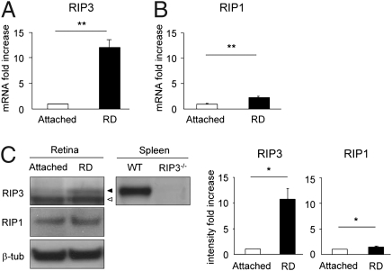
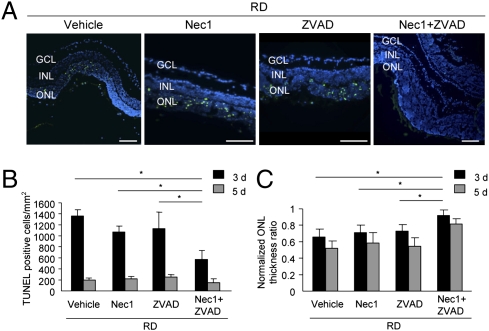
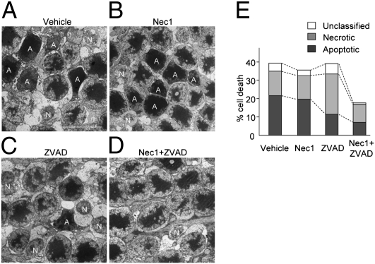
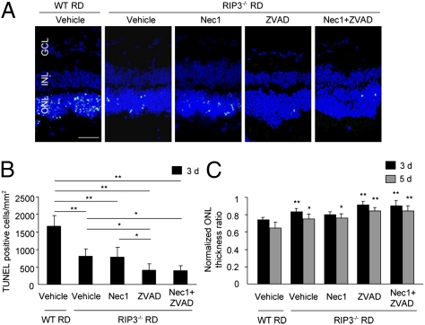
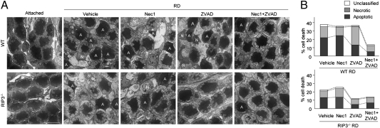
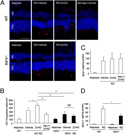
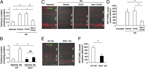
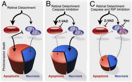
Apoptosis has been shown to be a significant form of cell loss in many diseases. Detachment of photoreceptors from the retinal pigment epithelium, as seen in various retinal disorders, causes photoreceptor loss and subsequent vision decline. Although caspase-dependent apoptotic pathways are activated after retinal detachment, caspase inhibition by the pan-caspase inhibitor Z-VAD fails to prevent photoreceptor death; thus, we investigated other pathways leading to cell loss. Here, we show that receptor interacting protein (RIP) kinase-mediated necrosis is a significant mode of photoreceptor cell loss in an experimental model of retinal detachment and when caspases are inhibited, RIP-mediated necrosis becomes the predominant form of death. RIP3 expression, a key activator of RIP1 kinase, increased more than 10-fold after retinal detachment. Morphological assessment of detached retinas treated with Z-VAD showed decreased apoptosis but significantly increased necrotic photoreceptor death. RIP1 kinase inhibitor necrostatin-1 or Rip3 deficiency substantially prevented those necrotic changes and reduced oxidative stress and mitochondrial release of apoptosis-inducing factor. Thus, RIP kinase-mediated programmed necrosis is a redundant mechanism of photoreceptor death in addition to apoptosis, and simultaneous inhibition of RIP kinases and caspases is essential for effective neuroprotection and may be a novel therapeutic strategy for treatment of retinal disorders.


Conflict of interest statement

Conflict of interest statement: G.T. and D.G.V. have a provisional patent filling.

Figures

Fig. 1.
Fig. 1.
Increases in RIP3 and RIP1 expression after retinal detachment. Quantitative real-time PCR analysis for RIP3 (A) and RIP1 (B) in control retina without retinal detachment and in retina 3 d after retinal detachment (n = 9 each); **P < 0.01. (C) Western blot analysis for RIP3 and RIP1 after retinal detachment (n = 4 each). Lane-loading differences were normalized by levels of β-tubulin. For RIP3 analysis, spleen samples from WT and Rip3−/− animals were used as positive and negative controls, respectively. Black arrowhead indicates RIP3; white arrowhead indicates nonspecific band. The bar graphs indicate the relative level of RIP3 and RIP1 to β-tubulin by densitometric analysis, reflecting the results from four independent experiments (*P < 0.05).
Fig. 2.
Fig. 2.
Nec-1 combined with Z-VAD prevents photoreceptor loss after retinal detachment. (A) TUNEL (green) and DAPI (blue) staining in detached retina treated with vehicle, Nec-1, Z-VAD, or Nec-1 plus Z-VAD on day 3 after retinal detachment. Quantification of TUNEL-positive photoreceptors (B) and ONL thickness ratio (C) on day 3 (vehicle, n = 12; Nec-1, n = 6; Z-VAD, n = 12; Nec-1 plus Z-VAD, n = 12) and day 5 (vehicle, n = 8; Nec-1, n = 6; Z-VAD, n = 8; Nec-1 plus Z-VAD, n = 8) after retinal detachment (*P < 0.05). GCL, ganglion cell layer, INL, inner nuclear layer. (Scale bar, 100 μm.)
Fig. 3.
Fig. 3.
Involvement of programmed necrosis during retinal detachment-induced photoreceptor death. (AD) TEM photomicrographs in the ONL on day 3 after retinal detachment in retina treated with vehicle (A), Nec-1 (B), Z-VAD (C), or Nec-1 plus Z-VAD (D). A, apoptotic cell; N, necrotic cell. (E) Quantification of apoptotic and necrotic photoreceptor death after retinal detachment (n = 4 each). Z-VAD treatment increased necrotic cells whereas decreased apoptotic cells (P < 0.05 vs. vehicle). Nec-1 plus Z-VAD significantly suppressed the necrotic cell death (P < 0.01 vs. Z-VAD). (Scale bar, 5 μm.)
Fig. 4.
Fig. 4.
Rip3 deficiency provides neuroprotection after retinal detachment that is augmented by Z-VAD. (A) TUNEL (green) and DAPI (blue) staining in WT and Rip3−/− retina treated with vehicle, Nec-1, Z-VAD, or Nec-1 plus Z-VAD on day 3 after retinal detachment. Quantification of TUNEL-positive photoreceptors (B) and ONL thickness ratio (C) on day 3 (WT vehicle, n = 6; Rip3−/− vehicle, n = 7; Rip3−/− Nec-1, n = 8; Rip3−/− Z-VAD, n = 7; Rip3−/− Nec-1 plus Z-VAD, n = 6) and day 5 (WT vehicle, n = 7; Rip3−/− vehicle, n = 6; Rip3−/− Nec-1, n = 6; Rip3−/− Z-VAD, n = 6; Rip3−/− Nec-1 plus Z-VAD, n = 6) after retinal detachment (*P < 0.05; **P < 0.01). (Scale bar, 50 μm.)
Fig. 5.
Fig. 5.
Rip3 deficiency inhibits induction of programmed necrosis and prevents photoreceptor death after retinal detachment. (A) TEM photomicrographs in the ONL on day 3 after retinal detachment in WT and Rip3−/− retina treated with vehicle, Nec-1, Z-VAD, or Nec-1 plus Z-VAD. In untreated attached retina, the retinal morphology was similar in WT and Rip3−/− mice. A, apoptotic cell; N, necrotic cell. (B) Quantification of apoptotic and necrotic photoreceptor death after retinal detachment (n = 4 each). Rip3 deficiency inhibits the switch to necrotic cell death by Z-VAD treatment and prevents photoreceptor death after retinal detachment. (Scale bar, 5 μm.)
Fig. 6.
Fig. 6.
Effect of RIP pathways on inflammatory response after retinal detachment. (A and B) Immunofluorescence for CD11b (A) and quantification of CD11b-positive macrophage/microglia in WT and Rip3−/− retina after retinal detachment. In WT mice, Z-VAD treatment significantly increased infiltration of CD11b-positive cells compared with vehicle treatment (P < 0.05). This increase of CD11b-positive cells was significantly suppressed with Nec-1 plus Z-VAD treatment or Rip3 deficiency (P < 0.01). (Scale bar, 50 μm.) (C and D) ELISA for MCP-1 on day 3 after retinal detachment in retina treated with vehicle (n = 5), Z-VAD (n = 5), or Nec-1 plus Z-VAD (n = 6) (C) and in WT and Rip3−/− retina (n = 5 each) (D). The retinas without retinal detachment were used as controls. *P < 0.05; **P < 0.01; NS, not significant.
Fig. 7.
Fig. 7.
RIP kinase inhibition prevents ROS production and AIF nuclear translocation after retinal detachment. (A and B) ELISA to detect carbonyl contents on day 3 after retinal detachment in retina treated with vehicle, Z-VAD or Nec-1 plus Z-VAD (n = 7 each) (A) and in WT and Rip3−/− retina (n = 10 each) (B). The retinas without retinal detachment were used as controls (n = 6–8). Double staining for AIF and TUNEL (C and E) and quantification of AIF/TUNEL double-positive photoreceptors (D and F) on day 3 after retinal detachment in retina treated with vehicle, Z-VAD, or Nec-1 plus Z-VAD (n = 6 each; C and D) and in WT and Rip3−/− retina (n = 5 each; E and F). IS, inner segment. *P < 0.05; **P < 0.01. (Scale bar, 50 μm.)
Fig. 8.
Fig. 8.
Proposed mechanism of photoreceptor loss after retinal detachment. (A) After retinal detachment, photoreceptor death is caused mainly by apoptosis. (B) Caspase inhibition by Z-VAD decreases apoptosis but promotes RIP-mediated programmed necrosis. (C) Blockade of both caspases and RIP kinases is essential for effective prevention of photoreceptor loss.

References

    1. Dunaief JL, Dentchev T, Ying GS, Milam AH. The role of apoptosis in age-related macular degeneration. Arch Ophthalmol. 2002;120:1435–1442. - PubMed
    1. Barber AJ, et al. Neural apoptosis in the retina during experimental and human diabetes. Early onset and effect of insulin. J Clin Invest. 1998;102:783–791. - PMC - PubMed
    1. Cook B, Lewis GP, Fisher SK, Adler R. Apoptotic photoreceptor degeneration in experimental retinal detachment. Invest Ophthalmol Vis Sci. 1995;36:990–996. - PubMed
    1. Arroyo JG, Yang L, Bula D, Chen DF. Photoreceptor apoptosis in human retinal detachment. Am J Ophthalmol. 2005;139:605–610. - PubMed
    1. Campo RV, et al. Pars plana vitrectomy without scleral buckle for pseudophakic retinal detachments. Ophthalmology. 1999;106:1811–1815. - PubMed

Publication types

MeSH terms