. 2010 Dec 14;107(50):21743-8.
doi: 10.1073/pnas.1010243107. Epub 2010 Nov 22.

Recognition of tandem PxxP motifs as a unique Src homology 3-binding mode triggers pathogen-driven actin assembly

Affiliations

Recognition of tandem PxxP motifs as a unique Src homology 3-binding mode triggers pathogen-driven actin assembly

Olli Aitio et al. Proc Natl Acad Sci U S A. .

Abstract

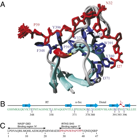
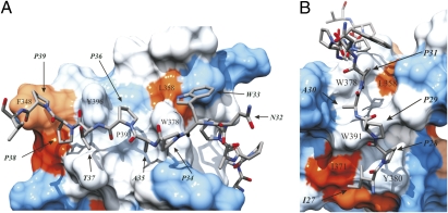
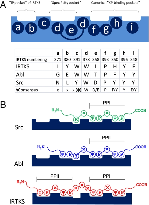
Src homology 3 (SH3) domains are globular protein interaction modules that regulate cell behavior. The classic SH3 ligand-binding site accommodates a hydrophobic PxxP motif and a positively charged specificity-determining residue. We have determined the NMR structure of insulin receptor tyrosine kinase substrate (IRTKS) SH3 domain in complex with a repeat from Escherichia coli-secreted protein F-like protein encoded on prophage U (EspF(U)), a translocated effector of enterohemorrhagic E. coli that commandeers the mammalian actin assembly machinery. EspF(U)-IRTKS interaction is among the highest affinity natural SH3 ligands. Our complex structure reveals a unique type of SH3 interaction based on recognition of tandem PxxP motifs in the ligand. Strikingly, the specificity pocket of IRTKS SH3 has evolved to accommodate a polyproline type II helical peptide analogously to docking of the canonical PxxP by the conserved IRTKS SH3 proline-binding pockets. This cooperative binding explains the high-affinity SH3 interaction and is required for EspF(U)-IRTKS interaction in mammalian cells as well as the formation of localized actin "pedestals" beneath bound bacteria. Importantly, tandem PxxP motifs are also found in mammalian ligands and have been shown to contribute to IRTKS SH3 recognition similarly.


Conflict of interest statement

The authors declare no conflict of interest.

Figures

Fig. 1.
Fig. 1.
(A) Ribbon presentation of the ensemble of 20 superimposed NMR structures of IRTKS SH3:EspFU R475 complex. The heavy atoms of R475 (residues 27–40) and IRTKS SH3 domain residues interacting with R475 are shown in red and blue, respectively. (B) Secondary structure elements of IRTKS SH3, with residues interacting with R475 underlined and colored in blue. (C) Sequence of R475, with amino acid numbering from 1 to 47, corresponding to residues 268–314 in full-length EspFU. The ‘H’ and ‘P’ regions important for the EspFU-WASP GTPase-binding domain (GBD) and EspFU-IRTKS SH3 interactions are highlighted.
Fig. 2.
Fig. 2.
Close-ups of C-terminal and N-terminal R475 binding sites on IRTKS SH3 as a hydrophobicity surface presentation. (A) C-terminal binding region. Residues forming the conserved proline-binding pockets are labeled on the surface. Heavy atoms of the C-terminal part of R475 residues N32APTPP39 are highlighted as a stick model: nitrogen (blue), oxygen (red), and carbon (gray). Surface coloring is according to the scale of Kyte and Doolittle (37). Blue corresponds to the most hydrophilic, white to intermediate, and red to the most hydrophobic. (B) N-terminal PPII helix of EspFU R475. The two hydrophobic clefts formed by L358, W378, W391, Y380, and I371 and the heavy atom of R475 residues I27PPAP31 occupying the clefts are shown.
Fig. 3.
Fig. 3.
Fine mapping of IRTKS SH3-binding preferences. Peptide array technology was used to define the residues critical for IRTKS SH3 binding in the EspFU R475 and in a panel of cellular proteins reported or suspected to interact with IRTKS/IRSp53. (A) Sixteen residues encompassing the region of R475 observed to contact IRTKS SH3 were systematically replaced with Ala. At positions naturally occupied by Ala, an uncharged polar amino acid (Asn) was used instead. Binding signals were quantified and classified into four categories. Strong indicates >85%, medium indicates 30–85%, modest indicates 5–29%, and weak/none indicates <5% of the average signal from triplicate dots printed with the corresponding unmodified peptide. (B) N- and C-terminal ends and the linker region (shaded in yellow) of 16-mer IRTKS SH3-binding peptide were altered as indicated (red font) and examined as above. (C) IRTKS binding of the 16-mer EspFU peptide was compared with that of similar peptides from cellular proteins. Human (h) and mouse (m) Eps8 protein sequence differ in this region and have both been included. N-terminal truncated versions of Shank2/3 peptides (*) were included because an earlier study suggested the minimal IRSp53 SH3-binding site to reside within this sequence (24).
Fig. 4.
Fig. 4.
Tandem PxxP motifs are required for EspFU recruitment and pedestal formation by EHEC. FLCs were infected for 3.5 h with strain EPEC KC12 expressing the EspFU HP (R475), PHP, PHPP28/31A, and PHPP36/39A variants and were visualized using fluorescent microscopy after staining with DAPI to localize bacteria (blue), FITC–anti-myc antibody was used to detect EspFU derivatives (green), and Alexa568-phalloidin was used to detect F-actin (red).
Fig. 5.
Fig. 5.
Schematic presentation of class I peptide binding to Src-, Abl-, and IRTKS-type SH3 domains. (A) Amino acids (a to i) forming the individual peptide-binding pockets of SH3 domains (numbering according to IRTKS SH3 in Fig. 1). (B) Peptide recognition is similar for all SH3 domains at the canonical “XP-binding pockets,” wherein each peptide adopts a left-handed PPII helical conformation. Selectivity is determined at the “specificity pocket,” wherein peptide conformation and peptide/SH3 domain interactions are different in each case. In IRTKS SH3, the specificity and “IP pockets” also accommodate PPII helical moiety.

References

    1. Mayer BJ. SH3 domains: Complexity in moderation. J Cell Sci. 2001;114:1253–1263. - PubMed
    1. Kaneko T, Li L, Li SS. The SH3 domain—A family of versatile peptide- and protein-recognition module. Front Biosci. 2008;13:4938–4952. - PubMed
    1. Yu H, et al. Structural basis for the binding of proline-rich peptides to SH3 domains. Cell. 1994;76:933–945. - PubMed
    1. Musacchio A, Saraste M, Wilmanns M. High-resolution crystal structures of tyrosine kinase SH3 domains complexed with proline-rich peptides. Nat Struct Biol. 1994;1:546–551. - PubMed
    1. Feng S, Chen JK, Yu H, Simon JA, Schreiber SL. Two binding orientations for peptides to the Src SH3 domain: Development of a general model for SH3-ligand interactions. Science. 1994;266:1241–1247. - PubMed

Publication types

MeSH terms

Associated data