. 2010 Dec 14;107(50):21830-5.
doi: 10.1073/pnas.0912793107. Epub 2010 Nov 22.

Biguanide metformin acts on tau phosphorylation via mTOR/protein phosphatase 2A (PP2A) signaling

Affiliations

Biguanide metformin acts on tau phosphorylation via mTOR/protein phosphatase 2A (PP2A) signaling

Eva Kickstein et al. Proc Natl Acad Sci U S A. .

Abstract

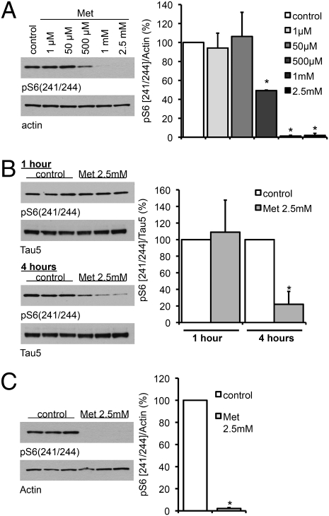
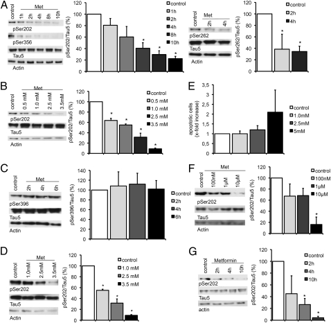
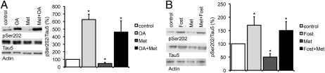
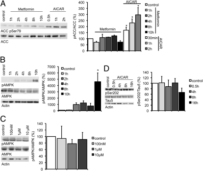
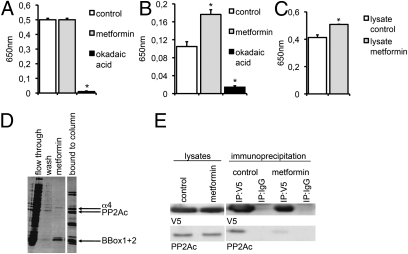
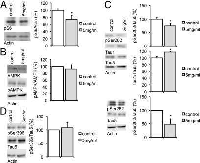
Hyperphosphorylated tau plays an important role in the formation of neurofibrillary tangles in brains of patients with Alzheimer's disease (AD) and related tauopathies and is a crucial factor in the pathogenesis of these disorders. Though diverse kinases have been implicated in tau phosphorylation, protein phosphatase 2A (PP2A) seems to be the major tau phosphatase. Using murine primary neurons from wild-type and human tau transgenic mice, we show that the antidiabetic drug metformin induces PP2A activity and reduces tau phosphorylation at PP2A-dependent epitopes in vitro and in vivo. This tau dephosphorylating potency can be blocked entirely by the PP2A inhibitors okadaic acid and fostriecin, confirming that PP2A is an important mediator of the observed effects. Surprisingly, metformin effects on PP2A activity and tau phosphorylation seem to be independent of AMPK activation, because in our experiments (i) metformin induces PP2A activity before and at lower levels than AMPK activity and (ii) the AMPK activator AICAR does not influence the phosphorylation of tau at the sites analyzed. Affinity chromatography and immunoprecipitation experiments together with PP2A activity assays indicate that metformin interferes with the association of the catalytic subunit of PP2A (PP2Ac) to the so-called MID1-α4 protein complex, which regulates the degradation of PP2Ac and thereby influences PP2A activity. In summary, our data suggest a potential beneficial role of biguanides such as metformin in the prophylaxis and/or therapy of AD.


Conflict of interest statement

The authors declare no conflict of interest.

Figures

Fig. 1.
Fig. 1.
Metformin decreases the phosphorylation of S6 ribosomal protein. (A) Primary cortical neurons of wild-type mice were treated with metformin (Met) at the indicated concentrations for 16 h. (B) Primary cortical neurons of wild-type mice were treated with or without 2.5 mM metformin for 1 h and 4 h. (C) H4 cells were treated with or without 2.5 mM metformin for 16 h. Cell lysates were analyzed by Western blotting using an antibody to phosphorylated S6 (241/244). Band intensities were normalized to either actin or total tau (Tau5). In each case a representative blot is shown along with the quantification of three independent experiments (n = 3). *P < 0.01.
Fig. 2.
Fig. 2.
Metformin induces dephosphorylation of tau at PP2A-sensitive sites. Representative Western blots and quantifications of three or four independent experiments are shown in A–D, F, and G. Band intensities were normalized to actin and compared with total tau (Tau 5). (A) Primary cortical neurons of wild-type mice were treated with 2.5 mM metformin (Met) over increasing time intervals. Cell lysates were analyzed by Western blot using antibodies detecting phosphorylation at specific PP2A-sensitive tau sites (Ser202, Ser262, Ser356; n = 3). (B) Primary cortical neurons from wild-type mice were treated with increasing concentration of metformin over 4 h. Cell lysates were analyzed by Western blot using an antibody detecting phosphorylation at Ser202 (n = 4). (C) Primary cortical neurons of wild-type mice were treated with 2.5 mM metformin over increasing time intervals. Cell lysates were analyzed by Western blot using an antibody detecting phosphorylation at the PP2A-insensitive tau site Ser396 (n = 3). (D) Primary cortical neurons of transgenic mice expressing human tau instead of murine tau were treated with increasing concentration of metformin over 4 h. Cell lysates were analyzed by Western blot using an antibody detecting phosphorylation at Ser202 (n = 3). (E) TUNEL assay of primary cortical neurons of wild-type mice treated with increasing concentrations of metformin. Percentages of apoptotic cells are shown. (F) Primary cortical neurons of wild-type mice were treated with low concentrations of metformin over 24 h. Cell lysates were analyzed by Western blot using an antibody detecting phosphorylation at Ser202 (n = 3). (G) Primary cortical neurons of wild-type mice were treated with 2.5 mM metformin over increasing time intervals in medium without insulin. Cell lysates were analyzed by Western blot using an antibody detecting phosphorylation at Ser202 (n = 3). *P < 0.05.
Fig. 3.
Fig. 3.
Inhibition of PP2A blocks the metformin effect on the phosphorylation of tau. Representative Western blots and quantifications of three or four independent experiments are shown in the subsequent experiments. Band intensities were normalized to actin and compared with total tau (Tau 5). (A) Primary cortical neurons of wild-type mice were treated with either OA (10 nM), only metformin (Met, 2.5 mM), or with OA (10 nM) before metformin (2.5 mM) for 4 h. Cell lysates were analyzed by Western blot using an antibody detecting phosphorylation at a specific PP2A-sensitive tau site (n = 4). *P < 0.05. (B) Primary cortical neurons of wild-type mice were treated with either fostriecin (Fost, 1 μM), only metformin (Met, 2.5 mM), or with fostriecin (1 μM) before metformin (2.5 mM) over 4 h. Cell lysates were analyzed by Western blot using an antibody detecting phosphorylation at a specific PP2A-sensitive tau site (n = 3). *P < 0.05.
Fig. 4.
Fig. 4.
Metformin effects on the phosphorylation of tau are AMPK independent. Representative Western blots and quantifications of three to five independent experiments are shown in the subsequent experiments. (A) Primary cortical neurons of wild-type mice were treated with either 2.5 mM of metformin (Met) or with 5 mM of AICAR over increasing time intervals. Cell lysates were analyzed by Western blot using an antibody detecting phosphorylation of the AMPK target ACC. Band intensities were compared with total ACC (n = 4). (B) Primary cortical neurons of wild-type mice were treated with 2.5 mM metformin over increasing time intervals. Cell lysates were analyzed by Western blot using an antibody detecting phosphorylated AMPK (pThr 172). Band intensities were compared with total AMPK (n = 3). (C) Primary cortical neurons of wild-type mice were treated with increasing concentrations of metformin over 24 h. Cell lysates were analyzed by Western blot using an antibody detecting phosphorylation of AMPK (pThr 172). Band intensities were normalized to AMPK total (n = 3). (D) Primary cortical neurons of wild-type mice were treated with 5 mM of AICAR over increasing time intervals. Cell lysates were analyzed by Western blot using an antibody detecting phosphorylation at a specific PP2A-sensitive tau site. Band intensities were normalized to actin and compared with total tau (Tau 5; n = 5). *P < 0.05.
Fig. 5.
Fig. 5.
Metformin increases PP2A activity in vitro. Three independent experiments were measured in A–C. (A) Purified PP2Ac was incubated with or without metformin (10 mM) or OA (10 nM). PP2A activity was determined in a malachite green assay. (B) Immunoprecipitated PP2A holoenzymes were incubated with or without metformin (10 mM) or OA (10 nM). PP2A activity was determined in a malachite green assay. (C) Cells were treated with or without metformin (2.5 mM) and lysed. PP2A activity was determined in a malachite green assay (n = 3). *P < 0.05. (D) Assembly of the PP2Ac- MID1-α4 protein complex is influenced by metformin. Affinity chromatography analysis shows the binding of immobilized PP2Ac to the MID1 B-box domains and α4. The flow-through, the washing fraction, the fraction obtained with a buffer containing 100 μM metformin, and the final elution fraction were collected and analyzed on SDS gels. Gels were stained with Coomassie. (E) Association between PP2Ac and α4 is influenced by metformin. Coimmunoprecipitations of PP2Ac and V5-tagged α4 in the presence or absence of metformin were performed and analyzed on Western blots using PP2Ac- or V5-specific antibodies. This experiment has been repeated three times.
Fig. 6.
Fig. 6.
Metformin decreases tau phosphorylation in vivo. Pairs of wild-type mice were fed with or without 5 mg/mL of metformin in the drinking water for 16–24 d. Representative Western blots and quantification of three technical replicates are shown in the subsequent experiments. (A) Brains were lysed and analyzed via Western blots for phosphorylation of S6. Band intensities were compared with actin. (B) Brains were lysed and analyzed via Western blots for phosphorylation of AMPK (pThr 172). Band intensities were compared with AMPK total. (C) Brains were lysed and analyzed via Western blots for phosphorylation of Tau. pSer202 was detected using a phospho-specific pSer202 antibody and a tau1 antibody, which detects dephosphorylated Ser202. Phosphorylation at position Ser262 was detected using a phospho-specific Ser262 antibody. Band intensities were normalized to actin and compared with total tau (Tau5). (D) Brains were lysed and analyzed via Western blots for phosphorylation of the PP2A-insensitive site pSer396. Band intensities were normalized to actin and compared with total tau (Tau5). *P < 0.05.

References

    1. Gong CX, Liu F, Grundke-Iqbal I, Iqbal K. Post-translational modifications of tau protein in Alzheimer's disease. J Neural Transm. 2005;112:813–838. - PubMed
    1. Alonso AD, Grundke-Iqbal I, Barra HS, Iqbal K. Abnormal phosphorylation of tau and the mechanism of Alzheimer neurofibrillary degeneration: Sequestration of microtubule-associated proteins 1 and 2 and the disassembly of microtubules by the abnormal tau. Proc Natl Acad Sci USA. 1997;94:298–303. - PMC - PubMed
    1. Hanger DP, Betts JC, Loviny TL, Blackstock WP, Anderton BH. New phosphorylation sites identified in hyperphosphorylated tau (paired helical filament-tau) from Alzheimer's disease brain using nanoelectrospray mass spectrometry. J Neurochem. 1998;71:2465–2476. - PubMed
    1. Goedert M, Jakes R, Qi Z, Wang JH, Cohen P. Protein phosphatase 2A is the major enzyme in brain that dephosphorylates tau protein phosphorylated by proline-directed protein kinases or cyclic AMP-dependent protein kinase. J Neurochem. 1995;65:2804–2807. - PubMed
    1. Sontag E, Nunbhakdi-Craig V, Lee G, Bloom GS, Mumby MC. Regulation of the phosphorylation state and microtubule-binding activity of Tau by protein phosphatase 2A. Neuron. 1996;17:1201–1207. - PubMed

Publication types

MeSH terms