. 2011 Jan 27;117(4):1301-7.
doi: 10.1182/blood-2010-08-302505. Epub 2010 Nov 22.

Immunoglobulin light chains activate nuclear factor-κB in renal epithelial cells through a Src-dependent mechanism

Affiliations

Immunoglobulin light chains activate nuclear factor-κB in renal epithelial cells through a Src-dependent mechanism

Wei-Zhong Ying et al. Blood. .

Abstract

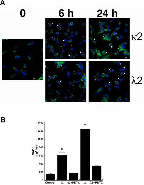
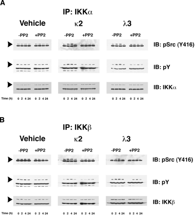
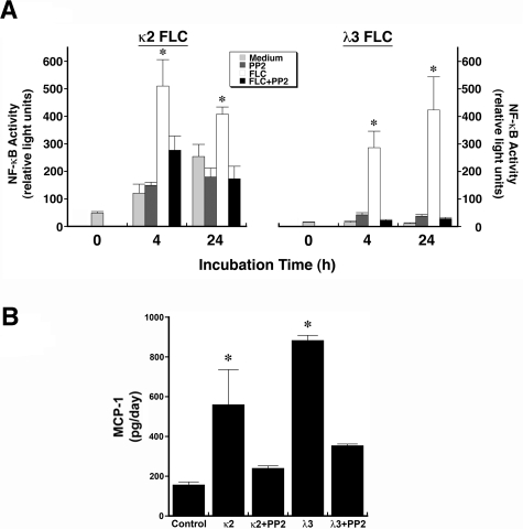
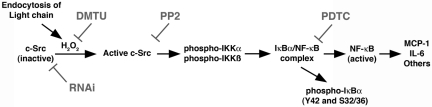
One of the major attendant complications of multiple myeloma is renal injury, which contributes significantly to morbidity and mortality in this disease. Monoclonal immunoglobulin free light chains (FLCs) are usually directly involved, and tubulointerstitial renal injury and fibrosis are prominent histologic features observed in myeloma. The present study examined the role of monoclonal FLCs in altering the nuclear factor κ light chain enhancer of activated B cells (NF-κB) activity of renal epithelial cells. Human proximal tubule epithelial cells exposed to 3 different human monoclonal FLCs demonstrated Src kinase-dependent activation of the NF-κB pathway, which increased production of monocyte chemoattractant protein-1 (MCP-1). Tyrosine phosphorylation of inhibitor of κB kinases (IKKs) IKKα and IKKβ and a concomitant increase in inhibitor of κB (IκB) kinase activity in cell lysates were observed. Time-dependent, Src kinase-dependent increases in serine and tyrosine phosphorylation of IκBα and NF-κB activity were also demonstrated. Proteasome inhibition partially blocked FLC-induced MCP-1 production. These findings fit into a paradigm characterized by FLC-induced redox-signaling events that activated the canonical and atypical (IKK-independent) NF-κB pathways to promote a proinflammatory, profibrotic renal environment.


Figures

Figure 1
Figure 1
Incubation of human proximal tubular epithelial cells with 2 different monoclonal FLCs (κ2 and λ2) increases the production of MCP-1 through activation of the NF-κB pathway. (A) Confocal laser scanning microscopy using the LSM 710 confocal microscope (Carl Zeiss MicroImaging) and the accompanying LSM 710 ZEN software demonstrated nuclear localization (arrowheads) of RelA (p65) in cells within 6 hours of exposure to κ2 and λ2 FLCs (1 mg/mL). The white bar represents 10 μm. (B) Increase in MCP-1 production induced by 24-hour incubation of renal epithelial cells with the FLCs was inhibited by PDTC, an inhibitor of NF-κB. *P < .05 compared with samples incubated in medium alone (control) and in medium containing the corresponding FLCs and PDTC; n = 6 experiments in each group.
Figure 2
Figure 2
FLCs promote a time-dependent coimmunoprecipitation of IKKα and IKKβ with activated c-Src, which induces tyrosine phosphorylation of these IκB kinases. Renal epithelial cells were incubated with 2 different FLCs (κ2 and λ3) and vehicle. At different time points (0, 2, 4, and 24 hours), the experiments were stopped and cell lysates were produced to immunoprecipitate either IKKα (A) or IKKβ (B). These experiments demonstrated a time-dependent association with activated (phosphorylated) c-Src (top panels) in lysates obtained from cells exposed to the monoclonal FLCs but not medium alone. The approximate 2- to 4-fold increase in association of activated c-Src with the IκB kinases was prevented by the addition of PP2. The middle panels of (A) and (B) demonstrate a contemporaneous 2- to 4-fold increase in tyrosine phosphorylation of the IκB kinases in those cells exposed to the FLCs but not vehicle. The bottom panels demonstrated the presence of similar amounts of IKKα (A) or IKKβ (B) in the immune precipitate. The other major band in the gels represents the heavy-chain component of the antibody used to precipitate the protein complexes.
Figure 3
Figure 3
FLCs (κ2 and λ3) induce Src kinase-dependent serine and tyrosine phosphorylation of IκBα in renal epithelial cells. Renal epithelial cells were incubated with 2 different FLCs (κ2 and λ3) and vehicle. At different time points (0, 2, 4, and 24 hours), the experiments were stopped and cell lysates were produced to immunoprecipitate IκBα. Incubation of cells with the FLCs κ2 and λ3, but not the vehicle, promoted a > 2-fold increase in serine phosphorylation of IκBα (top panels), indicating activation of the canonical NF-κB pathway. This effect, which was observed as early as after the first 2 hours of incubation, was inhibited by the Src kinase inhibitor PP2. Both FLCs, but not the vehicle, generated a 4-fold increase in tyrosine phosphorylation of IκBα (middle panels); this was also inhibited by PP2. The bottom panels demonstrate the presence of IκBα in all of the samples studied. The other major band in the gels represents the heavy-chain component of the antibody used to precipitate the protein complexes.
Figure 4
Figure 4
Incubation of renal epithelial cells with 2 different FLCs (κ2 and λ3) increase nuclear NF-κB activity and promote MCP-1 production in an Src kinase–dependent fashion. (A) Nuclear lysates were obtained from HK-2 cells after 4 and 24 hours of incubation in medium containing 2 different FLCs (κ2 and λ3). Nuclear NF-κB activity was quantified using a filter plate assay, which consisted initially of incubating nuclear extracts with a biotinylated NF-κB–specific DNA-binding sequence. This NF-κB DNA complex was captured on a filter plate to remove the unbound probes and was then denatured and hybridized onto precoated microwells. Bound NF-κB DNA complexes were detected with streptavidin–horseradish peroxidase and quantified using a luminometer. Results, expressed in relative light units, showed an increase in nuclear NF-κB activity at 4 and 24 hours of incubation of renal epithelial cells with κ2 and λ3 FLCs compared with medium alone. The addition of PP2 to the medium inhibited the FLC-induced increase in activity. *P < .05 compared with contemporaneous samples incubated in medium alone, PP2, and corresponding FLCs and PP2; n = 6-12 experiments in each group. (B) Increase in production of MCP-1 after overnight incubation of renal epithelial cells with the κ2 and λ3 FLCs; the addition of PP2 prevented this FLC-induced increase in MCP-1. *P < .05 compared with control and samples treated with PP2 and corresponding FLCs; n = 6 experiments in each group
Figure 5
Figure 5
Exposure of renal epithelial cells to PS-341 in concentrations < 22nM does not produce cytotoxicity. To determine the sensitivity of HK-2 cells to PS-341, cells were incubated overnight in medium containing PS-341 in concentrations between 0.8 and 200nM. Although concentrations up to 7mM were well tolerated, cell loss was observed at 22 and 200nM.
Figure 6
Figure 6
Overnight coincubation of PS-341 (7nM) with FLCs (κ2 and λ2) inhibits renal epithelial cell production of MCP-1. HK-2 cells were incubated overnight in medium that contained PS-341 (7mM) and 2 different FLCs (κ2 and λ2). While PS-341 did not affect MCP-1 production by cells incubated in medium alone, FLC-induced MCP-1 production was partially inhibited. *P < .05 compared with sample containing the corresponding FLCs; n = 6 in each group
Figure 7
Figure 7
Diagram depicting the proposed signal transduction pathway that is initiated after endocytosis of FLCs into renal epithelial cells and leads to activation of the canonical and atypical NF-κB pathways. Data supporting this pathway have been published previously, and are also found in the present study. RNAi, RNA interference; DMTU, 1,3-dimethyl-2-thiourea.

References

    1. Greenlee RT, Murray T, Bolden S, Wingo PA. Cancer statistics, 2000. CA Cancer J Clin. 2000;50(1):7–33. - PubMed
    1. Kyle RA, Gertz MA, Witzig TE, et al. Review of 1027 patients with newly diagnosed multiple myeloma. Mayo Clin Proc. 2003;78(1):21–33. - PubMed
    1. Abbott KC, Agodoa LY. Multiple myeloma and light chain-associated nephropathy at end-stage renal disease in the United States: patient characteristics and survival. Clin Nephrol. 2001;56(3):207–210. - PubMed
    1. Sanders PW. Myeloma and secondary involvement of the kidney in dysproteinemias. In: Wilcox CS, editor. Therapy in nephrology and hypertension: a companion to Brenner and Rector's the kidney. 5th ed. London, United Kingdom: WB Saunders; 2008. pp. 461–468.
    1. Batuman V, Dreisbach AW, Cyran J. Light-chain binding sites on renal brush-border membranes. Am J Physiol. 1990;258(5 Pt 2):F1259–F1265. - PubMed

Publication types

MeSH terms