. 2011 Feb 11;286(6):4357-63.
doi: 10.1074/jbc.M110.170134. Epub 2010 Nov 23.

RNA polymerase 1-driven transcription as a mediator of BDNF-induced neurite outgrowth

Affiliations

RNA polymerase 1-driven transcription as a mediator of BDNF-induced neurite outgrowth

Cynthia Gomes et al. J Biol Chem. .

Abstract

Neurite outgrowth is essential for development of the nervous system. Neurotrophins including BDNF are among extracellular signals that regulate neurite outgrowth. The ERK1/2 pathway contributes to intracellular signaling networks transducing the pro-neuritic effects of BDNF. In the nucleolus, RNA polymerase-1 (Pol1)-mediated transcription regulates ribosomal biogenesis, enabling cellular protein synthesis and growth. Hence, we tested the possibility that Pol1 is an effector for pro-neuritic signals such as BDNF. We report that Pol1-mediated nucleolar transcription was increased by BDNF in an ERK1/2-dependent manner in rat forebrain neurons. Conversely, in cultured hippocampal neurons, knockdown of a Pol1 coactivator, transcription initiation factor 1A (TIF1A), attenuated BDNF- or ERK1/2-induced neurite outgrowth. Also, upon overexpression, a constitutively active mutant of TIF1A strongly promoted neurite outgrowth, including increases in total neurite length and branching. Finally, overexpression of wild-type TIF1A enhanced the pro-neuritic effects of ERK1/2 activation. These observations indicate that the Pol1-mediated nucleolar transcription regulates neurite outgrowth and serves as a major pro-neuritic effector of the BDNF-activated ERK1/2 pathway. Thus, development of the nervous system appears critically dependent on the nucleolus.


Figures

FIGURE 1.
FIGURE 1.
BDNF activates nucleolar transcription. A and B, BDNF stimulation increased the 45 S pre-rRNA/18 S rRNA ratio. In A, DIV6 cortical neuron cultures were used; averages of three independent experiments are shown. In B, postnatal day 7 rat pups (four animals/condition) received BDNF injections into the left lateral ventricle; after 4 h, ipsilateral cortices and hippocampi were dissected and analyzed. C, blocking endogenous BDNF signaling with the soluble TrkB-Fc peptide reduced 45 S pre-rRNA levels. DIV6 cortical neurons were treated with 0.5 μg of TrkB-Fc or IgG control for 6 h; averages of three independent experiments are depicted. D, BDNF activated the rDNA promoter. DIV4 cortical neurons were cotransfected with the rDNA promoter-luciferase reporter construct and the pEF1αLacZ plasmid (0.2 + 0.2 μg of plasmid DNAs/5 × 105 neurons, respectively). Two days later, cells were treated with BDNF as indicated. The activity of the rDNA promoter was determined by the activity ratio of luciferase to β-gal. Data represent four sister cultures from three independent experiments. E and F, increased nascent RNA levels in neuronal nucleoli after BDNF stimulation of cortical cultures. At DIV6, BDNF was added to the cells as indicated, followed by a 1-h incubation with the RNA precursor 5-EU (1 mm). After fixation and co-immunofluorescence for the neuronal marker MAP2 and the nucleolar marker B23, nascent RNA was visualized using click chemistry (see “Experimental Procedures” for more details). In MAP2-positive neurons, BDNF increased the ratio between nucleolar and nuclear nascent RNA levels. The nucleolar compartment was defined by B23 immunofluorescence. In F, data are the means of at least 75 randomly selected individual neurons/condition from three independent experiments. In all graphs, error bars indicate S.E. *, p < 0.05; **, p < 0.01; ***, p < 0.001.
FIGURE 2.
FIGURE 2.
Pol1-driven transcription is required for BDNF-induced morphogenesis. DIV6 hippocampal neurons were cotransfected with expression plasmids for GFP (pmaxGFP) and shTIF1A or control Renilla luciferase shRNA (shLuc; 0.1 + 0.6 μg of plasmid DNAs/2 × 105 neurons, respectively). The expression plasmid for the dominant-negative mutant of p53 (CMV-p53-DD; 0.2 μg of DNA/2 × 105 cells) was added to all transfections to prevent apoptotic consequences of blocking neuronal Pol1 (18). The next day, neurons were treated with BDNF for 24 h. Transfected neurons were visualized by GFP immunostaining. A, representative images of transfected neurons. B–D, BDNF-induced increases in total neurite length, neurite branching, and perikaryal volume, respectively, were reduced by shTIF1A. E, the number of primary neurites was unaffected with or without BDNF. Data are means ± S.E. of at least 45 randomly selected individual neurons/condition from three independent experiments. ***, p < 0.001; NS (not significant), p > 0.05.
FIGURE 3.
FIGURE 3.
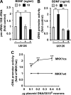
BDNF regulates Pol1-driven transcription through the ERK1/2 signaling pathway. A, pharmacological inhibition of the ERK1/2 signaling pathway attenuated the BDNF-induced increase in 45 S pre-rRNA levels. DIV6 cortical neurons were treated with BDNF and the MKK1/2 inhibitor U0126 (50 μm) or its vehicle control (0.2% Me2SO). After 8 h, cells were lysed, and 45 S pre-rRNA/18 S rRNA ratios were determined by quantitative RT-PCR. Our previously published studies validated the effectiveness and specificity of 50 μm U0126 as an ERK1/2 pathway inhibitor in cultured cortical neurons (46). B, pharmacological inhibition of the ERK1/2 signaling pathway attenuated BDNF-induced activation of the rDNA promoter. DIV4 cortical neurons were cotransfected with the β-gal expression plasmid (pEF1αLacZ) and the rDNA promoter-luciferase construct (0.2 + 0.2 μg of plasmid DNAs/5 × 105 cells, respectively). Two days after transfection, neurons were stimulated as described for A. C, selective activation of the ERK1/2 signaling pathway with MKK1ca was sufficient to stimulate the rDNA promoter. DIV4 cortical neurons were transfected as described for B. In addition, MKK1ca or MKK1wt was added to the transfection mixtures as indicated. At 48 h post-transfection, cells were washed twice with serum-free medium and then placed in serum-free medium containing the NMDA receptor antagonist MK801 (1 μm). This treatment was used to reduce the basal levels of the ERK1/2 pathway activity. Four hours later, cells were lysed, and rDNA promoter activity was determined. In A–C, data from three independent experiments are presented; four sister cultures from each experiment were analyzed in B and C. Error bars are S.E. **, p < 0.01; ***, p < 0.001.
FIGURE 4.
FIGURE 4.
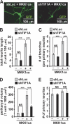
Pol1-driven transcription is required for the morphogenic effects of the ERK1/2 signaling pathway. DIV6 hippocampal neurons were cotransfected with expression plasmids for dominant-negative p53, GFP, MKK1ca, and shTIF1A or control Renilla luciferase shRNA (shLuc; 0.2 + 0.1 + 0.4 + 0.6 μg of plasmid DNAs/2 × 105 neurons, respectively). The empty expression vector pCEP4 was used as a negative control for MKK1ca. After 48 h, cells were fixed. GFP-positive neurons were analyzed morphometrically. A, representative images of transfected neurons. B–D, MKK1ca-induced increases in total neurite length, neurite branching, and perikaryal volume, respectively, were reduced by shTIF1A. E, the number of primary neurites was unaffected by shTIF1A or MKK1ca. Data are means ± S.E. of at least 45 randomly selected individual neurons/condition from three independent experiments. ***, p < 0.001; NS, p > 0.05.
FIGURE 5.
FIGURE 5.
Selective activation of Pol1-driven transcription induces neuronal morphogenesis. A, the constitutively active mutant form of TIF1A that contains phosphomimetic substitutions of the ERK1/2-regulated sites (S633D/S649D; TIF1Aca) increased rDNA promoter activity. DIV4 cortical neurons were cotransfected with the rDNA promoter-luciferase construct together with expression plasmids for β-gal (pEF1αLacZ) and TIF1Aca (0.2 + 0.2 + 0.6 μg of plasmid DNAs/5 × 105 neurons, respectively). TIF1Awt or empty expression vector (EV) served as a control for TIF1Aca. The activity of the rDNA promoter was determined 48 h post-transfection; means ± S.E. of four sister cultures from three independent experiments are presented. B–F, DIV6 hippocampal neurons were cotransfected with expression plasmids for enhanced GFP and TIF1Aca or its controls (0.02 + 0.6 μg of plasmid DNAs/2 × 105 neurons, respectively). Fixation and analysis were as performed as described for Fig. 3. B, representative images of transfected neurons. TIF1Aca increased total neurite length (C), neurite branching (D), and perikaryal volume (E) but not the number of primary neurites (F). Data are means ± S.E. of at least 45 randomly selected individual neurons/condition from three independent experiments. ***, p < 0.001; NS, p > 0.05.
FIGURE 6.
FIGURE 6.
Enhanced morphogenic effects upon coexpression of MKK1ca and TIF1Awt. DIV6 hippocampal neurons were cotransfected with expression plasmids for GFP, TIF1Awt, and/or MKK1ca (0.02 + 0.3 + 0.1 μg of plasmid DNAs/2 × 105 neurons, respectively) as indicated. In addition, empty cloning vectors were used as negative controls for the TIF1Awt and MKK1ca constructs. To avoid saturation of the morphogenic response induced by MKK1ca, its dose was 4-fold lower than in Fig. 3. Cell fixation and analysis were as performed as described for Fig. 3. Coexpression of TIF1A and MKK1ca increased total neurite length (A), neurite branching (B), and perikaryal volume (C) but not the number of primary neurites (D). Data are means ± S.E. of at least 45 randomly selected individual neurons/condition from three independent experiments. *, p < 0.05; **, p < 0.01; ***, p < 0.001; NS, p > 0.05.

Similar articles

Cited by

References

    1. Parrish J. Z., Emoto K., Kim M. D., Jan Y. N. (2007) Annu. Rev. Neurosci. 30, 399–423 - PubMed
    1. Miller F. D., Kaplan D. R. (2003) Curr. Opin. Neurobiol. 13, 391–398 - PubMed
    1. Dijkhuizen P. A., Ghosh A. (2005) Prog. Brain Res. 147, 17–27 - PubMed
    1. Redmond L., Kashani A. H., Ghosh A. (2002) Neuron 34, 999–1010 - PubMed
    1. Wayman G. A., Impey S., Marks D., Saneyoshi T., Grant W. F., Derkach V., Soderling T. R. (2006) Neuron 50, 897–909 - PubMed

Publication types

MeSH terms

Substances

LinkOut - more resources