. 2011 Feb 11;286(6):4341-8.
doi: 10.1074/jbc.M110.179747. Epub 2010 Nov 23.

Caveolin-1 assembles type 1 inositol 1,4,5-trisphosphate receptors and canonical transient receptor potential 3 channels into a functional signaling complex in arterial smooth muscle cells

Affiliations

Caveolin-1 assembles type 1 inositol 1,4,5-trisphosphate receptors and canonical transient receptor potential 3 channels into a functional signaling complex in arterial smooth muscle cells

Adebowale Adebiyi et al. J Biol Chem. .

Abstract

Physical coupling of sarcoplasmic reticulum (SR) type 1 inositol 1,4,5-trisphosphate receptors (IP(3)R1) to plasma membrane canonical transient receptor potential 3 (TRPC3) channels activates a cation current (I(Cat)) in arterial smooth muscle cells that induces vasoconstriction. However, structural components that enable IP(3)R1 and TRPC3 channels to communicate locally are unclear. Caveolae are plasma membrane microdomains that can compartmentalize proteins. Here, we tested the hypothesis that caveolae and specifically caveolin-1 (cav-1), a caveolae scaffolding protein, facilitate functional IP(3)R1 to TRPC3 coupling in smooth muscle cells of resistance-size cerebral arteries. Methyl-β-cyclodextrin (MβCD), which disassembles caveolae, reduced IP(3)-induced I(Cat) activation in smooth muscle cells and vasoconstriction in pressurized arteries. Cholesterol replenishment reversed these effects. Cav-1 knockdown using shRNA attenuated IP(3)-induced vasoconstriction, but did not alter TRPC3 and IP(3)R1 expression. A synthetic peptide corresponding to the cav-1 scaffolding domain (CSD) sequence (amino acids 82-101) also attenuated IP(3)-induced I(Cat) activation and vasoconstriction. A cav-1 antibody co-immunoprecipitated cav-1, TRPC3, and IP(3)R1 from cerebral artery lysate. ImmunoFRET indicated that cav-1, TRPC3 channels and IP(3)R1 are spatially co-localized in arterial smooth muscle cells. IP(3)R1 and TRPC3 channel spatial localization was disrupted by MβCD and a CSD peptide. Cholesterol replenishment re-established IP(3)R1 and TRPC3 channel close spatial proximity. Taken together, these data indicate that in arterial smooth muscle cells, cav-1 co-localizes SR IP(3)R1 and plasma membrane TRPC3 channels in close spatial proximity thereby enabling IP(3)-induced physical coupling of these proteins, leading to I(Cat) generation and vasoconstriction.


Figures

FIGURE 1.
FIGURE 1.
Caveolae are required for IP3-induced ICat activation in cerebral artery smooth muscle cells. A, representative recordings illustrating that MβCD (5 mm) inhibits IP3-induced ICat and reversal of this effect by further addition of Chol/MβCD (100 μg/ml). B, mean ICat density (at −120 mV) at baseline (n = 9) and IP3-induced ICat density in control (n = 6), MβCD (n = 7), and Chol/MβCD (n = 6)-treated cerebral artery smooth muscle cells. *, p < 0.05, when compared with IP3 and IP3 + Chol/MβCD; #, p < 0.05 when compared with the baseline.
FIGURE 2.
FIGURE 2.
Caveolae are required for myogenic tone and IP3-induced vasoconstriction in cerebral arteries. A, representative recordings illustrating that MβCD (10 mm) attenuates myogenic tone and reversal of this effect by Chol/MβCD (100 μg/ml). B, mean myogenic tone in control (n = 6), MβCD (n = 6), and Chol/MβCD (n = 4)-treated cerebral arteries. C, representative traces illustrating that MβCD (10 mm) inhibits Bt-IP3 (10 nm)-induced vasoconstriction and reversal of this effect by Chol/MβCD (100 μg/ml). D, mean Bt-IP3-induced vasoconstriction in control arteries (n = 6) or arteries treated with MβCD (n = 6), or MβCD followed by Chol/MβCD (n = 4). *, p < 0.05 when compared with the control and Chol/MβCD.
FIGURE 3.
FIGURE 3.
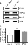
Cav-1 knockdown does not alter TRPC3 and IP3R1 expression in cerebral arteries. A, representative Western blot illustrating that cav-1shV induces knockdown of cav-1, but does not alter IP3R1 or TRPC3 expression. B, mean data illustrating effects of cav-1shV on TRPC3 and IP3R1 protein (n = 4 for each; *, p < 0.05).
FIGURE 4.
FIGURE 4.
Cav-1 knockdown attenuates myogenic tone and IP3-induced vasoconstriction in cerebral arteries. A, representative recordings illustrating that cav-1 knockdown attenuates myogenic tone in endothelium-denuded arteries. B, mean myogenic tone in cav-1scrm (n = 4)- and cav-1shV (n = 5)-treated arteries. C, representative traces illustrating Bt-IP3 (10 nm)-induced vasoconstriction in cav-1scrm and cav-1shV-treated cerebral arteries. D, mean Bt-IP3-induced vasoconstriction in cav-1scrm (control; n = 4)- or cav-1shV (n = 5)-treated arteries. *, p < 0.05 when compared with cav-1scrm.
FIGURE 5.
FIGURE 5.
CSD peptide attenuates IP3-induced ICat activation in cerebral artery smooth muscle cells. A, C terminus of TRPC3 and N and C termini of IP3R1 contain CSD binding sequences (highlighted in gray). Bold letters indicate tryptophan, phenylalanine, or tyrosine. B, fluorescent images indicating successful introduction of FITC-labeled ANT peptide into smooth muscle cells of cerebral arteries. C, representative recordings illustrating that ANT-CSD peptide (50 μm) attenuates IP3 (10 μm)-induced ICat activation in arterial smooth muscle cells. D, mean IP3 -induced ICat activation in ANT (control; n = 5)- and ANT-CSD (n = 6) peptide-treated cells. *, p < 0.05 when compared with ANT. Scale bar, 50 μm.
FIGURE 6.
FIGURE 6.
CSD peptide attenuates IP3-induced vasoconstriction in pressurized cerebral arteries. A, representative traces illustrating that ANT-CSD peptide attenuates Bt-IP3 (10 nm)-induced vasoconstriction. B, mean Bt-IP3-induced vasoconstriction in arteries treated with ANT (n = 6) and ANT-CSD (n = 5) peptides. *, p < 0.05 when compared with ANT.
FIGURE 7.
FIGURE 7.
Cav-1 antibody co-immunoprecipitates cerebral artery cav-1 (∼22 kDa), IP3R1 (∼270 kDa), and TRPC3 (∼90 kDa). Lysate supernatant (∼40 μg of protein) was used as the input control. Negative control (-ve ctrl.) received the same concentration of cav-1 antibody except that the coupling resin was replaced with control agarose resin that is not amine-reactive.
FIGURE 8.
FIGURE 8.
Spatial co-localization of cav-1, IP3R1, and TRPC3 and modulation by caveolae disruption and CSD peptide in cerebral artery smooth muscle cells. A, confocal images illustrating localization of cav-1 and TRPC3 channels in an arterial smooth muscle cell. Shown are fluorescent images generated by Alexa 546- and 488-conjugated antibodies, pixel overlay, and N-FRET in the same cell. B, confocal images illustrating cellular locations of IP3R1 and TRPC3 in a control cell. Images show the overlay and FRET generated by secondary antibodies bound to these proteins in the same control cell. The inset in panel 4 is a magnified view of the boxed area of the cell showing FRET localization to the plasma membrane. C, mean N-FRET data for IP3R1 and TRPC3 for control (n = 16), MβCD (5 mm, 30 min, n = 11), reversal of MβCD effect by Chol/MβCD (100 μg/ml, 1 h, n = 13), ANT (50 μm, 1 h, n = 8), and ANT-CSD peptide (50 μm, 1 h, n = 10). *, p < 0.05 when compared with control; #, p < 0.05 when compared with ANT. Scale bars, 10 μm.

Similar articles

Cited by

References

    1. Berridge M. J. (1993) Nature 361, 315–325 - PubMed
    1. Sanders K. M. (2001) J. Appl. Physiol. 91, 1438–1449 - PubMed
    1. Albert A. P., Saleh S. N., Large W. A. (2009) Curr. Med. Chem. 16, 1158–1165 - PubMed
    1. Adebiyi A., Zhao G., Narayanan D., Thomas-Gatewood C. M., Bannister J. P., Jaggar J. H. (2010) Circ. Res. 106, 1603–1612 - PMC - PubMed
    1. Xi Q., Adebiyi A., Zhao G., Chapman K. E., Waters C. M., Hassid A., Jaggar J. H. (2008) Circ. Res. 102, 1118–1126 - PMC - PubMed

Publication types

MeSH terms

LinkOut - more resources