. 2010 Dec;137(24):4135-45.
doi: 10.1242/dev.060483.

The Hippo tumor suppressor pathway regulates intestinal stem cell regeneration

Affiliations

The Hippo tumor suppressor pathway regulates intestinal stem cell regeneration

Phillip Karpowicz et al. Development. 2010 Dec.

Abstract

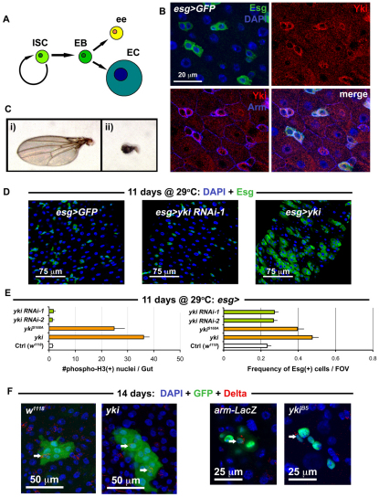
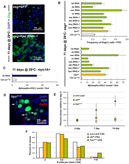
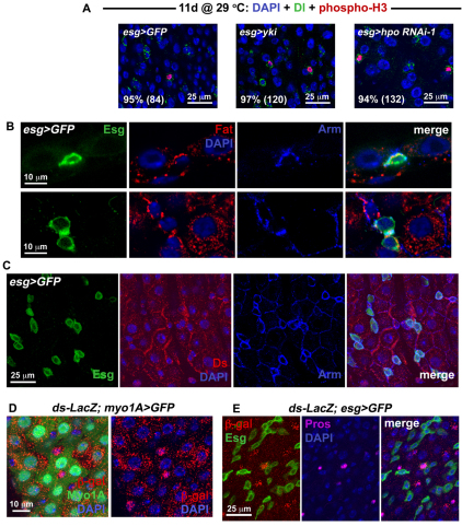
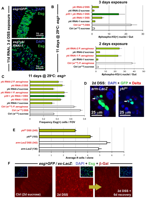
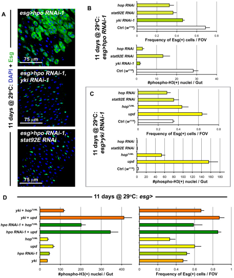
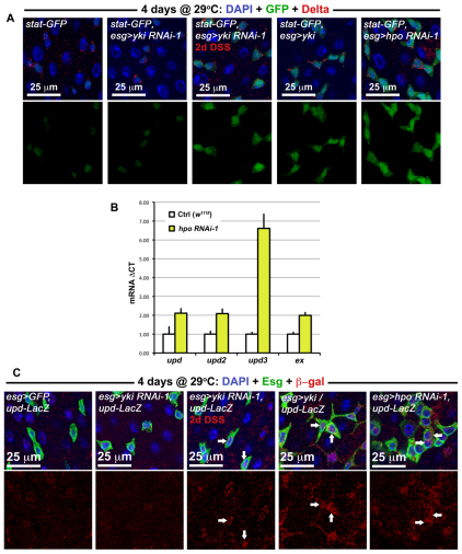
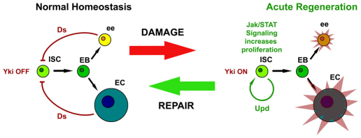
Identification of the signaling pathways that control the proliferation of stem cells (SCs), and whether they act in a cell or non-cell autonomous manner, is key to our understanding of tissue homeostasis and cancer. In the adult Drosophila midgut, the Jun N-Terminal Kinase (JNK) pathway is activated in damaged enterocyte cells (ECs) following injury. This leads to the production of Upd cytokines from ECs, which in turn activate the Janus kinase (JAK)/Signal transducer and activator of transcription (STAT) pathway in Intestinal SCs (ISCs), stimulating their proliferation. In addition, the Hippo pathway has been recently implicated in the regulation of Upd production from the ECs. Here, we show that the Hippo pathway target, Yorkie (Yki), also plays a crucial and cell-autonomous role in ISCs. Activation of Yki in ISCs is sufficient to increase ISC proliferation, a process involving Yki target genes that promote division, survival and the Upd cytokines. We further show that prior to injury, Yki activity is constitutively repressed by the upstream Hippo pathway members Fat and Dachsous (Ds). These findings demonstrate a cell-autonomous role for the Hippo pathway in SCs, and have implications for understanding the role of this pathway in tumorigenesis and cancer stem cells.


Figures

Fig. 1.
Fig. 1.
Yorkie induces ISC/Eb overproliferation in the gut epithelium. (A) The ISC lineage. (B) Esg(+) ISC/Eb cells in the wild-type posterior midgut epithelium stained with anti-Yki antibody. (C) Shown are: (i) wild type (w1118/+; MST1096-Gal4/+) versus (ii) Yorkie RNAi (MST1096-Gal4/+; yki RNAi-1/+) adult wings. (D) Confocal micrographs of wild type (first panel is esg>GFP: w1118/+; esg-Gal4, UAS-GFP, Tub-Gal80TS/+), RNAi against yki (middle panel is esg>yki RNAi-1: esg-Gal4, UAS-GFP, Tub-Gal80TS/+; yki RNAi-1/+) and overexpression of yki (left panel is esg>yki: esg-Gal4, UAS-GFP, Tub-Gal80TS/+; UAS-yki/+). The same hairpin that reduces wing size in C has no effect on ISC proliferation, whereas yki overexpression causes overproliferation. (E) Quantification of total mitoses in the entire midgut (left), and the frequency of Esg(+) cells in one field of view (FOV) in the posterior region of the midgut (right). All constructs were expressed in ISC/Eb cells. Both Yki and mutated Yki (YkiS168A) accelerated cell division, whereas RNAi constructs against yki produced no apparent changes when compared with wild type (Ctrl). Error bars indicate s.e.m. (P<0.05). (F) Confocal images showing Yki-overexpressing clones (shown on right, yki is hsFlp/+; UAS-GFP, act>CD2>Gal4/+; UAS-yki/+) and wild-type clones (left, w1118 is hsFlp/+; UAS-GFP, act>CD2>Gal4/+). Clones mutant for yki (ykiB5 is hsFlp, Tub-Gal4, UAS-nlsGFP/+; FRT42D, Tub-Gal80TS/FRT42D ykiB5) are no different from wild-type (arm-LacZ is hsFlp, Tub-Gal4, UAS-nlsGFP/+; FRT42D, Tub-Gal80TS/FRT42D arm-LacZ).
Fig. 2.
Fig. 2.
Perturbation of Hippo signaling causes hyperplasia in the gut epithelium. (A) RNAi against hpo leads to overproliferation (bottom panel is esg>hpo RNAi-1: esg-Gal4, UAS-GFP, Tub-Gal80TS/+; hpo RNAi-1/+) when compared with wild type (top panel is esg>GFP: w1118/+; esg-Gal4, UAS-GFP, Tub-Gal80TS/+). (B) Quantification of Esg(+) cell frequency (top) and mitoses (bottom). ds RNAi shows no difference compared with wild type (Ctrl), whereas every other Hippo pathway member tested shows overproliferation when expressed in ISC/Eb cells. Error bars indicate s.e.m. (P<0.05). (C) When ds is knocked down in ECs (Tub-Gal80TS/+; myo1A-Gal4, UAS-GFP/+; ds RNAi/+), it causes strong overproliferation. Error bars indicate s.e.m. (P<0.05). (D) Mosaic analysis shows that hpo mutation (hpo42-47 is hsFlp, Tub-Gal4, UAS-nlsGFP/+; FRT42D, Tub-Gal80TS/FRT42D hpo42-47) phenocopies yki overexpression (compare with Fig. 1F). (E) Quantification of clonal frequency reveals an expansion in clone number in hpo mutant clones, but not in yki mutant clones, which are similar to wild type (arm-LacZ). Genotypes are same as above, frequencies are shown as percentage normalized to the 4-day timepoint when each gut contained 5-15 clones in total (n=10-12 guts examined at each timepoint). Error bars indicate s.e.m. (P<0.05). (F) Histograms show that large clones are more frequent among hpo mutant clones than in wild type (arm-LacZ) or yki mutants. Brackets show total number of clones analyzed at this timepoint (14 days after clone induction).
Fig. 3.
Fig. 3.
Hippo signaling occurs in different cell types within the midgut epithelium. (A) Confocal images show wild type (left panel is esg>GFP: w1118/+; esg-Gal4, UAS-GFP, Tub-Gal80TS/+), overexpression of yki (center panel is esg>yki: esg-Gal4, UAS-GFP, Tub-Gal80TS/+; UAS-yki/+) and RNAi against hpo (right panel is esg>hpo RNAi-1: esg-Gal4, UAS-GFP, Tub-Gal80TS/+; hpo RNAi-1/+) stained for Dl (green) and phosphorylated Histone 3 (red). Nearly all mitotic cells are also Dl(+), as revealed by quantification of percentages (number of phospho-H3 cells counted are in brackets). (B) Fat staining is located in the basal region of wild-type posterior gut epithelium. Top panels show confocal sections through the epithelial wall, with an ISC labeled for Esg (green) located basally. The outside of the gut is just below the ISC. A crescent of Fat protein, coincident with Armadillo (Arm) which marks the cell cortex, is located between the ISC and its neighboring EC cells. Lower panels show confocal sections taken through the basal region of the epithelium, where ISC/Eb cells are located. Fat staining is present around the cortex of Esg(+) cells. (C) Confocal images show that the Ds protein, also coincident with Arm, is located at the edges of wild-type ECs – in some cases where the ECs appose Esg(+) cells. (D) Confocal images of a Tub-Gal80TS/ds-LacZ; myo1A-Gal4, UAS-GFP/+ posterior midgut. ds-LacZ is expressed in the EC cells which are GFP-labeled using the NP1 Gal4 driver. (E) Confocal images of a ds-LacZ/+; esg-Gal4, UAS-GFP, Tub-Gal80TS/+ midgut. Strong ds-LacZ expression is also present in Pros(+) ee cells.
Fig. 4.
Fig. 4.
Yki accelerates ISC division during regeneration. (A) The Yki-depleted midgut (bottom panel is esg>yki RNAi-1: esg-Gal4, UAS-GFP, Tub-Gal80TS/+; yki RNAi-1/+) contains fewer Esg(+) cells in response to DSS-induced damage, compared with the wild-type gut (top panel is esg>GFP: w1118/+; esg-Gal4, UAS-GFP, Tub-Gal80TS/+). (B) Quantification of mitoses shows that DSS (top) and Pseudomonas (bottom) exposure reduces the regenerative response of ISCs expressing yki RNAi constructs. Error bars indicate s.e.m. (P<0.05). (C) The frequency of Esg(+) cells in the posterior midgut is also reduced, when flies are fed either DSS or Pseudomonas, and Yki is concomitantly depleted. Error bars indicate s.e.m. (P<0.05). (D) Confocal images show a reduction in the size of yki mutant clones (ykiB5) when compared with wild type (arm-LacZ) when the midgut is injured, although both contain ISCs marked by Dl (arrows). (E) After injury, yki mutant clones are smaller than wild type (arm-LacZ). However, under baseline conditions, these are similar in size to wild type. Brackets show total number of clones analyzed at this timepoint (14 days after clone induction), error bars indicate s.e.m. (P<0.05). (F) Confocal images showing the esg>Gal4/ex-LacZ reporter line (esg-Gal4, UAS-GFP, Tub-Gal80TS/ex-LacZ) under normal conditions and injury conditions. Samples were prepared identically and images were taken using the same parameters.
Fig. 5.
Fig. 5.
JAK/STAT is integrated with Hippo signaling. (A) When hpo is depleted (top panel is esg>hpo RNAi-1: esg-Gal4, UAS-GFP, Tub-Gal80TS/+; hpo RNAi-1/+), cell division is increased. Knockdown of Yki (middle panel is esg>hpo RNAi-1, yki RNAi-1: esg-Gal4, UAS-GFP, Tub-Gal80TS/+; hpo RNAi-1/yki RNAi-1) and Stat92E (bottom panel is esg>hpo RNAi-1, stat92E RNAi: esg-Gal4, UAS-GFP, Tub-Gal80TS/+; hpo RNAi-1/stat92E RNAi) suppress this overproliferation. (B) Quantification of Esg(+) cell frequency (top) and mitosis (bottom). The depletion of yki, hop and stat92E suppress the overproliferation observed due to hpo depletion alone. Error bars indicate s.e.m. (P<0.05). (C) Quantification of Esg(+) cell frequency and mitosis, shows that when yki is depleted, JAK/STAT activation still results in increased proliferation. If both yki and hop, or yki and stat92E are depleted, no significant differences are observed from yki depletion alone. Error bars indicate s.e.m. (P<0.05). (D) Graphs show quantification of mitosis and Esg(+) cell frequency as above, genotypes examined are the same for both graph y-axes. The values for esg>yki and esg>hpo RNAi-1 are reproduced from Fig. 1E and Fig. 2B in order to compare the increase. Error bars indicate s.e.m. (P<0.05).
Fig. 6.
Fig. 6.
Yki activates the release of JAK/STAT cytokines. (A) The activity of the JAK/STAT pathway, using the stat-GFP reporter (stat-GFP is Tub-Gal80TS/+; esg-Gal4/10xSTAT-GFP and where present the overexpression constructs yki RNAi-1, yki or hpo RNAi-1 are heterozygous on the third chromosome). When either yki is overexpressed or hpo is depleted by RNAi, an increase in STAT activity is observed. DSS damage induces a similar increase even when yki is depleted. (B) qPCR of tested transcripts following hpo knockdown using the esg-Gal4 driver. Signal is normalized to the control gene rp49 and levels are shown normalized to esg>GFP controls; error bars are s.e.m. (C) Upd/Os expression is elevated in Esg(+) cells when either hpo is knoced down or when yki is overexpressed (esg>GFP, upd-LacZ refers to w1118/upd-LacZ; esg-Gal4, UAS-GFP, Tub-Gal80TS/+ and where present the overexpression constructs yki RNAi-1, yki or hpo RNAi-1 are heterozygous on the third chromosome). Normally signal is weak or non-existent in wild type Esg(+) cells (esg>GFP, upd-LacZ), but Esg(+) cells (arrows) become positive for upd-LacZ (red) during Yki-induced overgrowth (esg>yki, upd-LacZ) or Hpo knockdown (esg>hpo RNAi-1, upd-LacZ). Similar to the results above, Upd/Os signal still shows an increase (arrows) during DSS damage even when yki is depleted (esg>yki RNAi-1, upd-LacZ).
Fig. 7.
Fig. 7.
Hippo signaling restrains Yki-accelerated cell division in response to injury. Schematic shows the transition of ISCs between normal homeostasis, which occurs in the absence of injury, and acute regeneration following injury. Upon damage, the Hippo pathway ligand, Ds, no longer activates Hippo signaling in ISCs, allowing Yki to activate downstream targets. One of these targets is the Upd/Os cytokine, which stimulates proliferation of ISCs/Ebs through the JAK/STAT pathway. When ee and EC progeny are regenerated, Hippo signaling acts as a brake, cytokine production slows and the system returns to the homeostatic state.

Similar articles

Cited by

References

    1. Amcheslavsky A., Jiang J., Ip Y. T. (2009). Tissue damage-induced intestinal stem cell division in Drosophila. Cell Stem Cell 4, 49-61 - PMC - PubMed
    1. Apidianakis Y., Pitsouli C., Perrimon N., Rahme L. (2009). Synergy between bacterial infection and genetic predisposition in intestinal dysplasia. Proc. Natl. Acad. Sci. USA 106, 20883-20888 - PMC - PubMed
    1. Basler K., Struhl G. (1994). Compartment boundaries and the control of Drosophila limb pattern by hedgehog protein. Nature 368, 208-214 - PubMed
    1. Beebe K., Lee W. C., Micchelli C. A. (2010). JAK/STAT signaling coordinates stem cell proliferation and multilineage differentiation in the Drosophila intestinal stem cell lineage. Dev. Biol. 338, 28-37 - PubMed
    1. Benhamouche S., Curto M., Saotome I., Gladden A. B., Liu C. H., Giovannini M., McClatchey A. I. (2010). Nf2/Merlin controls progenitor homeostasis and tumorigenesis in the liver. Genes Dev. 24, 1718-1730 - PMC - PubMed

Publication types

MeSH terms