. 2010 Dec;137(24):4261-9.
doi: 10.1242/dev.051730.

Collagen XIXa1 is crucial for motor axon navigation at intermediate targets

Affiliations

Collagen XIXa1 is crucial for motor axon navigation at intermediate targets

Jona D Hilario et al. Development. 2010 Dec.

Abstract

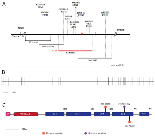
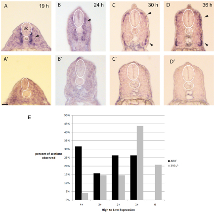
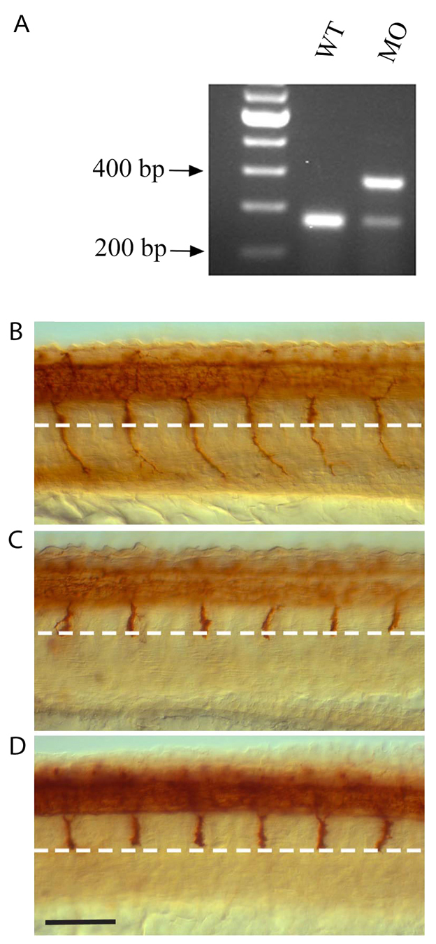
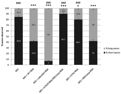
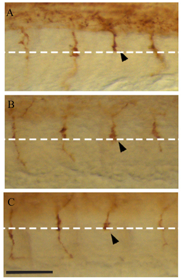
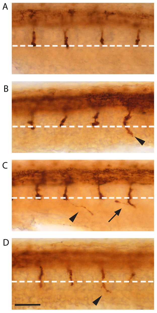
During development, motor axons navigate from the spinal cord to their muscle targets in the periphery using stereotyped pathways. These pathways are broken down into shorter segments by intermediate targets where axon growth cones are believed to coordinate guidance cues. In zebrafish stumpy mutants, embryonic development proceeds normally; however, as trunk motor axons stall at their intermediate targets, suggesting that Stumpy is needed specifically for motor axon growth cones to proceed past intermediate targets. Fine mapping and positional cloning revealed that stumpy was the zebrafish homolog of the atypical FACIT collagen collagenXIXa1 (colXIX). colXIX expression was observed in a temporal and spatial pattern, consistent with a role in motor axon guidance at intermediate targets. Knocking down zebrafish ColXIX phenocopied the stumpy phenotype and this morpholino phenotype could be rescued by adding back either mouse or zebrafish colXIX RNA. The stumpy phenotype was also partially rescued in mutants by first knocking down zebrafish ColXIX and adding back colXIX RNA, suggesting that the mutation is acting as a dominant negative. Together, these results demonstrate a novel function for a FACIT collagen in guiding vertebrate motor axons through intermediate targets.


Figures

Fig. 1.
Fig. 1.
Positional cloning of the stumpy gene. (A) The stumpyb393 mutation was mapped to chromosome 13 (Chr 13) and further mapped to BAC BX322620. The solid black line represents the relevant genomic region on Chr 13; lines beneath indicate individual BACs that were mapped to this region with the stumpy BAC in red; primer names and number of recombinants are listed above the line. The asterisk denotes the location of the stumpy gene coincident with the location of ColXIX. (B) Intron-exon structure of the ColXIX gene. The gene consists of 50 exons and spans ∼300 kb. (C) ColXIX protein structure with stumpyb393 mutations indicated. ColXIX is composed of an N terminus head consisting of a signal peptide and a LamG/TSPN domain (red). The C-terminal tail has five collagenous domains (Col1-Col5) ranging from 72 amino acids to 186 amino acids in length interrupted by five non-collagenous domains (NC1-NC5) ranging from 18 amino acids to 158 amino acids in length.
Fig. 2.
Fig. 2.
colXIX RNA is dynamically expressed during motor axon outgrowth. (A-D′) RNA in situ hybridization using colXIX anti-sense (A-D) and sense (A′-D′) riboprobes that encompasses exons 1-27 of the zebrafish colXIX cDNA at (A,A') 19 hpf, (B,B′) 24 hpf, (C,C′) 30 hpf and (D,D′) 36 hpf. Sections were from the mid-trunk region. (E) Histogram of colXIX RNA in situ hybridization intensity at 19 hpf for wild-type (n=8 embryos; ∼40 sections) and stumpy mutant embryos (n=8 embryos; ∼40 sections). The x-axis reflects the intensity of the colXIX expression. Spinal cord (sc) and notochord (nc) are indicated by white borders. Arrows show localization of colXIX transcripts in the myotome. Scale bar: 20 μm.
Fig. 3.
Fig. 3.
ColXIX knock down phenocopies the stumpy mutation. (A) RT-PCR shows inclusion of a 109 bp fragment in splice-blocking colXIX MO injected (MO) compared to wild-type (wt) embryos. (B-D) CaP axon phenotypes as visualized using znp-1 antibody in (B) uninjected wild-type embryos, (C) wild-type embryos injected with 9 ng colXIX MO and (D) stumpyb393–/– mutant embryos. White dashed line indicates the horizontal myoseptum. Scale bar: 70 μm.
Fig. 4.
Fig. 4.
ColXIX rescues the morphant phenotype. Wild-type mouse ColXIX RNA (WT RNA, 250 pg), wild-type zebrafish colXIX RNA (350 pg) and mouse ColXIX RNA (250 pg) double or single mutations were co-injected with 4.5 ng colXIX MO. P-values were calculated for % short axons observed versus MO-injected with significance indicated by a black asterisk (*), and against MO + WT mouse RNA (#): * or #P<0.05, ** or ##P<0.001, *** or ###P<0.0001.
Fig. 5.
Fig. 5.
Mutant ColXIX acts as a dominant negative. (A-C) Examples of CaP axon defects observed in embryos injected with mutant ColXIX RNA. White dashed line indicates the horizontal myoseptum. Arrowhead denotes Stumpy-like CaP axons.
Fig. 6.
Fig. 6.
Rescue of stumpyb393–/– mutants with mouse ColXIX RNA. (A) stumpyb393–/– embryo. (B-D) Representative images of stumpyb393–/– embryos co-injected with translation blocking colXIX MO and full-length mouse ColXIX RNA. Arrowheads indicate rescued CaP axons. An abnormal axon is indicated by the arrow. White dashed line indicates the horizontal myoseptum. Scale bar: 70 μm.

Similar articles

Cited by

References

    1. Beattie C. E. (2000). Control of motor axon guidance in the zebrafish embryo. Brain Res. Bull. 53, 489-500 - PubMed
    1. Beattie C. E., Eisen J. S. (1997). Notochord alters the permissiveness of myotome for pathfinding by an identified motoneuron in embryonic zebrafish. Development 124, 713-720 - PubMed
    1. Beattie C. E., Raible D. W., Henion P. D., Eisen J. S. (1999). Early pressure screens. Methods Cell Biol. 60, 71-86 - PubMed
    1. Beattie C. E., Melancon E., Eisen J. S. (2000). Mutations in the stumpy gene reveal intermediate targets for zebrafish motor axons. Development 127, 2653-2662 - PubMed
    1. Beattie C. E., Granato M., Kuwada J. Y. (2002). Cellular, genetic, and molecular mechanisms of axonal guidance in the zebrafish. Results Probl. Cell Differ. 40, 252-269 - PubMed

Publication types