. 2011 Jan 15;17(2):302-9.
doi: 10.1158/1078-0432.CCR-10-0999. Epub 2010 Nov 23.

Plectin-1 as a novel biomarker for pancreatic cancer

Affiliations

Plectin-1 as a novel biomarker for pancreatic cancer

Dirk Bausch et al. Clin Cancer Res. .

Abstract

Purpose: We are in great need of specific biomarkers to detect pancreatic ductal adenocarcinoma (PDAC) at an early stage, ideally before invasion. Plectin-1 (Plec1) was recently identified as one such biomarker. However, its suitability as a specific biomarker for human pancreatic cancer, and its usability as an imaging target, remain to be assessed.

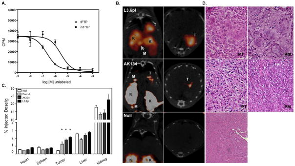
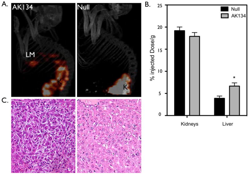
Experimental design: Specimens of human PDAC, chronic pancreatitis, and normal pancreata were evaluated by immunohistochemistry and Western blot analysis. To validate Plec1 as an imaging target, Plec1-targeting peptides (tPTP) were used as a contrast agent for single photon emission computed tomography in an orthotopic and liver metastasis murine model of PDAC.

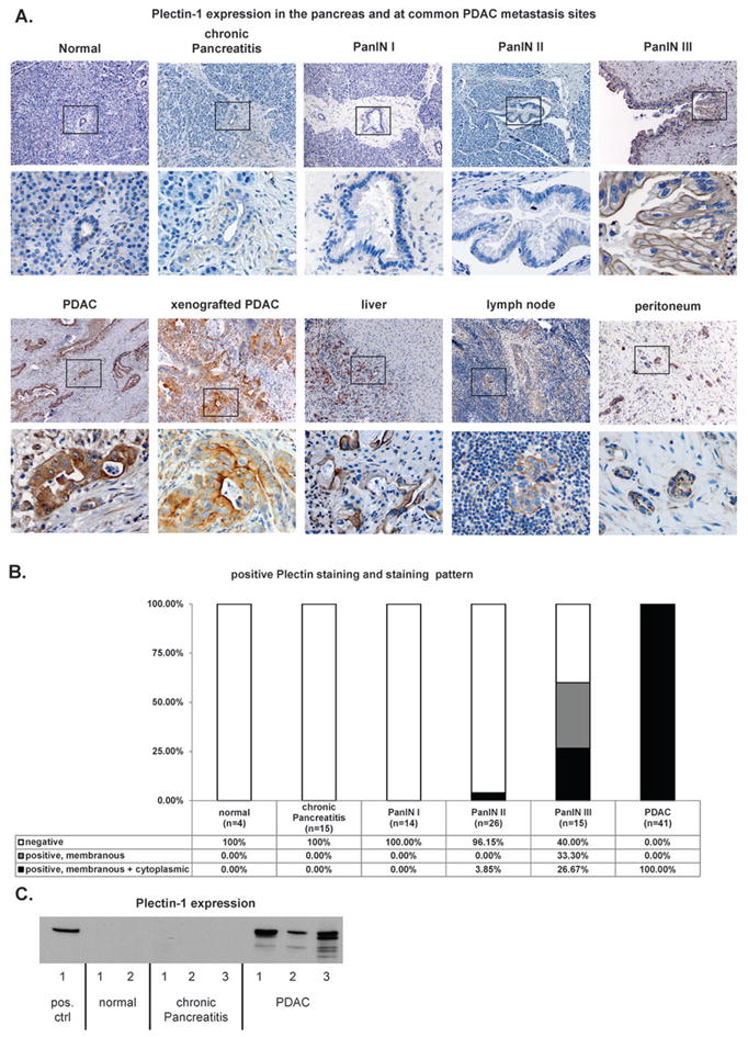
Results: Plec1 expression was noted to be positive in all PDACs but negative in benign tissues. Plec1 expression increases during pancreatic carcinogenesis. It was found to be misexpressed in only 0% to 3.85% of early PDAC precursor lesions (PanIN I/II) but in 60% of PanIN III lesions. Plec1 expression was further noted to be retained in all metastatic foci assayed and clearly highlighted these metastatic deposits in lymph nodes, liver, and peritoneum. In vivo imaging using tPTP specifically highlighted the primary and metastatic tumors. Biodistribution studies performed after imaging show that the primary pancreatic tumors and liver metastases retained 1.9- to 2.9-fold of tPTP over normal pancreas and 1.7-fold over normal liver.

Conclusions: Plec1 is the first biomarker to identify primary and metastatic PDAC by imaging and may also detect preinvasive PanIN III lesions. Strategies designed to image Plec1 could therefore improve detection and staging.


Figures

Figure 1
Figure 1. Plectin-1 Immunohistochemistry and Western Blot
A) Representative images of the evaluated normal pancreata, chronic pancreatitis, PanIN, PDAC, xenografted PDAC and PDAC metastasis sites (liver, lymph node and peritoneum). Overview (upper panel) and detailed view of the black box (lower panel). Chronic pancreatitis and normal pancreas do not express Plectin-1. PanIN III has a membranous staining pattern. PDAC and PDAC xenograft tissue stain moderately to strongly cytoplasmic and membranous for Plectin-1. Common PDAC metastasis sites do not show significant Plectin-1 expression, while the tumor cells stain intensely for Plec1. B) Distribution of staining intensity and staining pattern in the specimens. All PDAC cases were Plec1-positive, whereas normal pancreas and chronic pancreatitis did not express Plectin-1. All PanIN I and most II lesions were Plec1-negative, while the majority of PanIN III lesions were Plec1-positive. The cellular localization of Plec1 also changes during carcinogenesis. The protein is found only in the membrane in 33% of PanIN III lesions, while 27% of PanIN III and all PDAC show membranous and cytoplasmic PLec1 expression. C) Quantitative Western Blot for Plec1 from 50 mg of pancreatic tissue (snap-frozen surgical specimens). No Plec1 was detected in the normal pancreas and CP, while it was present in each PDAC.
Figure 2
Figure 2. Plectin-1 expression in normal and malignant human tissue: Immunohistochemistry of a tissue microarray evaluated for Plectin-1 expression
A) Most normal tissue shows only weak Plectin-1 expression. A clear difference in Plectin-1 expression distinguishing normal from malignant disease is observed in the pancreas, esophagus, stomach and lung. Common PDAC metastasis sites (lymph node, liver) do not express Plec1.
Figure 3
Figure 3. In vivo imaging of Plec1 in orthotopic PDAC
A) In vitro validation of tPTP. L3.6pl cells were plated on a 96-well plate and incubated with 111In-tPTP and increasing log concentrations of unlabeled tPTP or negative control tetramer. B) Mice bearing tumors from orthotopically implanted L3.6pl, AK134 cells and mice without tumors (null) were injected with 111In-tPTP and imaged via SPECT/CT 4 hours post injection. Note the accumulation of tPTP in PDAC, allowing the in vivo imaging of tumor in the pancreas and in peritoneal metastases. Coronal (left) and axial (right) SPECT/CT slices through the tumor are presented. T-tumor, K-kidney, M-peritoneal metastasis. C) After SPECT/CT imaging, animals were sacrificed, organs harvested and gamma counts assessed. Null data is from both nu/nu and FVB/NJ animals that were injected in the pancreas with saline. D) Histology. Animals that had orthotopically implanted tumors or null animals were sacrificed and pancreas and regions of visible peritoneal metastases were removed, embedded, sectioned and stained with H&E (20x image). PT–primary tumor, PM–peritoneal metastasis
Figure 4
Figure 4. Plec1 imaging allows non-invasive detection of liver metastases
A) AK134 cells, or saline (null), were injected intrasplenically to produce liver metastases. Left panel: Mice bearing liver metastasis (LM) from AK134 injection. Right panel: Null animals without intrasplenic tumor cell injected. K-kidney B) Biodistribution studies were performed on indicated tissues subsequent to imaging experiments. * p<0.01. C) Histology: Left panel: Histologic confirmation of liver metastasis. Right panel: normal liver.

Comment in

References

    1. Jemal A, Siegel R, Ward E, Murray T, Xu J, Thun MJ. Cancer statistics, 2007. CA Cancer J Clin. 2007;57:43–66. - PubMed
    1. Koorstra JB, Hustinx SR, Offerhaus GJ, Maitra A. Pancreatic carcinogenesis. Pancreatology. 2008;8:110–25. - PMC - PubMed
    1. Yeo CJ, Cameron JL, Sohn TA, Lillemoe KD, Pitt HA, Talamini MA, et al. Six hundred fifty consecutive pancreaticoduodenectomies in the 1990s: pathology, complications, and outcomes. Annals of Surgery. 1997;226:248–57. discussion 57–60. - PMC - PubMed
    1. Furukawa H, Okada S, Saisho H, Ariyama J, Karasawa E, Nakaizumi A, et al. Clinicopathologic features of small pancreatic adenocarcinoma. A collective study. Cancer. 1996;78:986–90. - PubMed
    1. Krishna NB, Mehra M, Reddy AV, Agarwal B. EUS/EUS-FNA for suspected pancreatic cancer: influence of chronic pancreatitis and clinical presentation with or without obstructive jaundice on performance characteristics. Gastrointestinal Endoscopy. 2009;70:70–9. - PubMed

Publication types