. 2011 Feb;187(2):455-65.
doi: 10.1534/genetics.110.120725. Epub 2010 Nov 23.

The putative lipid transporter, Arv1, is required for activating pheromone-induced MAP kinase signaling in Saccharomyces cerevisiae

Affiliations

The putative lipid transporter, Arv1, is required for activating pheromone-induced MAP kinase signaling in Saccharomyces cerevisiae

Michelle L Villasmil et al. Genetics. 2011 Feb.

Abstract

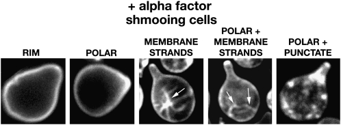
Saccharomyces cerevisiae haploid cells respond to extrinsic mating signals by forming polarized projections (shmoos), which are necessary for conjugation. We have examined the role of the putative lipid transporter, Arv1, in yeast mating, particularly the conserved Arv1 homology domain (AHD) within Arv1 and its role in this process. Previously it was shown that arv1 cells harbor defects in sphingolipid and glycosylphosphatidylinositol (GPI) biosyntheses and may harbor sterol trafficking defects. Here we demonstrate that arv1 cells are mating defective and cannot form shmoos. They lack the ability to initiate pheromone-induced G1 cell cycle arrest, due to failure to polarize PI(4,5)P(2) and the Ste5 scaffold, which results in weakened MAP kinase signaling activity. A mutant Ste5, Ste5(Q59L), which binds more tightly to the plasma membrane, suppresses the MAP kinase signaling defects of arv1 cells. Filipin staining shows arv1 cells contain altered levels of various sterol microdomains that persist throughout the mating process. Data suggest that the sterol trafficking defects of arv1 affect PI(4,5)P(2) polarization, which causes a mislocalization of Ste5, resulting in defective MAP kinase signaling and the inability to mate. Importantly, our studies show that the AHD of Arv1 is required for mating, pheromone-induced G1 cell cycle arrest, and for sterol trafficking.


Figures

F<sc>igure</sc> 1.—
Figure 1.—
Arv1 protein constructs. (A) A schematic of the various Arv1 protein constructs studied. Constructs are driven by the ARV1 promoter and are C-terminally tagged with 3HA in the vector pRS416. (B) Western analysis of cells expressing constructs described in A. Whole cell protein lysate was probed with anti-HA and anti-Pgk1.
F<sc>igure</sc> 2.—
Figure 2.—
arv1 cells harbor bilateral mating defects. Cells to be tested for mating efficiency were patched onto YEPD plates and grown for 1 day (WT and arv1). A total of 106 cells of the opposite mating partner that were grown to exponential phase in liquid YEPD media were spread onto YEPD plates and allowed to dry (MATα WT; MATa WT; MATα arv1; MATa arv1). Mating was performed at 30° for 3 hr. Diploid progeny were selected by replica plating onto the appropriate media and allowed to grow for 1 day at 30°.
F<sc>igure</sc> 3.—
Figure 3.—
arv1 cells harbor defects in shmoo formation. A total of 2 × 10⁷ cells/ml were incubated with 20 μg/ml of α-factor and cell aliquots were collected at 0, 1.5, 3, 4.5, and 7.5 hr. arv1 bar1 cells were transformed with the indicated Arv1 constructs which were C-terminally tagged with 3HA and on a pRS416 plasmid. Cells were collected at indicated time points and the numbers of shmoos/300 cells were counted for each sample. Arv1 full-length (solid circles), AHD (solid squares), ΔAHD (solid diamonds), empty vector (solid triangles) (mean ± SEM, n = 3, *P < 0.01, **P < 0.001).
F<sc>igure</sc> 4.—
Figure 4.—
arv1 cells harbor defects in pheromone-induced G1 cell cycle arrest. A total of 2 × 10⁷ cells/ml of cells were treated with 20 μg/ml of α-factor for the indicated times. MATa bar1 and arv1bar1 cells were transformed with plasmids containing full-length Arv1, AHD, and ΔAHD, as indicated; the constructs are under the control of the ARV1 promoter and are C-terminally tagged with 3HA in vector pRS416. Data are shown as the quantification of FACS profiles measuring propidium iodide-stained DNA. G1 phase (light shading), S phase (solid), G2 phase, (dark shading).
F<sc>igure</sc> 5.—
Figure 5.—
arv1 cells harbor defects in pheromone response pathway signaling. (A) Pheromone signaling is defective in arv1 cells treated with α-factor. MATa FUS1∷FUS1-lacZ∷LEU2 and arv1 FUS1∷FUS1-lacZ∷LEU2 cells were transformed with plasmids containing full-length Arv1, AHD, and ΔAHD; the constructs are under the control of the ARV1 promoter and are C-terminally tagged with 3HA in vector pRS416. FUS1-lacZ induction was measured after 2 hr of α-factor treatment (mean ± SEM, n = 3, *P < 0.02, **P < 0.006). (B) Ste12 overexpression rescues signaling defect in arv1 cells. pGAL1-STE12 was expressed in the strains described in A. FUS1-lacZ induction was measured 4 hr after galactose treatment (without α-factor) (mean ± SEM, n = 3).
F<sc>igure</sc> 6.—
Figure 6.—
Ste5 recruitment to plasma membrane is altered in arv1 cells. (A) Pheromone signaling is rescued by overexpression of Ste5Q59L. pGAL1-Ste5Q59L-GFP was expressed in strains described in Figure 5A. FUS1-lacZ induction was measured 4 hr after galactose treatment (without α-factor) (mean ± SEM, n = 3). (B) Expression of Ste5ΔN-CTM does not rescue signaling defect in arv1 or ΔAHD cells. pGAL1-Ste5ΔN-CTM-GFP was expressed in strains described in Figure 5A. FUS1-lacZ induction was measured 4 hr after galactose treatment (without α-factor) (mean ± SEM, n = 4, *P < 0.01). (C) Ste5-GFP was expressed in cells. Cells with polarized Ste5-GFP were counted after 1 hr of α-factor treatment (mean ± SEM, n = 4, *P < 0.001). (D) Cells with pGAL1-GST-GFP-PHPLCδ were treated with galactose for 3 hr. α-Factor was added during the last 1 hr of galactose treatment. Cells with polarized PI(4,5)P2 were counted (mean ± SEM, n = 3, *P < 0.005, **P < 0.001).
F<sc>igure</sc> 7.—
Figure 7.—
Activated Ste11 mutants do not rescue signaling defect in arv1 cells. (A) Expression of Ste11-Cpr does not rescue signaling defect in arv1, AHD, or ΔAHD cells. pGAL1-Ste11-Cpr was expressed in MATa ste11∷ADE2 FUS1∷FUS1-lacZ∷LEU2 or MATa ste11∷ADE2 arv1∷KANr FUS1∷FUS1-lacZ∷LEU2 cells. FUS1-lacZ induction was measured 4 hr after galactose treatment (without α-factor) (mean ± SEM, n = 3, *P < 0.02, **P < 0.001). (B) Ste11ΔN expression does not rescue signaling defect in arv1 or ΔAHD cells. pGAL1-Ste11ΔN was expressed in strains described in Figure 5A. FUS1-lacZ induction was measured 4 hr after galactose treatment (without α-factor) (mean ± SEM, n = 4, *P < 0.01, **P < 0.001).
F<sc>igure</sc> 8.—
Figure 8.—
Ste4 overexpression does not rescue arv1 signaling defect. pGAL1-Ste4-GFP was expressed in MATa ste4∷ADE2 FUS1∷FUS1-lacZ∷LEU2 or MATa ste4∷ADE2 arv1∷KANr FUS1∷FUS1-lacZ∷LEU2 cells. FUS1-lacZ induction was measured 4 hr after galactose treatment (without α-factor) (mean ± SEM, n = 4, *P < 0.02, **P < 0.01).
F<sc>igure</sc> 9.—
Figure 9.—
Fluorescence microscopic visualization of the lipid microdomains in cells during vegetative growth and α-factor treatment. The various lipid microdomains indicated in Table 3 are represented. Arrows indicate location of membrane strands.

Similar articles

Cited by

References

    1. Bagnat, M., and K. Simons, 2002. a Cell surface polarization during yeast mating. Proc. Natl. Acad. Sci. USA 99 14183–14188. - PMC - PubMed
    1. Bagnat, M., and K. Simons, 2002. b Lipid rafts in protein sorting and cell polarity in budding yeast Saccharomyces cerevisiae. Biol. Chem. 383 1475–1480. - PubMed
    1. Beh, C. T., and J. Rine, 2004. A role for yeast oxysterol-binding protein homologs in endocytosis and in the maintenance of intracellular sterol-lipid distribution. J. Cell Sci. 117 2983–2996. - PubMed
    1. Berlin, V., J. Brill, J. Trueheart, J. D. Boeke and G. R. Fink, 1991. Genetic screens and selections for cell and nuclear fusion mutants. Methods Enzymol. 194 774–792. - PubMed
    1. Bidlingmaier, S., and M. Snyder, 2004. Regulation of polarized growth initiation and termination cycles by the polarisome and Cdc42 regulators. J. Cell Biol. 164 207–218. - PMC - PubMed

Publication types

MeSH terms