. 2010 Nov 23;3(149):ra84.
doi: 10.1126/scisignal.2001148.

BRAF gene amplification can promote acquired resistance to MEK inhibitors in cancer cells harboring the BRAF V600E mutation

Affiliations

BRAF gene amplification can promote acquired resistance to MEK inhibitors in cancer cells harboring the BRAF V600E mutation

Ryan B Corcoran et al. Sci Signal. .

Abstract

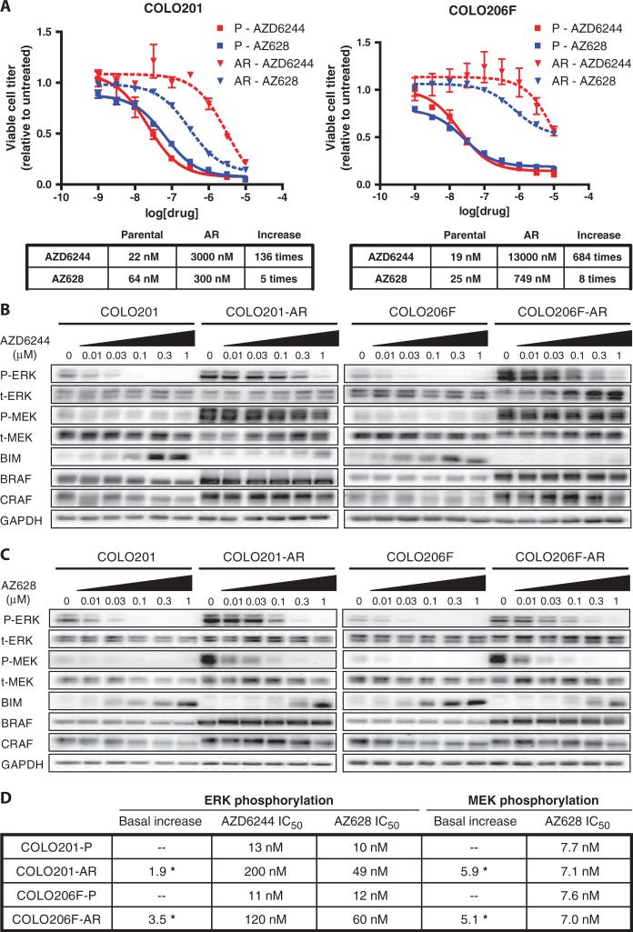
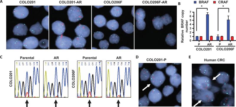
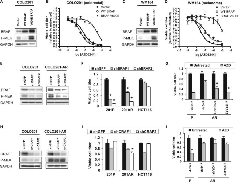
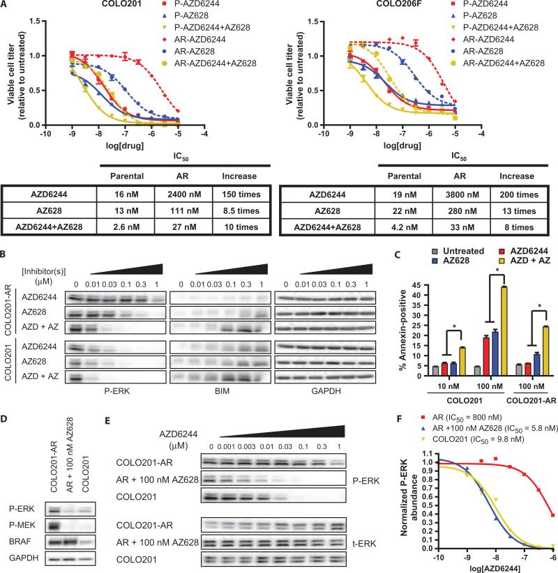
Oncogenic BRAF mutations are found in several tumor types, including melanomas and colorectal cancers. Tumors with BRAF mutations have increased mitogen-activated protein kinase pathway activity and heightened sensitivity to BRAF and MEK (mitogen-activated or extracellular signal-regulated protein kinase kinase) inhibitors. To identify potential mechanisms of acquired drug resistance, we generated clones resistant to the allosteric MEK inhibitor AZD6244 from two BRAF V600E mutant colorectal cancer cell lines that are highly sensitive to MEK or BRAF inhibition. These AZD6244-resistant (AR) clones, which exhibited cross-resistance to BRAF inhibitors, acquired resistance through amplification of the BRAF gene. A small percentage of treatment-naïve parental cells showed preexisting BRAF amplification. We observed similar amplification in a subset of cells in a BRAF-mutant colorectal cancer. In cell lines, BRAF amplification increased the abundance of phosphorylated MEK and impaired the ability of AZD6244 to inhibit ERK (extracellular signal-regulated kinase) phosphorylation. The ability of AZD6244 to inhibit ERK phosphorylation in AR cells was restored by treatment with a BRAF inhibitor at low concentrations that reduced the abundance of phosphorylated MEK to amounts observed in parental cells. Combined MEK and BRAF inhibition fully overcame resistance to MEK or BRAF inhibitors alone and was also more effective in parental cells compared to treatment with either inhibitor alone. These findings implicate BRAF amplification as a mechanism of resistance to both MEK and BRAF inhibitors and suggest combined MEK and BRAF inhibition as a clinical strategy to overcome, or possibly prevent, this mechanism of resistance.


Figures

Fig. 1
Fig. 1
AR clones are resistant to MEK and BRAF inhibition. (A) Parental (solid lines) COLO201 and COLO206F cells and AR (dashed lines) COLO201-AR and COLO206F-AR cells were treated in triplicate with the indicated concentrations of drug for 72 hours. Viable cell titer was determined, and the average values are shown relative to untreated controls for each cell line. Error bars represent the SD for each measurement. For each cell line, the IC50s for each inhibitor are shown in tabular form along with the increase in IC50 in AR cells relative to parental cells. (B and C) Western blots of RAF-MEK pathway components and effectors in parental and AR cells treated with the indicated concentrations of AZD6244 (B) or AZ628 (C) for 24 hours. (D) Tabular representation of chemiluminescent signal intensities from the blots in (B) and (C) showing IC50s for inhibition of ERK and MEK phosphorylation (full dose-response relationships are shown in fig. S2A). The statistically significant increases in basal phospho-ERK and phospho-MEK in AR cells relative to parental cells (average of at least three independent measurements) are also shown. *P < 0.01.
Fig. 2
Fig. 2
AR cells have amplification of BRAF. (A) FISHimages showingincrease in BRAF copy number in AR cells relative to parental cells. BRAF probes are red, centromere 7 is aqua, and nuclear stain is blue. (B) Quantitative PCR for BRAF and CRAF from genomic DNA from parental (P) and AR cells. Values are shown relative to the parental cell line. *P < 0.001. (C) Sequencing chromatograms for BRAF from parental and AR cells, with the nucleotide color scheme: A (blue), C (gold), G (black), T (red). The T→A nucleotide substitution encoding the V600E mutation occurs at position 1799, which is indicated by an arrow. The wild-type allele (GTG) is represented by the red (T) peak at position 1799, and the mutant allele (GAG) is represented by the blue (A) peak. (D) FISH image showing an example of a cell (arrow) with preexisting low-level amplification of BRAF from the parental COLO201 cell line. (E) FISH image of a human colorectal cancer harboring a V600E BRAF mutation. Two cells with preexisting high-level BRAF amplification are indicated by arrows.
Fig. 3
Fig. 3
Increased BRAF V600E is responsible for AZD6244 resistance in AR cells. (A and C) Western blots of COLO201 (A) and WM164 (C) cells infected with retroviruses encoding wild-type (WT) BRAF, mutant V600E BRAF, or vector control. (B and D) COLO201 (B) or WM164 (D) cells expressing WT BRAF, V600E BRAF, or vector control were treated with the indicated concentrations of AZD6244 for 72 hours and viable cell titer was determined. (E and H) Western blots of COLO201 and COLO201-AR cells infected with control shRNA (shGFP) or two different shRNAs directed against either BRAF (E) or CRAF (H). After infection, cells were selected in puromycin for 96 hours. Experiments were performed at least three times. (F and I) COLO201 (201P), COLO201-AR (201AR), and the KRAS mutant HCT116 cell line were infected with the above shRNAs. Cell viability was determined after 5 days of puromycin selection. Viral titers were used that yielded about the same results in the presence or absence of puromycin. *P < 0.001 relative to HCT116 cells infected with corresponding shRNA. (G and J) COLO201 (P) and COLO201-AR (AR) cells infected with the indicated shRNAs were selected in puromycin for 48 hours and then treated with or without 100 nM AZD6244 (AZD) for 72 hours before determination of cell viability. Viable cell titer is shown relative to the untreated control for each shRNA. *P < 0.001 relative to shGFP-infected COLO201-AR cells treated with AZD6244.
Fig. 4
Fig. 4
Resistance to MEK and BRAF inhibition in AR cells can be overcome by combination treatment with AZD6244 and AZ628. (A) Parental (P, solid lines) and AR cells (dashed lines) were treated with the indicated concentrations of inhibitors for 72 hours and viable cell titer was determined. AZD6244 + AZ628 treatments contain the indicated concentrations of each inhibitor in combination. The IC50s for each inhibitor alone and in combination are shown in tabular form along with the increase in IC50 in AR cells relative to parental cells. (B) Western blot of COLO201 or COLO201-AR cells treated with the indicated concentrations of AZD6244 or AZ628 alone, or in combination (AZD + AZ) for 24 hours. (C) COLO201 or COLO201-AR cells were treated for 72 hours with the indicated concentrations of inhibitors. Percent of apoptotic cells was determined by annexin V staining. *P < 0.001 relative to treatment with either inhibitor alone. (D) Western blot of COLO201 cells and COLO201-AR cells treated with or without 100 nM AZ628 for 24 hours. Experiments were performed at least three times. (E) Western blot of COLO201 and COLO201-AR cells in the presence or absence of 100 nM AZ628 and treated with the indicated concentrations of AZD6244 for 24 hours. (F) Dose-response curves were generated by plotting normalized phospho-ERK (P-ERK) abundance versus AZD6244 concentration determined from chemiluminescence signal intensity measurements of the blots shown in (E). Phospho-ERK abundance was normalized to cells in the absence of AZD6244. Calculated IC50 values are also shown.

References

    1. Davies H, Bignell GR, Cox C, Stephens P, Edkins S, Clegg S, Teague J, Woffendin H, Garnett MJ, Bottomley W, Davis N, Dicks E, Ewing R, Floyd Y, Gray K, Hall S, Hawes R, Hughes J, Kosmidou V, Menzies A, Mould C, Parker A, Stevens C, Watt S, Hooper S, Wilson R, Jayatilake H, Gusterson BA, Cooper C, Shipley J, Hargrave D, Pritchard-Jones K, Maitland N, Chenevix-Trench G, Riggins GJ, Bigner DD, Palmieri G, Cossu A, Flanagan A, Nicholson A, Ho JW, Leung SY, Yuen ST, Weber BL, Seigler HF, Darrow TL, Paterson H, Marais R, Marshall CJ, Wooster R, Stratton MR, Futreal PA. Mutations of the BRAF gene in human cancer. Nature. 2002;417:949–954. - PubMed
    1. Solit DB, Garraway LA, Pratilas CA, Sawai A, Getz G, Basso A, Ye Q, Lobo JM, She Y, Osman I, Golub TR, Sebolt-Leopold J, Sellers WR, Rosen N. BRAF mutation predicts sensitivity to MEK inhibition. Nature. 2006;439:358–362. - PMC - PubMed
    1. McDermott U, Sharma SV, Dowell L, Greninger P, Montagut C, Lamb J, Archibald H, Raudales R, Tam A, Lee D, Rothenberg SM, Supko JG, Sordella R, Ulkus LE, Iafrate AJ, Maheswaran S, Njauw CN, Tsao H, Drew L, Hanke JH, Ma XJ, Erlander MG, Gray NS, Haber DA, Settleman J. Identification of genotype-correlated sensitivity to selective kinase inhibitors by using high-throughput tumor cell line profiling. Proc. Natl. Acad. Sci. U.S.A. 2007;104:19936–19941. - PMC - PubMed
    1. Rinehart J, Adjei AA, Lorusso PM, Waterhouse D, Hecht JR, Natale RB, Hamid O, Varterasian M, Asbury P, Kaldjian EP, Gulyas S, Mitchell DY, Herrera R, Sebolt-Leopold JS, Meyer MB. Multicenter phase II study of the oral MEK inhibitor, CI-1040, in patients with advanced non-small-cell lung, breast, colon, and pancreatic cancer. J. Clin. Oncol. 2004;22:4456–4462. - PubMed
    1. Adjei AA, Cohen RB, Franklin W, Morris C, Wilson D, Molina JR, Hanson LJ, Gore L, Chow L, Leong S, Maloney L, Gordon G, Simmons H, Marlow A, Litwiler K, Brown S, Poch G, Kane K, Haney J, Eckhardt SG. Phase I pharmacokinetic and pharmacodynamic study of the oral, small-molecule mitogen-activated protein kinase kinase 1/2 inhibitor AZD6244 (ARRY-142886) in patients with advanced cancers. J. Clin. Oncol. 2008;26:2139–2146. - PMC - PubMed

Publication types

MeSH terms