. 2010 Dec;120(12):4520-31.
doi: 10.1172/JCI43895. Epub 2010 Nov 22.

LFA-1-specific therapy prolongs allograft survival in rhesus macaques

Affiliations

LFA-1-specific therapy prolongs allograft survival in rhesus macaques

Idelberto R Badell et al. J Clin Invest. 2010 Dec.

Abstract

Outcomes in transplantation have been limited by suboptimal long-term graft survival and toxicities associated with current immunosuppressive approaches. T cell costimulation blockade has shown promise as an alternative strategy to avoid the side effects of conventional immunosuppressive therapies, but targeting CD28-mediated costimulation alone has proven insufficient to prevent graft rejection in primates. Donor-specific memory T (TM) cells have been implicated in costimulation blockade-resistant transplant rejection, due to their enhanced effector function and decreased reliance on costimulatory signaling. Thus, we have tested a potential strategy to overcome TM cell-driven rejection by targeting molecules preferentially expressed on these cells, such as the adhesion molecule lymphocyte function-associated antigen 1 (LFA-1). Here, we show that short-term treatment (i.e., induction therapy) with the LFA-1-specific antibody TS-1/22 in combination with either basiliximab (an IL-2Rα-specific mAb) and sirolimus (a mammalian target of rapamycin inhibitor) or belatacept (a high-affinity variant of the CD28 costimulation-blocker CTLA4Ig) prolonged islet allograft survival in nonhuman primates relative to control treatments. Moreover, TS-1/22 masked LFA-1 on TM cells in vivo and inhibited the generation of alloproliferative and cytokine-producing effector T cells that expressed high levels of LFA-1 in vitro. These results support the use of LFA-1-specific induction therapy to neutralize costimulation blockade-resistant populations of T cells and further evaluation of LFA-1-specific therapeutics for use in transplantation.


Figures

Figure 1
Figure 1. LFA-1/CD11a is upregulated on rhesus TM cells relative to TN cells.
(A) PFC analysis of naive rhesus peripheral blood was used to define and characterize the following T cell subsets: CD28+CD95 (TN), CD28+CD95+ (TCM), and CD28CD95+ (TEM). (B) Assessment of untreated macaque lymphocytes (n = 6) for LFA-1/CD11a MFI on CD4+ and CD8+ subsets showed increased expression on TM (TCM + TEM) compared with that of TN cells. (C) Representative T cells have an inverse CD3 and CD11a bimodal distribution, where CD3hiCD11alo cells are largely CD28+ TN and CD3loCD11ahi cells are predominantly CD28 TEM. TCM segregate less clearly by these parameters. Plot numbers represent the percentage of TN, TCM, and TEM cells. PFC plots depicted are of CD3+CD8+ T cells, representative of both CD4+ and CD8+ cells. Data represent mean ± SEM.
Figure 2
Figure 2. TS-1/22 inhibits the generation of CD11ahi alloreactive T cells.
CFSE-labeled responder T cells were allostimulated in MLCs and analyzed after 5 days of culture using PFC. (A) Progressive generations of alloreactive T cells lose CFSE intensity with each division, allowing for their distinction from undivided cells (generation 0) and the evaluation of each individual generation for CD11a expression. (B) Increasing generations of alloresponsive CD4+ and CD8+ T cells progressively upregulated LFA-1/CD11a (n = 5). (C) CD4+ and (D) CD8+ T cell alloreactivity was blunted in the presence of TS-1/22 or belatacept in comparison with reactivity without blockade. The lower bar graphs depict dose-dependent inhibition of the percentage of dividing alloreactive T cells (n = 3). Data represent mean ± SEM.
Figure 3
Figure 3. TS-1/22 inhibits T cell differentiation into CD11ahi cytokine-producing effectors.
CFSE MLCs were used to determine the relationship between LFA-1 and cytokine-secreting T cells, using intracellular staining techniques and PFC. (A) Allostimulation of responder lymphocytes demonstrated that CD4+ and CD8+ T cell IFN-γ production mirrored an increase in CD11a surface density, as IFN-γ+ responder lymphocytes expressed large amounts of CD11a on their surfaces. This was in contrast to IFN-γ cells (n = 5). (B) Lymphocytes from monkeys immunized with VV and boosted with MVA upon VV lysate challenge displayed impaired differentiation of T cells into IFN-γ– and/or TNF-α–secreting cells in the presence of TS-1/22. This was in contrast to untreated and belatacept-treated samples. CD3+CD8+ PFC plots shown are representative of CD4+ and CD8+ T cells. Plot numbers represent the percentage of cells in each corresponding quadrant. (C) This effect was dose dependent and greatest on dual (IFN-γ+TNF-α+) cytokine-producers (data are representative of 2 independent experiments). The percentage maximum was calculated relative to an untreated control. Data represent mean ± SEM.
Figure 4
Figure 4. TS-1/22 plus basiliximab and sirolimus prolongs islet allograft survival.
(A) The experimental design is shown. Diabetic rhesus monkeys were transplanted with allogeneic islets and received TS-1/22 induction alone, basiliximab and sirolimus, or TS-1/22 plus basiliximab and sirolimus. After islet engraftment, rejection was defined as FBG of more than 130 mg/dl on 2 consecutive days. (B) Recipients treated with TS-1/22 induction plus basiliximab and sirolimus (filled squares) experienced significantly prolonged allograft function compared with that of the TS-1/22–treated (open circles) animals or the basiliximab/sirolimus–treated (asterisks) animals. TS-1/22 was discontinued on POD 35 (white arrow), and sirolimus was discontinued on POD 180 (black arrow). Statistical analysis using the log-rank test for graft survival among groups showed the superiority of combined therapy as compared with that of TS-1/22 alone (P = 0.0082) and basiliximab and sirolimus (P = 0.0046). (CE) Representative FBG graphs of (C) combined TS-1/22 plus basiliximab and sirolimus, (D) TS-1/22 alone, and (E) basiliximab and sirolimus islet recipients show immediate resolution of hyperglycemia on the day of transplant, followed by variable periods of euglycemia. Again, TS-1/22 was discontinued on POD 35 (white arrow), and sirolimus was discontinued on POD 180 (black arrow). Individual group member survival times (in days) are listed in the top right corner of each corresponding representative graph.
Figure 5
Figure 5. Long-term survivor islet allografts remained rejection free.
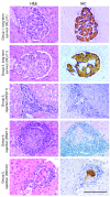
Representative histologic liver sections harvested at necropsy from animals in each treatment group are shown. Sections were stained using standard H&E (left column) and immunohistochemical (IHC; right column) methods. Group 1 and 2 long-term survivors had pristine donor islets embedded in the liver, free of any lymphocytic infiltrates and strong insulin (brown) and glucagon (blue) staining. In contrast, all group 3, 4, and 5 islet recipients exhibited dense lymphocytic infiltration and destruction of transplanted islets, with little to no insulin or glucagon staining, characteristic of allograft rejection. Scale bar: 100 μm.
Figure 6
Figure 6. Combined LFA-1/CD28 blockade extends islet allograft survival.
(A) The experimental design is shown. Diabetic rhesus monkeys were transplanted with allogeneic islets and received TS-1/22 induction alone, belatacept maintenance monotherapy, or a combination of both agents. After islet engraftment, rejection was defined as a FBG of more than 130 mg/dl on 2 consecutive days. (B) Recipients treated with TS-1/22 plus belatacept (filled squares) experienced significantly prolonged allograft function compared with that of TS-1/22–treated (open circles) animals or belatacept-treated (asterisks) animals. TS-1/22 was discontinued on POD 59 (white arrow). Statistical analysis using the log-rank test for graft survival among groups showed the superiority of the combined therapy as compared with TS-1/22 (P = 0.0082) and belatacept monotherapy (P = 0.0042). (CE) Representative FBG graphs of (C) combined TS-1/22 plus belatacept, (D) TS-1/22 alone, and (E) belatacept monotherapy islet recipients show immediate resolution of hyperglycemia on the day of transplant, followed by variable periods of euglycemia (the representative TS-1/22 monotherapy graph is the same as in Figure 4D). Again, TS-1/22 was discontinued on POD 59 (white arrow). Individual group member survival times (in days) are listed in the top right corner of each corresponding representative graph.
Figure 7
Figure 7. TS-1/22 achieves LFA-1/CD11a receptor occupancy in vivo.
Pharmacodynamic monitoring of serially sampled peripheral lymphocytes from anti–LFA-1–treated animals was done using PFC. A fluorochrome-conjugated antibody competitive with TS-1/22 for CD11a was used to determine the degree of LFA-1 receptor occupancy. (A) Representative PFC plots of recipient CD3+ lymphocytes before transplant (Pre-Tx, red) and while on TS-1/22 (blue) are shown. T cell CD11a was highly detectable before transplant and transitioned to undetectable levels during anti–LFA-1 treatment as TS-1/22 occupied LFA-1. (B) Staining CD3+ lymphocytes for the mouse IgG1 tail of TS-1/22 showed therapeutic antibody coating target cell surfaces. (C) CD4+ and CD8+ subsets (TN, TCM, and TEM) experienced equivalent dose-dependent CD11a receptor occupancy, despite differential expression at baseline and after the discontinuation of TS-1/22 (dosing is depicted by the black bars).
Figure 8
Figure 8. TS-1/22 induces a peripheral lymphocytosis.
Complete blood counts and PFC were serially performed on all islet allograft recipient peripheral blood. Animals receiving anti–LFA-1 therapy experienced an increase in the absolute number of circulating leukocytes, including T and B cells. Longitudinal blood counts on an anti–LFA-1–treated group 1 recipient compared with a group 4 animal not receiving LFA-1–specific therapy represent this dose-dependent TS-1/22-associated lymphocytosis. Counts normalized to pretransplant levels after TS-1/22 discontinuation (dosing is depicted by the gray bars).
Figure 9
Figure 9. TS-1/22 plus belatacept prevents DSA formation.
Donor lymphocytes were incubated with corresponding group 1 (TS-1/22, basiliximab, and sirolimus) and 2 (TS-1/22 and belatacept) islet recipient sera to test for the presence of alloantibodies using PFC. Before transplant and terminal after transplant (Post-Tx) time points are depicted. (A and B) Three out of four evaluable group 1 recipients formed allospecific antibodies, as indicated by the increase in anti-donor IgG after transplant relative to before transplant values. One long-term survivor (RLo11) did not develop alloantibodies. (C and D) In contrast, DSAs were not detected in any group 2 recipients, irrespective of allograft rejection (RDp11, RNw11, and RHu11) or survival (RLn11 and RGm11).
Figure 10
Figure 10. LFA-1–based regimens do not restrict NHP growth.
Group 1 and 2 primates receiving TS-1/22 exhibited posttransplant weight retention and growth, following diabetes induction. Apart from one group 1 animal (RWi11) receiving high-dose STZ, observed weight loss was limited to the pretransplant period. Anti–LFA-1–treated animals resumed weight gain and growth relative to their weights at the time of transplant.

References

    1. Bretscher P, Cohn M. A theory of self-nonself discrimination. Science. 1970;169(950):1042–1049. doi: 10.1126/science.169.3950.1042. - DOI - PubMed
    1. Lafferty KJ, Cunningham AJ. A new analysis of allogeneic interactions. Aust J Exp Biol Med Sci. 1975;53(1):27–42. - PubMed
    1. Mescher MF, et al. Signals required for programming effector and memory development by CD8+ T cells. Immunol Rev. 2006;211:81–92. doi: 10.1111/j.0105-2896.2006.00382.x. - DOI - PubMed
    1. Valujskikh A, Li XC. Frontiers in nephrology: T cell memory as a barrier to transplant tolerance. J Am Soc Nephrol. 2007;18(8):2252–2261. doi: 10.1681/ASN.2007020151. - DOI - PubMed
    1. Adams AB, et al. Heterologous immunity provides a potent barrier to transplantation tolerance. J Clin Invest. 2003;111(12):1887–1895. - PMC - PubMed

Publication types

MeSH terms

Substances