. 2010 Dec;120(12):4466-77.
doi: 10.1172/JCI41717. Epub 2010 Nov 22.

Hepcidin as a therapeutic tool to limit iron overload and improve anemia in β-thalassemic mice

Affiliations

Hepcidin as a therapeutic tool to limit iron overload and improve anemia in β-thalassemic mice

Sara Gardenghi et al. J Clin Invest. 2010 Dec.

Abstract

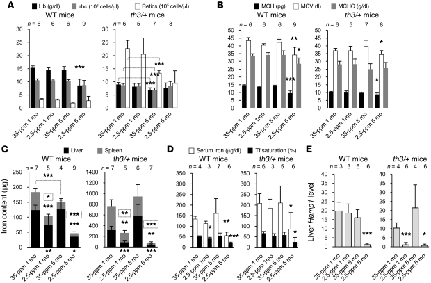
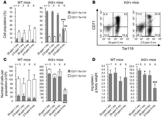
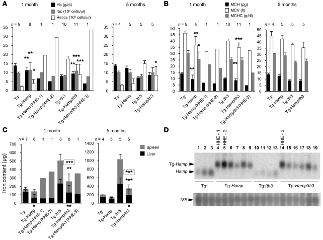
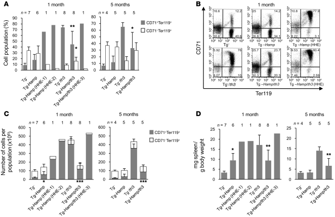
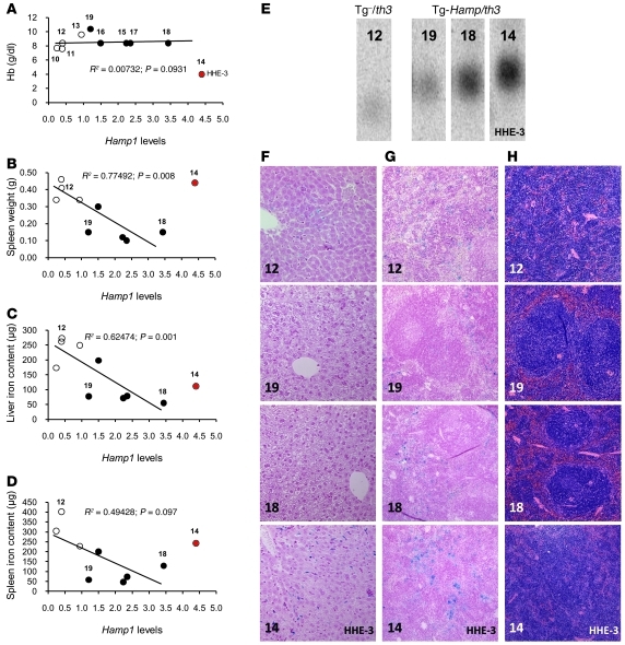
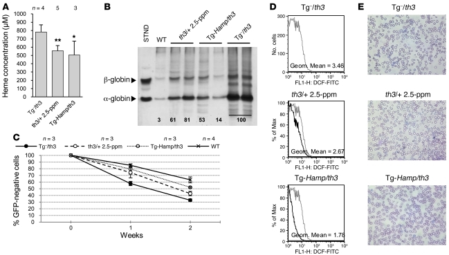
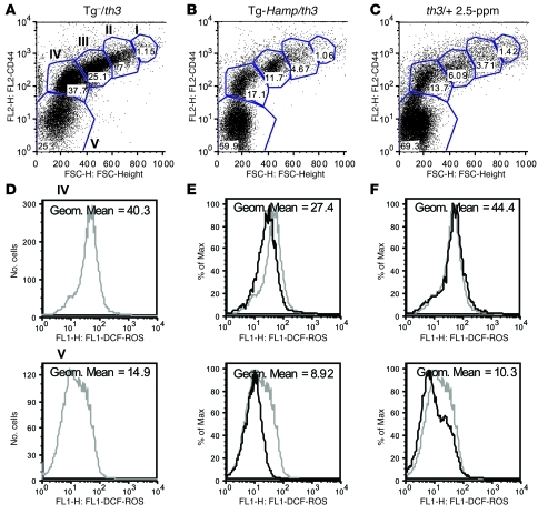
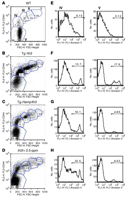
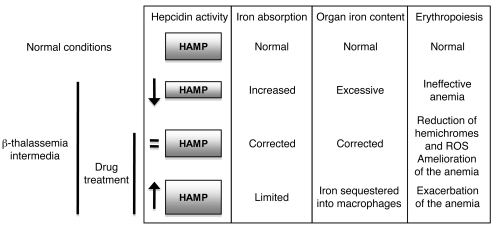
Excessive iron absorption is one of the main features of β-thalassemia and can lead to severe morbidity and mortality. Serial analyses of β-thalassemic mice indicate that while hemoglobin levels decrease over time, the concentration of iron in the liver, spleen, and kidneys markedly increases. Iron overload is associated with low levels of hepcidin, a peptide that regulates iron metabolism by triggering degradation of ferroportin, an iron-transport protein localized on absorptive enterocytes as well as hepatocytes and macrophages. Patients with β-thalassemia also have low hepcidin levels. These observations led us to hypothesize that more iron is absorbed in β-thalassemia than is required for erythropoiesis and that increasing the concentration of hepcidin in the body of such patients might be therapeutic, limiting iron overload. Here we demonstrate that a moderate increase in expression of hepcidin in β-thalassemic mice limits iron overload, decreases formation of insoluble membrane-bound globins and reactive oxygen species, and improves anemia. Mice with increased hepcidin expression also demonstrated an increase in the lifespan of their red cells, reversal of ineffective erythropoiesis and splenomegaly, and an increase in total hemoglobin levels. These data led us to suggest that therapeutics that could increase hepcidin levels or act as hepcidin agonists might help treat the abnormal iron absorption in individuals with β-thalassemia and related disorders.


Figures

Figure 1
Figure 1. Hematological and iron-related parameters in groups of WT and th3/+ mice fed diets with 35 and 2.5 ppm iron for 1 or 5 months.
Complete blood counts show (A) the Hb, rbc, and reticulocyte values, and the (B) MCH, MCV, and MCHC values. The figure also shows (C) total iron content of liver and spleen as measured by atomic absorption, (D) serum iron concentration and Tf saturation, and (E) Hamp1 mRNA expression levels relative to mouse Gapdh, obtained by Q-PCR of the liver using primers specific for mouse Hamp1. Error bars represent SD. P values were calculated using unpaired, 2-tailed Student’s t test. *P < 0.05; **P < 0.01; ***P < 0.001. Groups of mice on the 2.5-ppm diet were compared with corresponding groups on the 35-ppm diet fed for the same length of time unless otherwise indicated by brackets. In C, P values relative to comparisons between spleens are above the columns, while those between the livers are under the x axis. P values in the small rectangles above the columns refer to the total iron content of liver and spleen.
Figure 2
Figure 2. Effects of dietary iron on the size of the spleen (normalized to body weight) and the percentage of mature and immature erythroid cell populations in the spleens of WT and th3/+ mice.
(A) FACS analysis performed on splenic erythroid cells using CD71 (Tf receptor) and Ter119 (erythroid-specific) costaining. The graphs indicate the percentages of early erythroid precursors (CD71+Ter119+), which correspond mainly to basophilic erythroblasts and late basophilic and chromatophilic erythroblasts, and those of mature erythroid cells (CD71Ter119+), including both enucleated erythroid cells and orthochromatic erythroblasts. (B) FACS analysis of spleen cells from 2 representative th3/+ mice fed the 35-ppm (left panel) and 2.5-ppm (right panel) diets. (C) Number of CD71+Ter119+ and CD71Ter119+ cells in the spleen. (D) Spleen weights normalized to body weight. Groups of mice on the 2.5-ppm diet were compared with the corresponding groups on the 35-ppm diet fed for the same length of time. *P < 0.05; **P < 0.01; ***P < 0.001. Data are presented as mean ± SD.
Figure 3
Figure 3. Hematological and iron-related parameters in Tg-Hamp and Tg-Hamp/th3 mice fed a diet containing 35 ppm iron for 1 and 5 months.
(A) Hb, rbc, and reticulocyte values. (B) MCH, MCV, and MCHC values. (C) Total iron content of liver and spleen as measured by atomic absorption. Groups of Tg-Hamp mice were compared with Tg mice, and Tg-Hamp/th3 mice with Tg/th3 controls. (D) Northern blot analysis of the endogenous (bottom band) and transgenic (upper band) Hamp1 mRNA transcripts. 18S, ribosomal subunit 18S loading control. *P < 0.05; **P < 0.01; ***P < 0.001. Data are presented as mean ± SD.
Figure 4
Figure 4. Effects due to overexpression of Hamp1 on the percentage of mature and immature erythroid cell populations and on the spleen size (normalized to body weight) of Tg-Hamp and Tg-Hamp/th3 mice fed a diet containing 35 ppm iron for 1 and 5 months.
(A) FACS analysis of splenic erythroid cells using CD71 and Ter119 costaining. (B) FACS profiles from representative Tg, Tg-Hamp, and Tg-Hamp (HHE) mice (upper panels) and Tg/th3, Tg-Hamp/th3, and Tg-Hamp/th3 (HHE) mice (lower panels). (C) Number of CD71+Ter119+ and CD71Ter119+ cells in the spleen. (D) Spleen weights normalized to body weight. *P < 0.05; **P < 0.01; ***P < 0.001. Data are presented as mean ± SD.
Figure 5
Figure 5. Effects of different levels of transgenic Hamp1 expression on the distribution of iron in the liver and spleen, and on spleen size and morphology in th3/+ mice.
The numbers refer back to the Northern blot in Figure 3D. Correlations between transgenic Hamp1 expression (as determined by the band intensity of the Northern blot in Figure 3D) in Tg/th3 (white circles) and Tg-Hamp/th3 mice (black circles) and (A) Hb values, (B) spleen weight, (C) hepatic iron content, and (D) splenic iron content. The spleen specimen of a control mouse (number 11) was lost during the iron analysis. Histopathological examinations were performed on 1 Tg/th3 mouse (number 12), 2 Tg-Hamp/th3 mice with intermediate levels of Hamp1 expression (numbers 19 and 18), and the Tg-Hamp/th3 HHE mouse (number 14). (E) Northern blot from Figure 3D. Iron deposition in (F) the liver and (G) spleen. (H) Spleen morphology. Images were captured on a Nikon Eclipse E800 microscope, with a Retiga Exi camera (Qimaging), then acquired using the IPLab 3.65a software (Scanalytics Inc.). Brightness/contrast and color balance were adjusted using Adobe Photoshop 7.0.1 (Adobe System Inc.). Original magnification, ×200 (F); ×100 (G and H).
Figure 6
Figure 6. Overexpression of transgenic Hamp1 improves erythropoiesis in th3/+ mice.
(A) Quantification of the heme content in th3/+ mice fed the 2.5-ppm iron diet, Tg/th3, and Tg-Hamp/th3 mice, using the same number of rbc from each mouse. (B) rbc membrane fractions analyzed by TAU gel electrophoresis to determine the amount of bound globins. The membrane fractions prepared from the same number of rbc have been loaded in each lane. The number under each lane represents the α-globin band intensity expressed as percentage of the mean band intensity of the 2 th3/+ mice. (C) rbc life spans measured as a percentage of that of GFP-negative cells collected from GFP-positive mice injected with blood from WT mice, th3/+ mice fed the 2.5-ppm iron diet, Tg(-)/th3, and Tg-Hamp/th3 mice. Data are presented as mean ± SD. (D) ROS measured in the peripheral blood of 3 representative mice, a th3/+ fed the 2.5-ppm diet, a Tg/th3, and a Tg-Hamp/th3 using CM-H2DCFDA. Both the Tg-Hamp/th3 mouse and the th3/+ mouse on the 2.5-ppm diet showed reduced fluorescence compared with the Tg/th3 mouse (in gray). (E) Blood smears stained with May-Grunwald-Giemsa stain, showing the rbc morphology of representative mice, a th3/+ fed the 2.5-ppm iron diet, a Tg/th3, and a Tg-Hamp/th3. Original magnification, ×400. Data are presented as mean ± SD.
Figure 7
Figure 7. Different stages of erythroid differentiation in the spleens of th3/+ mice fed the 2.5-ppm iron diet or overexpressing Hamp1.
FACS analysis of representative mice (A) Tg/th3, (B) Tg-Hamp/th3, and (C) th3/+ on the 2.5-ppm diet mice. D, E, and F show representative analyses of ROS for stages IV (upper panel) and V (lower panel) using CM-H2DCFDA. Data are presented as mean ± SD.
Figure 8
Figure 8. Different stages of erythroid differentiation in the spleens of th3/+ mice fed the 2.5-ppm iron diet or overexpressing Hamp1.
FACS analysis of representative mice (A) WT, (B) Tg/th3, (C) Tg-Hamp/th3, and (D) th3/+ on the 2.5-ppm diet mice. Numbers indicate the percentage of cells measured in each distinct erythroid population. E, F, G, and H show representative analyses of membrane phosphatidylserine exposure for stages IV (left panel) and V (right panel) using annexin V. Data are presented as mean ± SD.
Figure 9
Figure 9. Potential effects of hepcidin agonists or activators on iron absorption under normal and β-thalassemic conditions.
=, normal; ↓, abnormally low; ↑, abnormally high hepcidin expression levels.

Comment in

References

    1. Steinberg MH, Forget BG, Higgs DR, Nagel RL.Disorders of hemoglobin: Genetics, Pathophysiology and Clinical Management . Cambridge, United Kingdom: Cambridge University Press; 2001.
    1. Heinrich HC, et al. Absorption of inorganic and food iron in children with heterozygous and homozygous beta-thalassemia. Z Kinderheilkd. 1973;115(1):1–22. doi: 10.1007/BF00438987. - DOI - PubMed
    1. Pippard MJ, Callender ST, Warner GT, Weatherall DJ. Iron absorption and loading in beta-thalassaemia intermedia. Lancet. 1979;2(8147):819–821. - PubMed
    1. Hershko C, Rachmilewitz EA. Mechanism of desferrioxamine-induced iron excretion in thalassaemia. Br J Haematol. 1979;42(1):125–132. doi: 10.1111/j.1365-2141.1979.tb03704.x. - DOI - PubMed
    1. Yang B, Kirby S, Lewis J, Detloff PJ, Maeda N, Smithies O. A mouse model for beta 0-thalassemia. Proc Natl Acad Sci U S A. 1995;92(25):11608–11612. doi: 10.1073/pnas.92.25.11608. - DOI - PMC - PubMed

Publication types

MeSH terms