. 2010 Dec;120(12):4445-52.
doi: 10.1172/JCI43796. Epub 2010 Nov 22.

αv Integrin expression by DCs is required for Th17 cell differentiation and development of experimental autoimmune encephalomyelitis in mice

Affiliations

αv Integrin expression by DCs is required for Th17 cell differentiation and development of experimental autoimmune encephalomyelitis in mice

Mridu Acharya et al. J Clin Invest. 2010 Dec.

Abstract

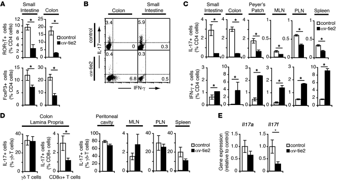
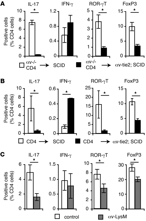
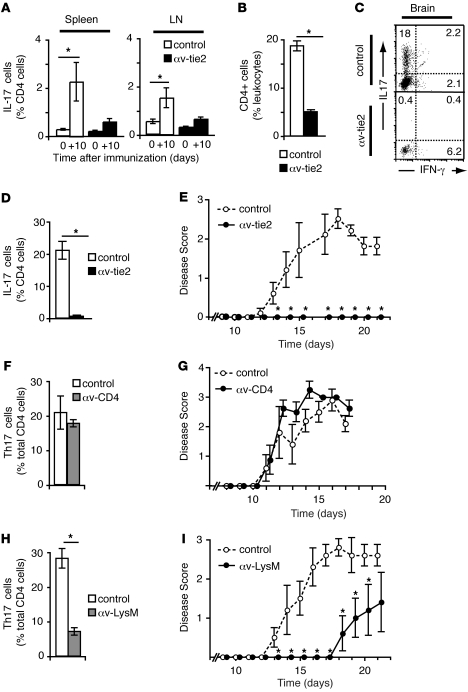
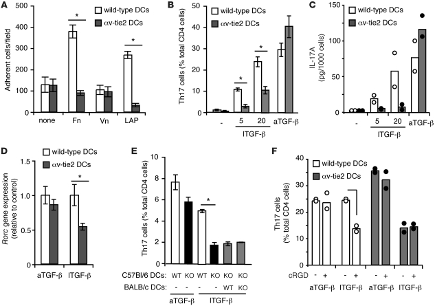
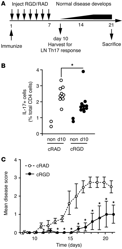
Th17 cells are a distinct lineage of T helper cells that protect the body from bacterial and fungal infection. However, Th17 cells also contribute to inflammatory and autoimmune disorders such as multiple sclerosis. Th17 cell generation requires exposure of naive T cells to the cytokine TGF-β in combination with proinflammatory cytokines. Here we show that differentiation of Th17 cells is also critically dependent on αv integrins. In mice, lack of integrin αv in the immune system resulted in loss of Th17 cells in the intestine and lymphoid tissues. It also led to protection from experimental autoimmune encephalomyelitis (EAE). Further analysis indicated that αv integrins on DCs activated latent TGF-β during T cell stimulation and thereby promoted differentiation of Th17 cells. Furthermore, pharmacologic inhibition of αv integrins using cyclic RGD peptides blocked TGF-β activation and Th17 cell generation in vitro and protected mice from EAE. These data demonstrate that activation of TGF-β by αv-expressing myeloid cells may be a critical step in the generation of Th17 cells and suggest that αv integrins could be therapeutic targets in autoimmune disease.


Figures

Figure 1
Figure 1. Lack of intestinal Th17 cells in αv-tie2 mice.
(A) Proportion of ROR-γT+ and FoxP3+ CD4+ cells in LP of control and αv-tie2 mice. (B and C) Proportion of IL-17–producing (Th17 cells) and IFN-γ–producing (Th1 cells) CD4+ T cells isolated from the LP of the small intestine and colon, Peyer’s patch, MLN, peripheral LN (PLN), and spleen of control and αv-tie2 mice. (B) Representative FACS data from one experiment. The plots are gated on CD4+ cells, and the numbers represent the percentage of CD4+ cells that stained for IL-17 or IFN-γ. (C) Mean ± SEM from at least 3 mice/group. (D) Proportion of IL-17–producing γδ and CD8α+ T cells isolated from the LP of the colon, and IL-17–producing γδ T cells from indicated tissues, of control and αv-tie2 mice. (E) Expression of Il17a and Il17f measured by QRT-PCR in RNA isolated from proximal colon of control and αv-tie2 mice. All graphs show mean ± SEM from at least 3 mice/group. *P < 0.05.
Figure 2
Figure 2. αv Expression by myeloid cells is required for Th17 cell differentiation.
(A and B) Tregs and Th17 and Th1 cells in the colonic LP of SCID control and αv-tie2/ SCID mice 6 weeks following adoptive transfer with splenic CD4+ T cells from αv-tie2 (A) or wild-type control (B) mice. Data are from 4 littermate recipients/group. (C) Th17 and Th1 cells and Tregs in the colonic LP of control and αv-LysM mice (12 weeks of age). Data are from 4 littermates/group. In all cases, similar differences were seen in at least 3 independent experiments. *P < 0.05, Student’s t test.
Figure 3
Figure 3. αv-tie2 mice are protected from EAE.
(A) Percentage of CD4+ T cells that express IL-17 in spleen and LNs of αv-tie2 and control mice before immunization (day 0) and 10 days after immunization with MOG peptide in CFA (+10). (BD) Percentage of CD4+ cells (B) or IL-17– and IFN-γ–producing cells (C and D) in leukocytes isolated from brain 21 days after immunization. (C) representative FACS data gated on CD4+ cells. (D) Percentage of CD4+ cells that expressed IL-17. (E) Progression of EAE in control and αv-tie2 mice. Similar data were seen in 3 independent experiments. (F and H) IL-17–producing T cells in the brain of αv-CD4 mice (F) and αv-LysM mice (H) 21 days after induction of EAE. (G and I) Development of EAE in control and αv-CD4 mice (G) or αv-LysM mice (I). Similar results were seen in 3 (αv-tie2 data) or 2 (αv-LysM) independent experiments. For all graphs, data are presented as mean ± SEM from at least 4 mice/group. *P < 0.05, Student’s t test.
Figure 4
Figure 4. Expression of αv integrins on DCs is required for T cell responses to latent TGF-β.
(A) DCs bound LAP though αv integrins. The number of control and αv-knockout DCs binding to untreated plates or plates coated with LAP, fibronectin (Fn), or vitronectin (Vn) is shown. Data are mean ± SD of 3 wells. (B) Proportion of IL-17–producing T cells generated after in vitro culture of control T cells with DCs from control and αv-tie2 mouse spleens in the presence of anti-CD3, IL-6, and active TGF-β (aTGF-β) or latent TGF-β (lTGF-β) (5 or 20 ng/ml). Data are from 3 separate DC preparations; histograms show the mean ± SEM. Similar results were seen in at least 5 independent experiments. (C and D) Production of IL-17A protein in culture supernatant (C) and mRNA expression of Rorc (D) in T cells stimulated as in C. (E) Contact with DCs was required for T cells to respond to TGF-β activated by αv. Proportion of IL-17–producing T cells generated after culture of T cells from OT2 TCR transgenic mice incubated with the indicated combinations of DCs from C57BL/6 and BALB/c background mice in the presence of OVA peptide, IL-6, and active or latent TGF-β. (F) cRGD peptides inhibited TGF-β activation by DCs. Experimental conditions were the same as in B, with T cells cultured with DCs in the presence of anti-CD3, IL-6, aTGF-β, lTGF-β, and cRGD peptides. Dots represent DC cultures from independent control or αv-tie2 mice. Data are from 2 separate DC preparations from either control or αv-tie2 mice. Similar results were seen in 3 independent experiments.
Figure 5
Figure 5. αv Blockade inhibits Th17 cell differentiation and protects from EAE in vivo.
(A) Schematic of experiments to assess effects of cRGD on EAE progression. Mice were immunized (day 1) and given daily injections of cRGD or control peptide for 7 days, and their immune response was assessed at day 10, with paralysis monitored until day 21. (B) Percentage of LN CD4+ T cells that expressed IL-17 ten days after immunization (d10), or non-immunized mice (non) that also received cRAD/cRGD for 7 days. Each point represents an individual mouse, and data are combined from 2 independent experiments. *P < 0.05, Student’s t test (n = 9 cRAD, n = 10 cRGD). (C) Progression of EAE in cRGD- and cRAD-treated mice. Data are mean ± SEM from at least 5 mice/group. *P < 0.05, Student’s t test.

Comment in

Similar articles

Cited by

References

    1. Harrington LE, et al. Interleukin 17-producing CD4+ effector T cells develop via a lineage distinct from the T helper type 1 and 2 lineages. Nat Immunol. 2005;6(11):1123–1132. doi: 10.1038/ni1254. - DOI - PubMed
    1. Park H, et al. A distinct lineage of CD4 T cells regulates tissue inflammation by producing interleukin 17. Nat Immunol. 2005;6(11):1133–1141. doi: 10.1038/ni1261. - DOI - PMC - PubMed
    1. Korn T, Bettelli E, Oukka M, Kuchroo VK. IL-17 and Th17 cells. Annu Rev Immunol. 2009;27:485–517. doi: 10.1146/annurev.immunol.021908.132710. - DOI - PubMed
    1. Steinman L. A brief history of T(H)17, the first major revision in the T(H)1/T(H)2 hypothesis of T cell-mediated tissue damage. Nat Med. 2007;13(2):139–145. - PubMed
    1. Ivanov II, et al. The orphan nuclear receptor RORgammat directs the differentiation program of proinflammatory IL-17+ T helper cells. Cell. 2006;126(6):1121–1133. doi: 10.1016/j.cell.2006.07.035. - DOI - PubMed

Publication types

MeSH terms