. 2010 Dec;120(12):4375-87.
doi: 10.1172/JCI37649. Epub 2010 Nov 22.

Role of chronic ryanodine receptor phosphorylation in heart failure and β-adrenergic receptor blockade in mice

Affiliations

Role of chronic ryanodine receptor phosphorylation in heart failure and β-adrenergic receptor blockade in mice

Jian Shan et al. J Clin Invest. 2010 Dec.

Abstract

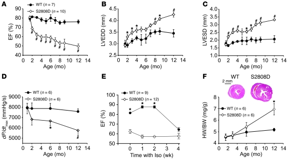
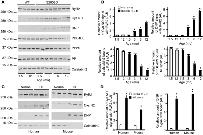
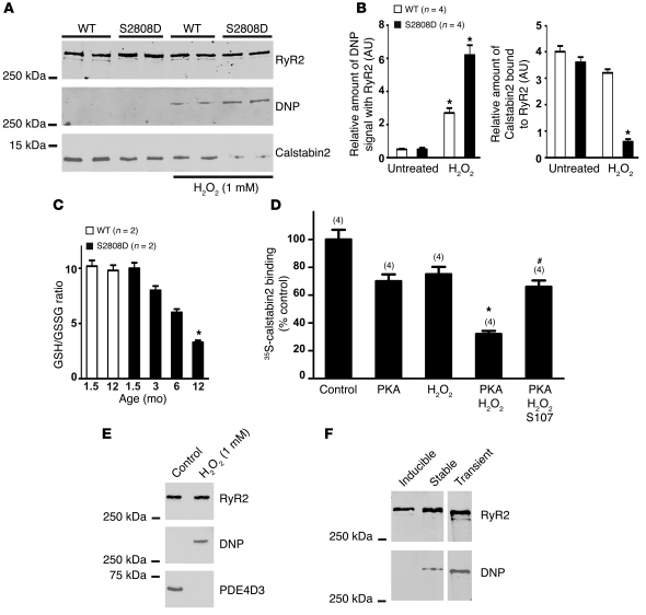
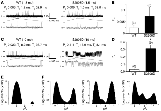
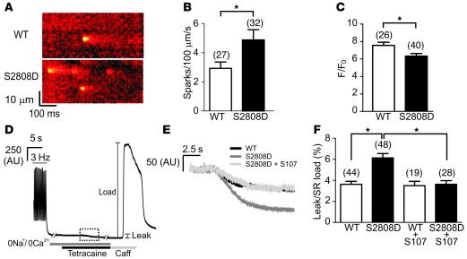
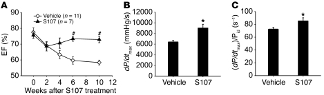
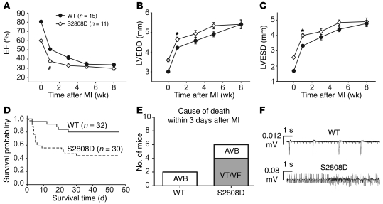
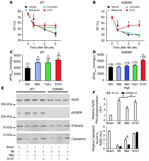
Increased sarcoplasmic reticulum (SR) Ca2+ leak via the cardiac ryanodine receptor/calcium release channel (RyR2) is thought to play a role in heart failure (HF) progression. Inhibition of this leak is an emerging therapeutic strategy. To explore the role of chronic PKA phosphorylation of RyR2 in HF pathogenesis and treatment, we generated a knockin mouse with aspartic acid replacing serine 2808 (mice are referred to herein as RyR2-S2808D+/+ mice). This mutation mimics constitutive PKA hyperphosphorylation of RyR2, which causes depletion of the stabilizing subunit FKBP12.6 (also known as calstabin2), resulting in leaky RyR2. RyR2-S2808D+/+ mice developed age-dependent cardiomyopathy, elevated RyR2 oxidation and nitrosylation, reduced SR Ca2+ store content, and increased diastolic SR Ca2+ leak. After myocardial infarction, RyR2-S2808D+/+ mice exhibited increased mortality compared with WT littermates. Treatment with S107, a 1,4-benzothiazepine derivative that stabilizes RyR2-calstabin2 interactions, inhibited the RyR2-mediated diastolic SR Ca2+ leak and reduced HF progression in WT and RyR2-S2808D+/+ mice. In contrast, β-adrenergic receptor blockers improved cardiac function in WT but not in RyR2-S2808D+/+ mice.Thus, chronic PKA hyperphosphorylation of RyR2 results in a diastolic leak that causes cardiac dysfunction. Reversing PKA hyperphosphorylation of RyR2 is an important mechanism underlying the therapeutic action of β-blocker therapy in HF.


Figures

Figure 1
Figure 1. Age-dependent cardiomyopathy in RyR2-S2808D+/+ mice.
(AC) Serial echocardiographic measurements were performed from 2 to 12 months of age in RyR2-S2808D+/+ (S2808D) mice and WT littermates. Compared with WT littermates, RyR2-S2808D+/+ mice exhibited progressive cardiac dysfunction and LV enlargement. LVEDD, LV end-diastolic diameter (WT, n = 7; RyR2-S2808D+/+, n = 10; #P < 0.01 versus WT; *P < 0.05 versus WT). (D) Cardiac catheterization also revealed age-dependent cardiac dysfunction (*P < 0.05 versus WT; #P < 0.01 versus WT). (E) Chronic Iso treatment in RyR2-S2808D+/+ mice and WT littermates. At 4 months of age, RyR2-S2808D+/+ and WT mice were treated with Iso (30 mg/kg/d) for 4 weeks. Cardiac function was monitored with echocardiography at baseline, 1, 2, and 4 weeks (*P < 0.05 versus RyR2-S2808D+/+ mice). (F) Representative histology of age-matched RyR2-S2808D+/+ and WT littermates (top). Cross section at papillary muscle level. Scale bar: 2 mm. Heart weight to body weight (HW/BW) ratio (bottom). Filled circles represent WT mice; open diamonds represent RyR2-S2808D+/+ mice (*P < 0.05 versus WT).
Figure 2
Figure 2. RyR2 channel complex remodeling in RyR2-S2808D mice.
(A) Progressive oxidation, Cys-nitrosylation (Cys NO) of cardiac RyR2, and depletion of PDE4D3, calstabin2, PP2A, and PP1 from the cardiac RyR2 complex in the RyR2-S2808D mice. (B) Levels of proteins in the RyR2 complex and levels of oxidation and Cys-nitrosylation were normalized to the total amount of RyR2 (AU). *P < 0.05 versus WT at 1.5 months. (C) Oxidation and Cys-nitrosylation of RyR2 in HF samples. RyR2 immunoprecipitations were performed on hearts from mice with MI and sham-treated mice or on samples from HF and non-HF human heart tissue. (D) Levels of oxidation and Cys-nitrosylation were normalized to the total amount of RyR2 (AU). *P < 0.05 versus normal samples.
Figure 3
Figure 3. Combined effects of PKA phosphorylation and oxidation on calstabin2 binding to RyR2.
(A) CSR preparations were treated with 1 mM H2O2, and RyR2 was immunoprecipitated, size fractionated, and immunoblotted for oxidation (DNP) and calstabin2 in the RyR2 complex. (B) Levels of oxidation and calstabin2 in the RyR2 complex were normalized to the total amount of RyR2 (AU). *P < 0.05 untreated versus H2O2-treated samples. (C) GSH/GSSG ratios (n = 2) were compared between WT and RyR2-S2808D+/+ mice. *P < 0.05. (D) 35S-calstabin binding was measured in samples treated with PKA, H2O2, or a combination of the 2, in the presence or absence or S107. Radioactivity counts were normalized to the untreated control samples. Data are presented as mean ± SEM (n = 4). The numbers of replicates for each condition are indicated by the parenthetical numbers over each bar. *P < 0.05 compared with control; #P < 0.05 compared with PKA/H2O2 treatment without S107. (E) CSR was treated with 1 mM H2O2, and RyR2 was immunoprecipitated and immunoblotted for oxidation (DNP) and PDE4D3 in the RyR2 complex. (F) RyR2 was expressed in CHO cells using an inducible vector (tetracycline), transient transfection, or stable transfection and immunoblotted with anti-DNP antibody to determine oxidation of the channel.
Figure 4
Figure 4. Functional characterization of cardiac RyR2 channels from WT and RyR2-S2808D+/+ mice.
(A) Representative single channel current traces of cardiac RyR2 channels isolated from WT and RyR2-S2808D+/+ mice at 1.5 months of age. (B) Bar graph summarizing average Po in WT (n = 3) and RyR2-S2808D+/+ (n = 6) channels from 1.5-month-old mice (P = NS). The number of channels recorded from each sample is indicated by the parenthetical numbers over each bar. (C) Representative single channel current traces of cardiac RyR2 channels isolated from 10-month-old WT and RyR2-S2808D+/+ mice. (D) Bar graph summarizing average Po in WT (n = 6) and S2808D+/+ (n = 9) channels from 10-month-old mice (*P < 0.05). The number of channels recorded from each sample is indicated by the parenthetical numbers over each bar. Channel openings are shown as upward deflections; the open and closed (c) states of the channel are indicated by horizontal bars in the beginning of each trace. Examples of the channel activity are shown at 2 different time scales (5 s for upper trace and 500 ms for lower trace depicted by the thick gray bar) as indicated by dimension bars. The respective Po, To (average open time), and Tc (average closed time) are shown above each 5 second trace and correspond to that particular experiment. (E) Amplitude histogram of a representative WT cardiac RyR2 channel (at 10 months old), showing 2 distinct peaks corresponding to fully open (~4 pA) and closed (0 pA) states of the channel. (F) Samples of amplitude histograms from 3 different experiments, using RyR2-S2808D+/+ channels (10-month-old mice), showing subconductance states.
Figure 5
Figure 5. Reduced store content and increased diastolic Ca2+ leak in RyR2-S2808D+/+ mice.
(A) Representative line scans obtained from ventricular myocytes isolated from WT and RyR2-S2808D+/+ mice, showing an increased spark frequency in RyR2-S2808D myocytes. (B) Pooled data showing mean ± SEM spark frequency from the number of myocytes indicated parenthetically. *P < 0.05. (C) Pooled data showing mean ± SEM amplitude of caffeine-evoked signals from the number of cells indicated parenthetically. *P < 0.05. (D) Representative trace depicting the protocol for determining the level of SR Ca2+ leak in ventricular myocytes. After termination of 3-Hz pacing, cells were superfused with Na+- and Ca2+-free solution. Application of tetracaine (1 mM) reduced the baseline fluorescence (leak). Caffeine (Caff; 10 mM) was applied at the end of the protocol to assess the SR Ca2+ load. The box made of dashed lines indicates the region expanded in E. (E) Representative signals during tetracaine application from WT, RyR2-S2808D+/+, and S107-treated RyR2-S2808D+/+ myocytes. (F) Pooled data from the number of cells indicated parenthetically, showing the mean ± SEM leak/load relationship of WT and RyR2-S2808D+/+ myocytes in the presence and absence of S107 (1 μM). Values represent the magnitude of reduction due to tetracaine expressed as a percentage of the increase in signal in response to caffeine (*P < 0.05). Myocytes were prepared from 5 or 6 mice in each experimental group.
Figure 6
Figure 6. S107 improves cardiac function in RyR2-S2808D+/+ mice.
(A) Echocardiographic measurements during a 10-week treatment period, showing that the S107-treated (20 mg/kg/d via osmotic pump) group exhibited preserved cardiac function compared with the vehicle-treated group (#P < 0.01 versus vehicle-treated group). (B and C) Cardiac catheterization was performed at the end of study, and both dP/dtmax and (dP/dtmax)/Pid, where Pid indicates the instantaneous developed pressure in mmHg and (s–1) is the unit of measure for (dP/dtmax)/Pid, showed a significant improvement in S107-treated group (*P < 0.05 versus vehicle-treated group).
Figure 7
Figure 7. Increased mortality after MI in RyR2-S2808D+/+ mice.
(AC) Serial echocardiographic measurements after MI. (D) Kaplan-Meier survival curve of RyR2-S2808D+/+ mice (n = 30) and WT littermates (n = 32) after MI. The solid line represents WT mice, and the dashed line represents RyR2-S2808D+/+ mice. (E) Telemeters were implanted in WT (n = 8) and RyR2-S2808D (n = 8) mice and cause of death within 3 days after MI was determined. AVB, atrial-ventricular block; VF, ventricular fibrillation; VT, ventricular tachycardia. (F) Representative telemetry traces depicting atrial-ventricular block in a WT mouse and ventricular tachycardia in a RyR2-S2808D+/+ mouse after MI. #P < 0.01 versus WT; *P < 0.05 versus WT.
Figure 8
Figure 8. S107 but not metoprolol or carvedilol improves cardiac function in RyR-S2808D+/+ mice after MI.
(A and B) Echocardiographic measurements at baseline, 1, 3, and 5 weeks after MI. Treatment started 1 week after MI (arrows). (A) In the WT group, S107, metoprolol (Met), and carvedilol (Carv) showed a beneficial effect at the end of study (week 4 of treatment). (B) In the RyR2-S2808D+/+ group, only S107 inhibited HF progression. *P < 0.05 versus vehicle group. (C and D) Hemodynamic data were obtained by cardiac catheterization at the end of study. (C) In the WT group, S107, metoprolol, and carvedilol improved LV systolic function (dP/dtmax). (D) In the RyR2-S2808D+/+ group, only S107 improved LV systolic function. The number of mice in each treatment group is indicated by the parenthetical numbers over each bar. Black represents vehicle; light blue represents low-dose metoprolol (30 mg/kg/d); dark blue represents high-dose metoprolol (300 mg/kg/d); green represents carvedilol (10 mg/kg/d); and red represents S107 (30 mg/kg/d). *P < 0.05 versus vehicle group. (E) Representative immunoblots from in vivo studies. Equivalent amounts of RyR2 were immunoprecipitated from cardiac lysates using an anti-RyR2 antibody. In the WT group, MI increased RyR2-S2808 PKA phosphorylation and calstabin2 depletion from the RyR2 channel complex and metoprolol decreased RyR2-S2808 PKA phosphorylation and reduced depletion of calstabin2 from the cardiac RyR2 channel complex. In the RyR2-S2808D+/+ group, calstabin2 was depleted from RyR2 channel complex in both vehicle- and metoprolol-treated groups, whereas S107 reduced depletion of calstabin2 from the cardiac RyR2 channel complex. (F) Pooled data from 3 separate immunoblots. *P < 0.05 versus sham; #P < 0.05 versus metoprolol treated.

Comment in

References

    1. Lloyd-Jones D, et al. Heart disease and stroke statistics--2010 update: a report from the American Heart Association. Circulation. 2010;121(7):e46–e215. doi: 10.1161/CIRCULATIONAHA.109.192667. - DOI - PubMed
    1. Colucci WS, et al. Carvedilol inhibits clinical progression in patients with mild symptoms of heart failure. US Carvedilol Heart Failure Study Group. Circulation. 1996;94(11):2800–2806. - PubMed
    1. Packer M, et al. The effect of carvedilol on morbidity and mortality in patients with chronic heart failure. U.S. Carvedilol Heart Failure Study Group. N Engl J Med. 1996;334(21):1349–1355. doi: 10.1056/NEJM199605233342101. - DOI - PubMed
    1. Packer M, et al. Effect of carvedilol on survival in severe chronic heart failure. N Engl J Med. 2001;344(22):1651–1658. doi: 10.1056/NEJM200105313442201. - DOI - PubMed
    1. Reiken S, et al. beta-adrenergic receptor blockers restore cardiac calcium release channel (ryanodine receptor) structure and function in heart failure. Circulation. 2001;104(23):2843–2848. doi: 10.1161/hc4701.099578. - DOI - PubMed

Publication types

MeSH terms

Substances