. 2010 Dec;120(12):4332-41.
doi: 10.1172/JCI43918. Epub 2010 Nov 22.

Vancomycin-resistant Enterococcus domination of intestinal microbiota is enabled by antibiotic treatment in mice and precedes bloodstream invasion in humans

Affiliations

Vancomycin-resistant Enterococcus domination of intestinal microbiota is enabled by antibiotic treatment in mice and precedes bloodstream invasion in humans

Carles Ubeda et al. J Clin Invest. 2010 Dec.

Abstract

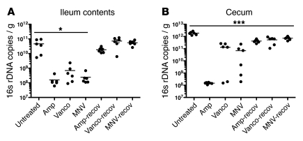
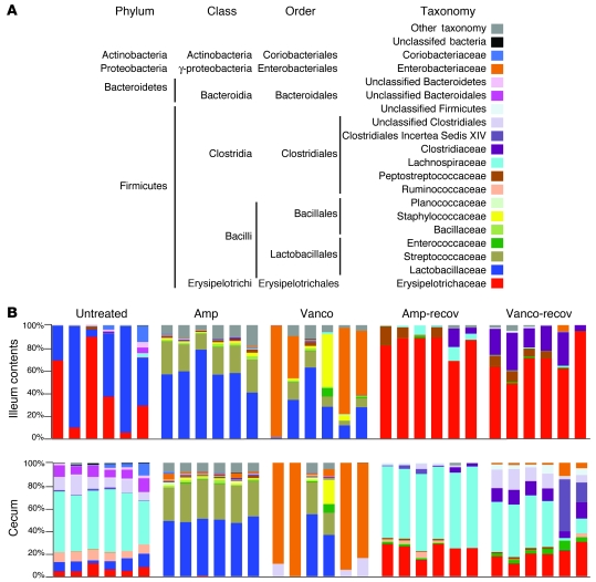
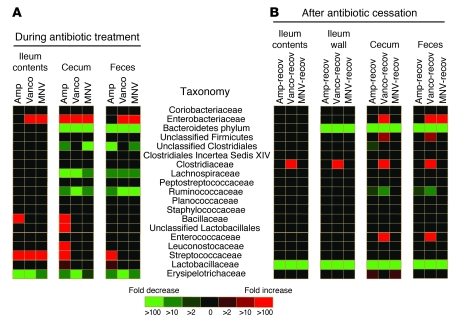
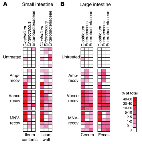
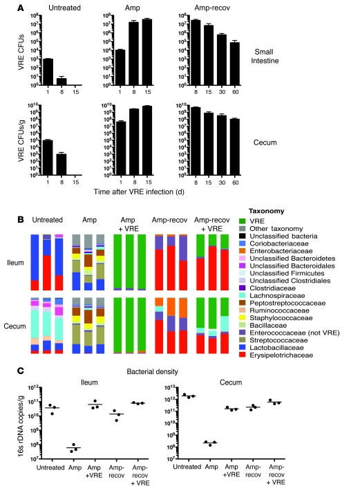
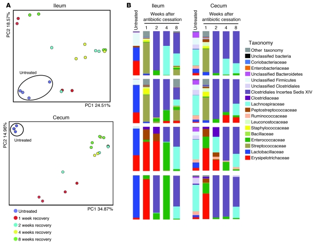
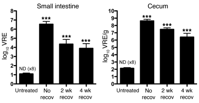
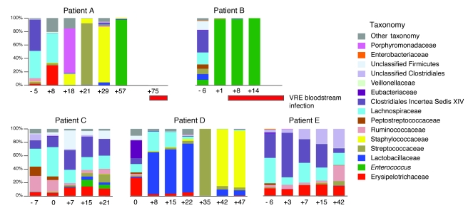
Bloodstream infection by highly antibiotic-resistant bacteria, such as vancomycin-resistant Enterococcus (VRE), is a growing clinical problem that increasingly defies medical intervention. Identifying patients at high risk for bacterial sepsis remains an important clinical challenge. Recent studies have shown that antibiotics can alter microbial diversity in the intestine. Here, we characterized these effects using 16s rDNA pyrosequencing and demonstrated that antibiotic treatment of mice enabled exogenously administered VRE to efficiently and nearly completely displace the normal microbiota of the small and large intestine. In the clinical setting, we found that intestinal domination by VRE preceded bloodstream infection in patients undergoing allogeneic hematopoietic stem cell transplantation. Our results demonstrate that antibiotics perturb the normal commensal microbiota and set the stage for intestinal domination by bacteria associated with hospital-acquired infections. Thus, high-throughput DNA sequencing of the intestinal microbiota could identify patients at high risk of developing bacterial sepsis.


Figures

Figure 1
Figure 1. Antibiotic treatment alters intestinal microbial density.
The number of 16s rDNA copies was determined by qPCR of (A) ileum or (B) cecum samples from untreated mice, mice treated with ampicillin (Amp), vancomycin (Vanco), or MNV, or mice 2 weeks after cessation (i.e., recovery [recov]) of antibiotic treatment with ampicillin, vancomycin, or MNV (6 mice per group). *P < 0.05, ***P < 0.001 versus untreated, 1-way ANOVA with Bonferroni correction.
Figure 2
Figure 2. Antibiotic treatment alters the composition of the intestinal microbiota.
(A) Color code for the most predominant bacterial taxa found in the murine intestine. (B) Phylogenetic classification of 16S rDNA frequencies in the ileum contents or cecum from samples collected from untreated mice, mice treated with ampicillin or vancomycin, or mice allowed to recover for 2 weeks from antibiotic treatment. Each bar represents the microbiota of an individual mouse.
Figure 3
Figure 3. Statistically significant changes in intestinal bacterial populations observed upon antibiotic treatment.
Heatmap showing statistically significant differences (P < 0.05) in intestinal bacterial populations between (A) untreated mice and mice treated for 1 week with ampicillin, vancomycin, or MNV, or (B) untreated mice and mice allowed to recover for 2 weeks from antibiotic treatment. Colors in the key indicate the observed fold changes (red, increase; green, decrease).
Figure 4
Figure 4. Antibiotic treatment promotes intestinal colonization by Clostridium, Enterococcus, and Enterobacteriaceae.
Heatmap showing the frequencies of 16s rDNA sequences classified as Clostridium, Enterococcus, or Enterobacteriaceae in the small (A) or large (B) intestine of untreated mice or mice allowed to recover for 2 weeks after ampicillin, vancomycin, or MNV treatment. Colors in key indicate the frequency of each group of bacteria in each sample. Each row represents an individual mouse.
Figure 5
Figure 5. Ampicillin treatment promotes VRE intestinal colonization in mice.
(A) The number of VRE CFUs in the small intestine or cecum of untreated mice, mice treated for 1 week with ampicillin before infection and kept on ampicillin after VRE infection, or mice switched back to antibiotic-free water 1 day after infection and allowed to recover. Mice were infected with 108 CFUs. Samples were harvested on different days after VRE infection. n ≥ 6 mice per group and day, except day 15 ampicillin (n = 3). (B) Phylogenetic classification of 16S rDNA frequencies or (C) number of 16s rDNA copies in the ileum and cecum of untreated mice as well as ampicillin-treated mice, infected or not with VRE after 1 week of treatment, and either treated for 8 days or allowed to recover starting at 1 day after infection. The most predominant bacterial populations identified are color coded as indicated. Each bar represents the microbiota composition of an individual mouse.
Figure 6
Figure 6. Incomplete recovery of the microbiota after ampicillin treatment.
Four groups of mice were treated with ampicillin and allowed to recover from ampicillin treatment. Each group of mice was housed in a separate cage. 1 mouse per cage was euthanized at weeks 1, 2, 4, or 8 after antibiotic withdrawal, and the ileum and cecum was harvested for microbiota analysis. As controls, 4 mice were euthanized before antibiotic treatment. (A) Unweighted UniFrac analysis of ileum or cecum samples. Each point represents the microbiota of an individual mouse. (B) Phylogenetic classification of 16S rDNA frequencies in the ileum and cecum. Each bar represents the microbiota of an individual mouse. The most predominant bacterial populations identified are color coded as indicated.
Figure 7
Figure 7. Incomplete recovery of the intestinal microbiota allows VRE intestinal colonization.
The number of VRE CFUs was determined 8 days after infection in the small intestine or cecum of untreated mice, mice treated with ampicillin for 1 week before infection and switched back to antibiotic-free water 1 day after infection (i.e., no recovery time), and mice treated with ampicillin for 1 week and allowed to recover for 2 or 4 weeks from ampicillin treatment before infection. If VRE was not detected (ND), the limit of detection was considered to be the number of CFUs in the sample. n = 9 per group, except 2-week recovery (n = 10). ***P < 0.001 versus untreated, 1-way ANOVA with Bonferroni correction on the log-transformed CFU values.
Figure 8
Figure 8. VRE dominates the intestinal microbiota in humans prior to invading the bloodstream.
Phylogenetic classification of 16S rDNA frequencies in stool samples collected from allo-HSCT patients. Samples were collected upon hospital admission and periodically during the transplant course; 5 patients were studied. Each bar represents the microbiota of 1 stool sample. The timing of sample collection relative to the day of transplant (day 0) is indicated below each bar. The most predominant bacterial populations identified are color coded as indicated. The timing of VRE bloodstream infection relative to analyses of the microbiota for patients A and B is indicated by red horizontal bars.

Comment in

  • Which species are in your feces?
    Zaph C. Zaph C. J Clin Invest. 2010 Dec;120(12):4182-5. doi: 10.1172/JCI45263. Epub 2010 Nov 22. J Clin Invest. 2010. PMID: 21099104 Free PMC article.

References

    1. Donskey CJ. Antibiotic regimens and intestinal colonization with antibiotic-resistant gram-negative bacilli. Clin Infect Dis. 2006;43(suppl 2):S62–S69. - PubMed
    1. Rice LB. Antimicrobial resistance in gram–positive bacteria. Am J Med. 2006;119(6 suppl 1):S11–S19. - PubMed
    1. Bartlett JG. Narrative review: the new epidemic of Clostridium difficile-associated enteric disease. Ann Intern Med. 2006;145(10):758–764. - PubMed
    1. Eckburg PB, et al. Diversity of the human intestinal microbial flora. Science. 2005;308(5728):1635–1638. doi: 10.1126/science.1110591. - DOI - PMC - PubMed
    1. Backhed F, Ley RE, Sonnenburg JL, Peterson DA, Gordon JI. Host-bacterial mutualism in the human intestine. Science. 2005;307(5717):1915–1920. doi: 10.1126/science.1104816. - DOI - PubMed

Publication types

MeSH terms