. 2010 Dec;120(12):4388-98.
doi: 10.1172/JCI32726. Epub 2010 Nov 22.

Phosphorylation of the ryanodine receptor mediates the cardiac fight or flight response in mice

Affiliations

Phosphorylation of the ryanodine receptor mediates the cardiac fight or flight response in mice

Jian Shan et al. J Clin Invest. 2010 Dec.

Abstract

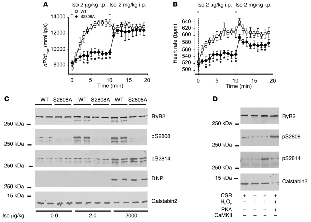
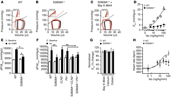
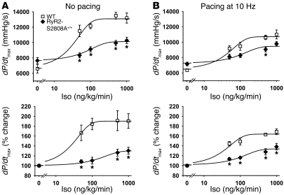
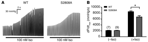
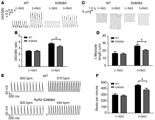
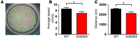
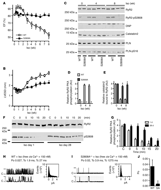
During the classic "fight-or-flight" stress response, sympathetic nervous system activation leads to catecholamine release, which increases heart rate and contractility, resulting in enhanced cardiac output. Catecholamines bind to β-adrenergic receptors, causing cAMP generation and activation of PKA, which phosphorylates multiple targets in cardiac muscle, including the cardiac ryanodine receptor/calcium release channel (RyR2) required for muscle contraction. PKA phosphorylation of RyR2 enhances channel activity by sensitizing the channel to cytosolic calcium (Ca²+). Here, we found that mice harboring RyR2 channels that cannot be PKA phosphorylated (referred to herein as RyR2-S2808A+/+ mice) exhibited blunted heart rate and cardiac contractile responses to catecholamines (isoproterenol). The isoproterenol-induced enhancement of ventricular myocyte Ca²+ transients and fractional shortening (contraction) and the spontaneous beating rate of sinoatrial nodal cells were all blunted in RyR2-S2808A+/+ mice. The blunted cardiac response to catecholamines in RyR2-S2808A+/+ mice resulted in impaired exercise capacity. RyR2-S2808A+/+ mice were protected against chronic catecholaminergic-induced cardiac dysfunction. These studies identify what we believe to be new roles for PKA phosphorylation of RyR2 in both the heart rate and contractile responses to acute catecholaminergic stimulation.


Figures

Figure 1
Figure 1. PKA phosphorylation of RyR2 mediates the chronotropic and inotropic response to β-adrenergic activation.
Cardiac contractility and heart rate were measured in vivo using left ventricular catheterization in response to low-dose (2 μg/kg i.p.) and high-dose (2 mg/kg i.p.) Iso. (A) Contractility reported as dP/dtmax. (B) Heart rate in bpm. Dashed lines indicate the points at which Iso was administered i.p. (WT, n = 5; RyR2-S2808A+/+, n = 6; *P < 0.05, versus WT). (C) Representative immunoblot analyses of RyR2 immunoprecipitates (from 100 μg of cardiac lysates), using phosphoepitope-specific antibodies that detect PKA-phosphorylated RyR2-Ser2808 (pS2808), CaMKII-phosphorylated RyR2-Ser2814 (pS2814), oxidation of RyR2 (DNP), and calstabin2 associated with the channel. (D) Representative immunoblot of CSR treated with hydrogen peroxide (H2O2) plus either PKA or CaMKII and levels of calstabin2 associated with the channel. S2808A, RyR2-S2808A+/+.
Figure 2
Figure 2. Blunted cardiac response to acute β-adrenergic stimulation in RyR2-S2808A+/+ mice.
(AC) Pressure-volume loops before (black) and during (red) Iso (100 ng/kg/min i.v.) infusion. (A) The steeper and left-shifted Ees line indicates increased contractility as evidenced in WT, which is blunted in (B) RyR2-S2808A+/+ mice. (C) Treatment with Bay K 8644 (100 μg/kg i.p.) restored the inotropic response in RyR2-S2808A+/+ mice. Insets show representative examples of end-systolic pressure-volume relationships (ESPVRs) during β-adrenergic stimulation; Ees is determined by the slope of the automated end-systolic pressure-volume relationship regression analysis through a series of end-systolic points (black dots). (D) Ees response to gradually increasing Iso (0–100 ng/kg/min) and sigmoid regression analysis (n = 10 for each genotype; *P < 0.05 each versus WT). (E) dP/dtmax of WT and RyR2-S2808A+/+ mice prior to and after db-cAMP infusion (*P < 0.05). (F) dP/dtmax prior to and after treatment with Iso (100 ng/kg/min i.v.), according to genotype as indicated (calstabin2, referred to herein as CLN2–/–; phospholamban, Pln–/–; and crossed mouse strains as indicated) (*P < 0.05 versus baseline; #P < 0.05 versus WT baseline; **P < 0.05). (G) Ees change normalized to WT after treatment with inotropic agonists Bay K 8644 (100 μg/kg i.p.), Ca2+ (30 mg/kg/min i.v.), or ouabain (0.1 mg/kg/min i.v.) (P = NS versus WT; n = 5). (H) Heart rates in response to gradually increasing Iso concentrations (0–100 ng/kg/min) and sigmoid regression analysis from D (n = 10 for each genotype; *P < 0.05).
Figure 3
Figure 3. Blunted inotropic responses to Iso in RyR2-S2808A+/+ mice in the absence and presence of 10 Hz pacing.
Iso dose-dependent enhancement of cardiac contractility in WT versus RyR2-S2808A+/+ mice, with or without heart rate control (via right ventricular pacing). Cardiac contractility measured in vivo by left ventricular catheterization in WT (n = 6) versus RyR2-S2808A+/+ mice (n = 7). (A) Cardiac contractility measured without RV pacing. The top panel shows dP/dtmax, and bottom panel shows the percentage change in dP/dtmax. (B) Cardiac contractility measured with RV pacing at 10 Hz (heart rate = 600 bpm). *P < 0.05 versus WT.
Figure 4
Figure 4. Blunted Iso-induced contractility in RyR2-S2808A+/+ ex vivo hearts.
Contractility measurements of WT (n = 7) and RyR2-S2808A+/+ (n = 9) isolated hearts in the absence and presence of 100 nM Iso. (A) Representative traces of left ventricular pressure before and during Iso infusion, demonstrating blunted developed pressure response to Iso in the RyR2-S2808A+/+ hearts (B) Quantitative summary of dP/dtmax (*P < 0.01).
Figure 5
Figure 5. Blunted responses to Iso in RyR2-S2808A+/+ ventricular myocytes and SANCs.
(A) Representative Ca2+ transients and (C) fractional shortening traces from WT and RyR2-S2808A+/+ ventricular myocytes loaded with Fura-2 paced at 3 Hz in the absence and presence of 100 nM Iso. The label "Δ 0.5” refers to a 0.5 change in the Fura-2 (340:380) ratio. (B) Summary of Ca2+ transient and (D) fractional shortening data, demonstrating that the RyR2-S2808A+/+ myocytes have blunted increases in Ca2+ transient and fractional shortening amplitude in response to Iso (*P < 0.05). (E) Representative SAPs generated from WT and RyR2-S2808A+/+ SANCs in the absence (left) and presence of 50 nM Iso (right). (F) Bar graph summarizing the rate of SAP generation in WT (n = 9) versus RyR2-S2808A+/+ SANCs (n = 8; *P < 0.05).
Figure 6
Figure 6. RyR2-S2808A+/+ mice exhibit reduced exercise capacity.
(A) Representative tracing of the computer generated swimming pattern of a mouse placed in a large water bath (diameter 122 cm) for 5 minutes. White squares represent starting and ending positions. (B and C) Graphical representation of the average (B) swimming velocity and (C) distance of WT and RyR2-S2808A+/+ mice over the course of 5 minutes (WT, n = 17; RyR2-S2808A+/+, n = 18; *P < 0.05).
Figure 7
Figure 7. Chronic β-adrenergic stimulation causes cardiac dysfunction and remodeling of the RyR2 channel complex.
(A) Echocardiography of left ventricular EF (A) and LVESD (B) in WT mice and RyR2-S2808A+/+ mice during chronic Iso treatment (n = 6 in both groups; mean ± SEM; *P < 0.05 versus WT; #P < 0.05 versus WT baseline). (C) Equivalent amounts of RyR2 were immunoprecipitated with RyR2-specific antibody followed by immunoblotting for relative PKA phosphorylation of RyR2 at Ser2808, oxidation (DNP), and of calstabin2 bound to RyR2. Immunoblotting of PLN- and PKA-phosphorylated PLN was performed on whole heart lysates from WT and RyR2-S2808A+/+ mice. (D and E) Quantification summaries. Mice were continuously treated with Iso for 56 days at a dose of 30 mg/kg/d (WT, n = 3; RyR2-S2808A+/+, n = 3; mean ± SD; *P < 0.05). (F) Aliquots of cardiac lysates from WT mice treated with Iso for 1 or 28 days were incubated at 37°C before the addition of phosphatase inhibitors at the indicated times (top) to stop the RyR2 dephosphorylation reaction. (G) Quantification of RyR2 PKA phosphorylation from F (*P < 0.05). (H) Cardiac RyR2 single-channel activity measured from microsomes from Iso-treated WT and (I) RyR2-S2808A+/+ mice. Channel openings are upward, “C” indicates closed state of the channel, and dotted lines indicate levels of partial openings or subconductance states. Top dash indicates the fully open 4 pA level. The top tracings are condensed time scale (5 s) and the bottom tracings are expanded time scale (500 ms). Amplitude histograms are shown for each channel. To, average open time; Tc, average closed time. (J) Summary of WT (n = 6 channels from 3 mice) and RyR2-S2808A+/+ (n = 8 channels from 3 mice) Po. *P < 0.05.

Comment in

Similar articles

Cited by

References

    1. Vatner SF. Sympathetic mechanisms regulating myocardial contractility in concious animals. In: Fozzard HA, Haber E, eds.The Heart and Cardiovascular System . New York, New York, USA: Raven Press, Ltd; 1992:1709–1728.
    1. Rohrer DK. Physiological consequences of beta-adrenergic receptor disruption. J Mol Med. 1998;76(11):764–772. doi: 10.1007/s001090050278. - DOI - PubMed
    1. Bers DM. Cardiac excitation-contraction coupling. Nature. 2002;415(6868):198–205. doi: 10.1038/415198a. - DOI - PubMed
    1. Fabiato A. Simulated calcium current can both cause calcium loading in and trigger calcium release from the sarcoplasmic reticulum of a skinned canine cardiac Purkinje cell. J Gen Physiol. 1985;85(2):291–320. doi: 10.1085/jgp.85.2.291. - DOI - PMC - PubMed
    1. Cannell MB, Berlin JR, Lederer WJ. Effect of membrane potential changes on the calcium transient in single rat cardiac muscle cells. Science. 1987;238(4832):1419–1423. doi: 10.1126/science.2446391. - DOI - PubMed

Publication types

MeSH terms

Substances