. 2010 Jul-Aug;2(4):252-7.
doi: 10.4161/isl.2.4.12556.

No non-redundant function of suppressor of cytokine signaling 2 in insulin producing β-cells

Affiliations

No non-redundant function of suppressor of cytokine signaling 2 in insulin producing β-cells

Ramona Puff et al. Islets. 2010 Jul-Aug.

Abstract

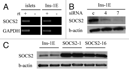
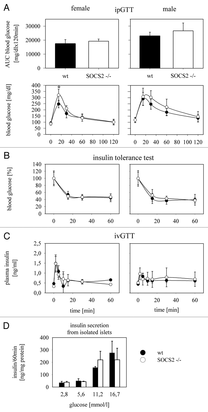
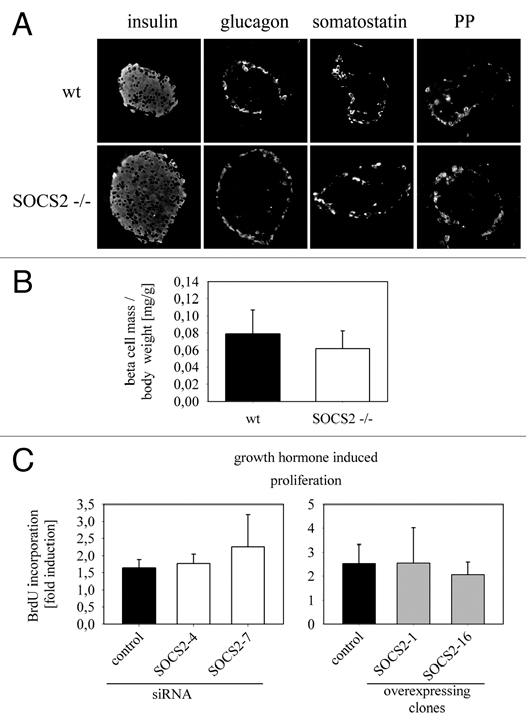
The members of the Suppressor of Cytokine Signaling (SOCS) protein family mainly modulate the Janus kinase (JAK)/signal transducer and activator of transcription (STAT) pathway. SOCS-1 and SOCS-3 have already been shown to influence growth and apoptosis of pancreatic beta cells. We hypothesized that SOCS-2, which is expressed in pancreatic islets, also contributes to β-cell physiology. We tested this hypothesis in vivo in SOCS-2-/- knockout mice and in vitro in Ins-1E rat insulinoma cells. We found that SOCS-2-/- mice have normal islet insulin secretion and unchanged glucose and insulin tolerance compared to wildtype controls. SOCS-2-/- are bigger than wildtype mice but body weight-corrected β-cell mass and islet morphology were normal. Growth hormone-induced proliferation of Ins-1E cells was not affected by either siRNA-mediated SOCS-2 knockdown or stable SOCS-2 overexpression. Interleukin-1β mediated cell death in vitro was unchanged after SOCS-2 knockdown. Similarly, autoimmune destruction of beta cells in vivo after multiple low-dose injections of streptozotocin (STZ) was not altered in SOCS-2-/- mice. In summary, SOCS-2-/- knockout mice have a normal function of insulin-producing pancreatic β-cells, a fully adapted beta cell mass and a normal morphology of the endocrine islets. Based on in vitro evidence, the increased β-cell mass in the mutants is likely due to indirect adaptive mechanisms and not the result of altered growth hormone signaling within the β-cells. Immune mediated β-cell destruction is also not affected by SOCS-2 ablation in vitro and in vivo.


Figures

Figure 1
Figure 1
SOCS-2 expression in mouse pancreatic islets and Ins-1E rat insulinoma cells; suppression by siRNA and overexpression in stably transfected cell clones. (A) SOCS-2 mRNA is detectable in isolated mouse islets and in Ins-1E rat insulinoma cells by rtPCR. (B and C) Western blots of whole cell lysates. (B) SOCS-2 protein is present in Ins-1E cells and is effectively suppressed by two different gene-specific siRNAs (numbers 4 and 7) 72 hours after transient transfection. (C) Two Ins-1E clones stably overexpressing SOCS-2 were studied (numbers 1 and 16).
Figure 2
Figure 2
Glucose metabolism in SOCS-2−/− knockout mice. (A) Intraperitoneal glucose tolerance tests were done with 2 g glucose/kg body weight after 6 hrs of fasting in SOCS-2−/− mice and matched wt controls. Female (n = 7) and male (n = 8) animals were analyzed separately. The area under the glucose curve as a measure of overall glucose tolerance is not different between wt and SOCS-2 null mice in both sexes. A difference in blood glucose is only seen in female mice 15 min after glucose injection when SOCS-2−/− animals show significantly higher values (p < 0.01; student's t-test). (B) Intraperitoneal insulin tolerance tests were done with 0.75 IU/kg body weight (n = 6 for female, n = 7 for male mice). Insulin sensitivity is comparable in SOCS-2−/− and wt animals of both sexes. (C) To test whether the higher early glucose values in the ipGTT in female SOCS-2−/− mice are the result of a reduced first phase insulin secretion, intravenous glucose tolerance tests with 1 g glucose/kg body weight were done (n = 2 for female and male mice, respectively). First phase insulin secretion is detected at a comparable magnitude in SOCS-2 null and wt mice, ruling out a defect in this β-cell function as a reason for the more extensive early rise in blood glucose in SOCS-2−/− female mice. (D) Glucose-induced insulin secretion from isolated SOCS2−/− and wt islets is also not significantly different.
Figure 3
Figure 3
Morphology of the SOCS-2−/− mouse pancreas, β-cell mass and growth hormone-induced proliferation of Ins-1E insulinoma cells in vitro. (A) Immunostainings with antibodies against the four major islet hormones were performed on paraffin sections from SOCS-2−/− and wt pancreas. The proportions of the different cell types and the distribution of the cells within the islet are comparable in knockout and wt tissue. (B) pancreatic β-cell mass corrected for body weight was determined in 11–17 week old SOCS-2−/− mice and wt controls (n = 6; male and female combined). No significant difference is seen between the two groups indicating a fully adapted β-cell mass in the knockout mice, which are generally bigger than their wt counterparts. (C) To determine whether SOCS-2 has a direct effect on growth hormone signaling in insulin-producing β-cells, Ins-1E cells were incubated for 24 hours with and without growth hormone (50 ng/ml; 2.2 nmol/l) in serum-free media. BrdU incorporation during that time period was quantified by immunostaining. Induction of proliferation by growth hormone is not significantly different in cells transfected with control or SOCS-2 specific siRNA (left) and also not in control and SOCS-2 overexpressing cells (right).
Figure 4
Figure 4
Interleukin-1β induced cell death in vitro and autoimmune mediated β-cell destruction in vivo. (A) To analyze whether SOCS-2 influences cytokine-mediated β-cell death, Ins-1E cells were incubated with and without 100 pg/ml recombinant mouse interleukin 1-β for 24 hours in serum-free media. Seventy-two hours earlier the cells were transfected with control or SOCS-2 specific siRNA. No effect of SOCS-2 knockdown on the interleukin-induced reduction of viability is seen (MTT assays). (B) The multiple low dose STZ regimen was used as a model of autoimmune diabetes and β-cell destruction in vivo. SOCS-2 and wt mice received five consecutive daily i.p. injections of STZ (40 mg/kg body weight; n = 3 for males; n = 2 for females). Fed blood glucose was recorded daily (except for days 12 and 13), starting on day 7 after the first injection. Male mice became diabetic with the same kinetics in both groups. Female mice, as expected for wt C57Bl/6, remained normoglycemic.

References

    1. Rico-Bautista E, Flores-Morales A, Fernandez-Perez L. Suppressor of cytokine signaling (SOCS) 2, a protein with multiple functions. Cytokine Growth Factor Rev. 2006;17:431–439. - PubMed
    1. Cottet S, Dupraz P, Hamburger F, Dolci W, Jaquet M, Thorens B. SOCS-1 protein prevents Janus Kinase/STAT-dependent inhibition of beta cell insulin gene transcription and secretion in response to interferongamma. J Biol Chem. 2001;276:25862–25870. - PubMed
    1. Karlsen AE, Heding PE, Frobose H, Ronn SG, Kruhoffer M, Orntoft TF, et al. Suppressor of cytokine signalling (SOCS)-3 protects beta cells against IL-1beta-mediated toxicity through inhibition of multiple nuclear factor-kappaB-regulated proapoptotic pathways. Diabetologia. 2004;47:1998–2011. - PubMed
    1. Chong MM, Chen Y, Darwiche R, Dudek NL, Irawaty W, Santamaria P, et al. Suppressor of cytokine signaling-1 overexpression protects pancreatic beta cells from CD8+ T cell-mediated autoimmune destruction. J Immunol. 2004;172:5714–5721. - PubMed
    1. Lindberg K, Ronn SG, Tornehave D, Richter H, Hansen JA, Romer J, et al. Regulation of pancreatic beta-cell mass and proliferation by SOCS-3. J Mol Endocrinol. 2005;35:231–243. - PubMed

Publication types

MeSH terms

LinkOut - more resources