Review
. 2010 Nov-Dec;4(6):453-8.
doi: 10.4161/chan.4.6.12889. Epub 2010 Nov 1.

Characterization of the gating brake in the I-II loop of CaV3 T-type calcium channels

Affiliations
Review

Characterization of the gating brake in the I-II loop of CaV3 T-type calcium channels

Edward Perez-Reyes. Channels (Austin). 2010 Nov-Dec.

Abstract

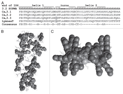
Our interest was drawn to the I-II loop of Cav3 channels for two reasons: one, transfer of the I-II loop from a high voltage-activated channel (Cav2.2) to a low voltage-activated channel (Cav3.1) unexpectedly produced an ultra-low voltage activated channel; and two, sequence variants of the I-II loop found in childhood absence epilepsy patients altered channel gating and increased surface expression of Cav3.2 channels. To determine the roles of this loop we have studied the structure of the loop and the biophysical consequences of altering its structure. Deletions localized the gating brake to the first 62 amino acids after IS6 in all three Cav3 channels, establishing the evolutionary conservation of this region and its function. Circular dichroism was performed on a purified fragment of the I-II loop from Cav3.2 to reveal a high α-helical content. De novo computer modeling predicted the gating brake formed a helix-loop-helix structure. This model was tested by replacing the helical regions with poly-proline-glycine (PGPGPG), which introduces kinks and flexibility. These mutations had profound effects on channel gating, shifting both steady-state activation and inactivation curves, as well as accelerating channel kinetics. Mutations designed to preserve the helical structure (poly-alanine, which forms α-helices) had more modest effects. Taken together, we conclude the second helix of the gating brake establishes important contacts with the gating machinery, thereby stabilizing a closed state of T-channels, and that this interaction is disrupted by depolarization, allowing the S6 segments to spread open and Ca (2+) ions to flow through.


Figures

Figure 1
Figure 1
Conservation of the gating brake. (A) Alignment of the three human CaV3 amino acid sequences to that determined for the freshwater pond snail, Lymnaea stagnalis. The location of helix-turn-helix of the gating brake is shown above, and secondary structure prediction by the SOPMA algorithm. Bottom line shows consensus sequence for residues that are identical in all four sequences. Amino acids are colored based on their physical properties to emphasize conservative amino acid changes. (B) Model of the CaV3.2 I–II loop gating brake with residues conserved in Lymnea displayed in Corey-Pauling-Koltun (CPK) space filling model. Model was originally reported by Arias et al. and modified according to the Lymnea sequence reported by Senatore and Spafford. The model is viewed from the side, with the plasma membrane on top and helix to the left. Note how conserved residues face away from the hydrophobic core, suggesting they form conserved protein-protein contacts. Substitutions in the core are conservative—hydrophobic for hydrophobic and the putative salt bridge is conserved. (C) Second view of the model is from the bottom looking up at the membrane. Note how the conserved “arginine fingers” jut out perpendicular to the plane of the gating brake. These residues are comprised of five Arg residues on one face and two on the other.
Figure 2
Figure 2
Two models of the gating brake. (A) The model of the gating brake (light blue) was superimposed on the crystal structure of the KV1.2–KV2.1 paddle chimera (PDB #2R9R). View is from the inside of the cell facing the membrane. The gating brake model begins with the glycine-serine motif in the middle of IS6—a conserved motif observed to form a hinge in the MthK crystal structure. It was then superimposed on the threonine-isoleucine motif in the KV1.2 channel (alignment by Jan and Jan). In this manner the orientation of the gating brake with respect to IS6 is the same as the extension of S6 in the crystal structure. IIS5 and S6 are highlighted in red to emphasize possible contacts with helix 2 of the gating brake. (B) Here the gating brake has been flipped 180°, resulting in possible helix 2 contacts with the IS4-S5 (red). (C and D) Location of the contact points between repeats IV, I and II as deduced from the same KV crystal structure.

Similar articles

Cited by

References

    1. Buraei Z, Yang J. The β-subunit of voltage-gated Ca2+ channels. Physiol Rev. 2010;90:1461–1506. - PMC - PubMed
    1. Zamponi GW, Bourinet E, Nelson D, Nargeot J, Snutch TP. Crosstalk between G proteins and protein kinase C mediated by the calcium channel a1 subunit. Nature. 1997;385:442–446. - PubMed
    1. Arias JM, Murbartián J, Vitko I, Lee JH, Perez-Reyes E. Transfer of β subunit regulation from high to low voltage-gated Ca2+ channels. FEBS Lett. 2005;579:3907–3912. - PubMed
    1. Raybaud A, Dodier Y, Bissonnette P, Simoes M, Bichet DG, Sauve R, et al. The role of the GX9GX3G motif in the gating of high voltage-activated Ca2+ channels. J Biol Chem. 2006;281:39424–39436. - PubMed
    1. Marksteiner R, Schurr P, Berjukow S, Margreiter E, Perez-Reyes E, Hering S. Inactivation determinants in segment IIIS6 of CaV3.1. J Physiol (Lond) 2001;537:27–34. - PMC - PubMed

Publication types

MeSH terms

LinkOut - more resources