. 2010 Dec 15;29(24):4132-45.
doi: 10.1038/emboj.2010.287. Epub 2010 Nov 19.

The poly A polymerase Star-PAP controls 3'-end cleavage by promoting CPSF interaction and specificity toward the pre-mRNA

Affiliations

The poly A polymerase Star-PAP controls 3'-end cleavage by promoting CPSF interaction and specificity toward the pre-mRNA

Rakesh S Laishram et al. EMBO J. .

Abstract

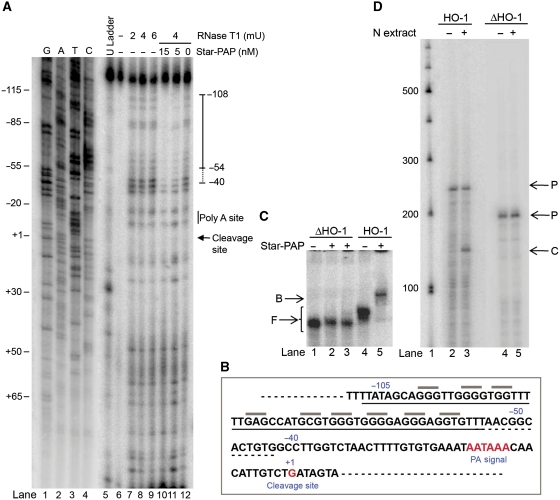
Star-PAP is a poly (A) polymerase (PAP) that is putatively required for 3'-end cleavage and polyadenylation of a select set of pre-messenger RNAs (mRNAs), including heme oxygenase (HO-1) mRNA. To investigate the underlying mechanism, the cleavage and polyadenylation of pre-mRNA was reconstituted with nuclear lysates. siRNA knockdown of Star-PAP abolished cleavage of HO-1, and this phenotype could be rescued by recombinant Star-PAP but not PAPα. Star-PAP directly associated with cleavage and polyadenylation specificity factor (CPSF) 160 and 73 subunits and also the targeted pre-mRNA. In vitro and in vivo Star-PAP was required for the stable association of CPSF complex to pre-mRNA and then CPSF 73 specifically cleaved the mRNA at the 3'-cleavage site. This mechanism is distinct from canonical PAPα, which is recruited to the cleavage complex by interacting with CPSF 160. The data support a model where Star-PAP binds to the RNA, recruits the CPSF complex to the 3'-end of pre-mRNA and then defines cleavage by CPSF 73 and subsequent polyadenylation of its target mRNAs.


Conflict of interest statement

The authors declare that they have no conflict of interest.

Figures

Figure 1
Figure 1
Star-PAP is required for HO-1 mRNA cleavage in vivo. (A) Schematic of 3′-RACE assay. (B) 3′-RACE assay of HO-1 (lanes 1–3) and GAPDH (lanes 6–8) with total RNA isolated from HeLa cells—resting with control RNAi (WT), treated with tBHQ (+tBHQ), and siRNA knockdown of Star-PAP (siStar-PAP). A RACE product (∼1000 bp) and DNA size markers (kilobases) is indicated. The western analysis of Star-PAP knockdown is shown in the right panel. (C) Schematic of primer extension analysis. (D) Primer extension analysis by reverse transcription reaction using a primer specific to HO-1 (lanes 1–4) or GAPDH (lanes 5–8) RNA that is 3′ of the cleavage site. The DNA size marker (lane 9), origin of the gel and the primer extension product are indicated.
Figure 2
Figure 2
Star-PAP specifically promotes cleavage of its target mRNA in a cell-free system. (A) Schematic of the HO-1 cleavage substrate. (B) Cleavage assay of radiolabelled HO-1 substrate reconstituted (+/− PI4,5P2) with nuclear extracts prepared from HeLa cells (+/− tBHQ treatment) (lanes 2–5). The control RNA (HO-1 RNA) (lane 1), pre-mRNA (P), cleaved RNA (C) are indicated. The approximate sizes of RNA are indicated by a DNA size marker. (C) Cleavage assay with poly A signal mutation (PAM) under the similar conditions as in B (lanes 1–4). Control—HO-1 RNA without treatment with nuclear extract. (D) Cleavage assay of HO-1 RNA with nuclear extracts from HeLa cells after the siRNA knockdown of Star-PAP (siStar-PAP) (lanes 6–9) and control HeLa cells (HeLa NE) (lanes 1–4). (E, F) Cleavage assay of Star-PAP non-target, GCLC (lanes 1–9) and GAPDH (lanes 1–8) UTRs with nuclear extracts prepared from control (HeLa NE) and siRNA Star-PAP knockdown (siStar-PAP) with or without tBHQ treatment (+/− tBHQ). Control—substrate RNA without treatment with nuclear extract. (G) Western blot analysis of nuclear fractionation, C, cytosolic fraction; N, nuclear fraction. (H) Western analysis of siRNA Star-PAP knockdown; cells transfected with control RNA, siRNA Star-PAP (siStar-PAP) and untransfected (extract WT) are indicated.
Figure 3
Figure 3
Recombinant Star-PAP rescues the cleavage defect and Star-PAP interacts with subunits of CPSF. (A) Cleavage assay of HO-1 RNA with nuclear extracts prepared from siRNA Star-PAP knockdown HeLa cells (siStar-PAP) (lanes 4–7), supplemented with 20 nM recombinant His-tagged Star-PAP (lanes 8–11) or recombinant His-tagged PAPα (lanes 12–15) as indicated. (B) Cleavage assay of poly A signal mutant (PAM) with supplemented His-Star-PAP (lanes 1–4) under similar conditions as in (A). (C) Cleavage assay of HO-1 with siRNA Star-PAP nuclear extract supplemented with increasing amounts of His-Star-PAP (0.01–20 nM) (lanes 4–10) as indicated. The control RNA, pre-mRNA (P) and cleaved RNA (C) are indicated. (D) Western analysis of siRNA Star-PAP knockdown in Hela cells. (E) Western blot analysis of Flag Star-PAP purification for the indicated CPSF subunits, CstF and PAPα; Sup, supernatant; FT, flow through; Eluate, elution fraction; W, wash. (F) GST pull-down assay with GST-tagged Star-PAP, GST (as indicated on the top) with overexpressed CPSF subunits in E. coli (indicated on the right). Input shows 20% of the lysates used for binding.
Figure 4
Figure 4
Star-PAP associates and interacts directly with HO-1 RNA. (A) Schematic of HO-1 3′-UTR for RIP analysis. (B) RIP analysis of HO-1 and GCLC UTR by IP with antibodies specific to RNA Pol II (RNAPII), Star-PAP and PAPα followed by reverse transcription–PCR with primers specific to HO-1 and GCLC UTR regions. (C) RNA EMSA of HO-1 (lanes 1–6) or GAPDH (lanes 7–12) (substrate for cleavage assays) with increasing concentrations of recombinant His-Star-PAP (0–20 nM; from lanes 2–6, respectively, 1, 2, 4, 10 and 20 nM). The control RNA (Probe), unbound RNA (F) and Star-PAP–RNA complex (B) are indicated. (D) EMSA of HO-1 RNA with 10 nM Star-PAP in the presence of increasing Star-PAP antibody (antibody super shift) (lanes 2–7) or (E) β-tubulin antibody (lanes 2–5). Unbound RNA (F), Star-PAP-HO-1 RNA binary complex (B) and antibody, Star-PAP and RNA ternary complexes (T) are indicated. (F) Cold competitions of HO-1 RNA binding with Star-PAP. EMSA of HO-1 RNA with increasing Star-PAP concentrations (lanes 2–5, respectively, 10, 20, 30 and 40 nM) (lanes 1–5) and 20-fold molar excess of the respective competitor RNAs as indicated (lanes 6–9). Specific (S) and non-specific (NS) competitions are indicated. Lanes 6–7 and 8–9 correspond to specific and non-specific competitions equivalent of reactions in lanes 4–5, respectively. (GJ) EMSA of HO-1 RNA with increasing amounts of various His-Star-PAP deletions as indicated (ΔZF, zinc-finger deletion; ΔRRM, RNA recognition motif deletion; ΔZF-RRM, combine deletion of both ZF and RRM; FL, full-length Star-PAP), or (K) His-ZF-RRM peptide. The control (Probe), unbound (F) and binary complexes (B) are indicated.
Figure 5
Figure 5
Determination of Star-PAP-binding site on HO-1 RNA. (A) Footprint of Star-PAP on HO-1 UTR RNA by RNase T1 probing. The digestion pattern with different RNase T1 concentrations (mU) in the absence of Star-PAP (lanes 7–9, 12), or in the presence of 5 or 15 nM Star-PAP (lanes 10–11), and the untreated HO-1 RNA (lane 6) are indicated. Sequencing ladder generated using the primer for primer extension from the template of HO-1 RNA (lanes 1–4) and a dideoxy U ladder generated by primer extension reaction (lane 5) are indicated. Numbers refer to the position of nucleotides with respect to cleavage site taken as +1. (B) The primary region of Star-PAP protection (solid line) on HO-1 RNA, the extended minor protection (dashed line) and the putative reactive sites protected (grey lines) (C) RNA EMSA of HO-1 RNA deleted for the Star-PAP protection site from −110 to −50 (lanes 1–3) and control EMSA with HO-1 UTR RNA (lanes 4–5). (D) Cleavage assay of HO-1 UTR deletion as in (C) with HeLa nuclear extract (lanes 4–5) and control HO-1 UTR RNA (lanes 2–3).
Figure 6
Figure 6
Star-PAP promotes CPSF binding and stabilizes the cleavage complex on HO-1 RNA. (A) RIP analysis using antibodies specific to RNAPII (lanes 2–3) and CPSF subunits (lanes 5–12), followed by detection of associated RNA by RT–PCR using primers specific to the UTR region encompassing the poly A signal of HO-1 and GAPDH. (B) RNA EMSA of HO-1 UTR (Figure 4A) with the CPSF 160 complex. CPSF 160 was IP'ed from HeLa cell with control RNAi lysates (+) (lanes 2–3) or lysates of Star-PAP RNAi (Si) (lanes 4–5), and the IP eluate was incubated with radiolabelled HO-1 RNA (see Figure 4A). The unbound RNA (F) and complex of the 3′-RNA with CPSF 160 (S) are indicated. (C) EMSA of HO-1 RNA with nuclear extracts from HeLa cells with (lanes 6–10) or without (lanes 1–5) Star-PAP knockdown for various incubation times. The unbound fraction (F) and specific processing complex (S) are indicated. (D) RNA EMSA of poly A signal mutated HO-1 RNA (lanes 1–5) with HeLa nuclear extract. (E) EMSA of GAPDH UTR RNA with nuclear extracts control (lanes 1–5) or Star-PAP knockdown (lanes 6–10). F, unbound RNA fraction; S, specific processing complex.
Figure 7
Figure 7
Star-PAP facilitates HO-1 cleavage by CPSF subunits, and PI4,5P2 stimulates coupled polyadenylation. (A) Cleavage reactions of HO-1 (lanes 1–5) and poly A mutation (lanes 6–8) with recombinant CPSF 160 (40 nM) and 73 (120 nM) with Star-PAP (20 nM) as indicated. The control HO-1 RNA, pre-mRNA (P) and cleaved RNA (C) are indicated. (B) Cleavage reaction as in (A) but in the presence of mutant CPSF 73 (lanes 3–4). (C) Cleavage assay of HO-1 RNA with individual proteins as indicated (D) Coupled cleavage and polyadenylation assay of HO-1 RNA (lanes 3–6) and poly A signal mutation (lanes 8–11) in the presence or absence of PI4,5P2 with HeLa nuclear extracts with or without tBHQ treatment as indicated. Polyadenylated (poly A) and pre-mRNA (P) are indicated. (E) Stable ternary complex formation on HO-1 RNA by EMSA (lanes 1–6) with CPSF 160–30 nM (lanes 2 and 4), 80 nM (lane 3) and Star-PAP (15 nM) (lanes 4 and 6). The control RNA (Probe), unbound RNA (F), binary complex of Star-PAP-HO-1 or CPSF 160-HO-1 (B) and ternary complex of CPSF, Star-PAP and HO-1 RNA (T) are indicated. (F) RNA EMSA experiment of HO-1 with increasing CPSF (0–120 nM) (lanes 1–6) (G) RNA EMSA of HO-1 with increasing CPSF (0–120 nM) in the presence of 15 nM Star-PAP (lanes 2–6). The approximate half-maximal binding was obtained from the mobility shifts. Symbols are as described in legend to (E). (H) EMSA of poly A signal mutant HO-1 RNA with increasing amount of His-CPSF 160 (left, lanes 1–3), or His-Star-PAP (right, lanes 4–6).
Figure 8
Figure 8
Model of Star-PAP-mediated cleavage of target RNA. Direct contact of Star-PAP with HO-1 RNA recruits CPSF 160 to the poly A signal and association of other cleavage factors in the complex is also indicated.

Comment in

Similar articles

Cited by

References

    1. Ansari A, Hampsey M (2005) A role for the CPF 3′-end processing machinery in RNAP II-dependent gene looping. Genes Dev 19: 2969–2978 - PMC - PubMed
    1. Auboeuf D, Dowhan DH, Dutertre M, Martin N, Berget SM, O'Malley BW (2005) A subset of nuclear receptor coregulators act as coupling proteins during synthesis and maturation of RNA transcripts. Mol Cell Biol 25: 5307–5316 - PMC - PubMed
    1. Barabino SM, Hubner W, Jenny A, Minvielle-Sebastia L, Keller W (1997) The 30-kD subunit of mammalian cleavage and polyadenylation specificity factor and its yeast homolog are RNA-binding zinc finger proteins. Genes Dev 11: 1703–1716 - PubMed
    1. Bardwell VJ, Wickens M, Bienroth S, Keller W, Sproat BS, Lamond AI (1991) Site-directed ribose methylation identifies 2′-OH groups in polyadenylation substrates critical for AAUAAA recognition and poly(A) addition. Cell 65: 125–133 - PubMed
    1. Barlow CA, Laishram RS, Anderson RA (2009) Nuclear phosphoinositides: a signaling enigma wrapped in a compartmental conundrum. Trends Cell Biol 20: 25–35 - PMC - PubMed

Publication types

MeSH terms