. 2011 Feb;25(2):331-40.
doi: 10.1038/leu.2010.261. Epub 2010 Nov 19.

Selective killing of Burkitt's lymphoma cells by mBAFF-targeted delivery of PinX1

Affiliations
Free PMC article

Selective killing of Burkitt's lymphoma cells by mBAFF-targeted delivery of PinX1

L Zhang et al. Leukemia. 2011 Feb.
Free PMC article

Abstract

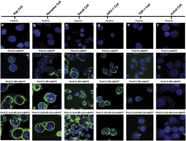
Increased expression of BAFF (B cell-activating factor belonging to the TNF family) and its receptors has been identified in numerous B-cell malignancies. A soluble human BAFF mutant (mBAFF), binding to BAFF receptors but failing to activate B-lymphocyte proliferation, may function as a competitive inhibitor of BAFF and may serve as a novel ligand for targeted therapy of BAFF receptor-positive malignancies. Pin2/TRF1-interacting protein X1 (PinX1), a nucleolar protein, potently inhibits telomerase activity and affects tumorigenicity. In this study, we generated novel recombinant proteins containing mBAFF, a polyarginine tract 9R and PinX1 (or its C/N terminal), to target lymphoma cells. The fusion proteins PinX1/C-G(4)S-9R-G(4)S-mBAFF and PinX1/C-9R-mBAFF specifically bind and internalize into BAFF receptor-positive cells, and subsequently induce growth inhibition and apoptosis. The selective cytotoxicity of the fusion proteins is a BAFF receptor-mediated process and depends on mBAFF, PinX1/C and 9R. Moreover, the fusion proteins specifically kill BAFF receptor-expressing Burkitt's lymphoma (BL) cells by inhibiting telomerase activity and the consequent shortening of telomeres. Therapeutic experiments using PinX1C-G(4)S-9R-G(4)S-mBAFF in severe combined immunodeficient (SCID) mice implanted with Raji cells showed significantly prolonged survival times, indicating the in vivo antitumor activity of the fusion protein. These results suggest the potential of PinX1/C-G(4)S-9R-G(4)S-mBAFF in targeted therapy of BL.


Figures

Figure 1
Figure 1
Schematic representation of the recombinant fusion proteins used in this work. PinX1, the 328-aa full-length PinX1; PinX1C, the 74-aa C-terminal fragment of PinX1; PinX1N, the 142-aa N-terminal fragment of PinX1; PinX1/C/N, PinX1 or PinX1C, or PinX1N; 9R, nine arginine residues; G4S, four glycine residues and one serine residue; mBAFF, a soluble human BAFF mutant, in which amino acids 217–224 are replaced by two glycine residues.
Figure 2
Figure 2
mBAFF mediates the specific binding and internalization of PinX1C-containing fusion proteins into BAFF receptor-positive cells. The BAFF receptor-positive cells including Raji (a), Namalwa (b), Daudi (c), JeKo-1 (d) and THP-1 (e), and BAFF receptor-negative Jurkat cells (f) were treated with 250-n fusion proteins for 1 h. The cells were then adhered onto a microscope slide and fixed in 4% paraformaldehyde, followed by a brief rinse with PBS. Subsequently, the cells were incubated with an anti-His monoclonal antibody. After a brief wash with PBS, cells were incubated with FITC-conjugated goat antimouse IgG. After washing with PBS, slides were mounted in mounting medium and then analyzed by laser scanning confocal microscopy.
Figure 3
Figure 3
The fusion proteins PinX1C–G4S–9R–G4S–mBAFF and PinX1C–9R–mBAFF selectively induce growth inhibition of BAFF receptor-expressing cells. (A) Cell proliferation assay was performed using the [3H]-thymidine incorporation method. BAFF receptor-expressing cells including Raji (a), Namalwa (b), Daudi (c), JeKo-1 (d) and THP-1 (e), and BAFF receptor-negative Jurkat cells (f) were seeded into a 96-well flat-bottom plate (1 × 10⁴ cells per well) and treated in triplicate with the fusion proteins at various concentrations. Thereafter, the cells were incubated for 5 days and [3H]-thymidine was added to the wells (1 μCi/well) during the last 16 h of incubation. Subsequently, the cells were collected on glass fiber filters, washed, dried and counted using standard scintillation methods, and percentages of the inhibition of proliferation were calculated. (B) Blocking assay was conducted to confirm the importance of the fused mBAFF for the activity of the fusion proteins. Raji cells were pretreated with 10 μg/ml of mBAFF for 1 h, followed by treatment with 250-n of the fusion proteins in quadruplicate wells. The cells were then incubated for 5 days, and proliferation assays measuring [3H]-thymidine incorporation were conducted. DHFR, which does not bind BAFF receptors, serves as a negative control. All data in A and B are expressed as mean±s.e.
Figure 4
Figure 4
Fusion proteins PinX1C–G4S–9R–G4S–mBAFF and PinX1C-9R-mBAFF selectively induce apoptosis of BAFF receptor-expressing cells. Raji (a) and Jurkat (b) cells were treated in triplicate with 250-n fusion proteins for 5 days, and apoptosis was then measured using flow cytometry after staining cells with annexin V-PI. Specific apoptosis in comparison with control cell death was calculated and presented as a percentage of control. The results are expressed as mean±s.e.
Figure 5
Figure 5
The fusion proteins inhibit telomerase activity and shorten telomeres of Raji cells. (a) PinX1/C–G4S–9R–G4S–mBAFF and PinX1/C–9R–mBAFF inhibit telomerase activity. Telomerase-containing extracts from Raji cells were incubated with various concentrations of fusion proteins for 10 min at 4 °C and then subjected to telomerase extension. Telomerase products were separated on 10% polyacrylamide gels, after which the gels were stained with chemiluminescence reagent. Telomerase activity was semiquantified by normalizing the band intensities of the characteristic 6-bp telomerase-specific ladder to that of the 216-bp internal control (IC) using NIH image software. Arrows indicate 216-bp IC. −, telomerase-containing extracts without treatment with any protein; Rnase, telomerase-containing extracts pretreated with RNase. (b) PinX1/C–G4S–9R–G4S–mBAFF and PinX1/C–9R–mBAFF shorten telomere length. Raji cells were continuously maintained in culture by splitting the cells and seeding them at 2 × 10⁵ per well in the presence of 100-n fusion proteins for 30 population doublings. Genomic DNA was isolated and digested with HinfI and RsaI, and Southern blot analysis was performed using a TTAGGG repeat as a probe. The average TRF length of Raji cells was quantified using ImageQuant. −, Raji cell without treatment with protein.
Figure 6
Figure 6
Antitumor effects of PinX1C–G4S–9R–G4S–mBAFF on disseminated Raji xenografts in SCID mice. A model of disseminated Raji xenografts was established by injecting 5 × 10⁶ Raji cells i.v. into SCID mice (n=6 per group). The mice were then given 50 μg of PinX1C–G4S–9R–G4S–mBAFF five times (on day 1, 4, 7, 10 and 13). The proportions of surviving mice are graphed over time. Animals were judged terminal if they died or if hindlimb paralysis occurred. Data were graphed as proportion surviving versus time. Statistical analysis was performed using the log-rank test, and the PinX1C–G4S–9R–G4S–mBAFF-treated group was compared with the PinX1C–mBAFF-, PinX1C- and mBAFF-alone-treated groups.

Similar articles

Cited by

References

    1. Lyu MA, Cheung LH, Hittelman WN, Marks JW, Aguiar RC, Rosenblum MG. The rGel/BLyS fusion toxin specifically targets malignant B cells expressing the BLyS receptors BAFF-R, TACI, and BCMA. Mol Cancer Ther. 2007;6:460–470. - PubMed
    1. Nimmanapalli R, Lyu MA, Du M, Keating MJ, Rosenblum MG, Gandhi V. The growth factor fusion construct containing B-lymphocyte stimulator (BLyS) and the toxin rGel induces apoptosis specifically in BAFF-R-positive CLL cells. Blood. 2007;109:2557–2564. - PubMed
    1. Wang T, Zhao J, Ren JL, Zhang L, Wen WH, Zhang R, et al. Recombinant immunoproapoptotic proteins with furin site can translocate and kill HER2-positive cancer cells. Cancer Res. 2007;67:11830–11839. - PubMed
    1. Zhang L, Zhao J, Wang T, Yu CJ, Jia LT, Duan YY, et al. HER2-targeting recombinant protein with truncated pseudomonas exotoxin A translocation domain efficiently kills breast cancer cells. Cancer Biol Ther. 2008;7:1226–1231. - PubMed
    1. Novak AJ, Grote DM, Stenson M, Ziesmer SC, Witzig TE, Habermann TM, et al. Expression of BLyS and its receptors in B-cell non-Hodgkin lymphoma: correlation with disease activity and patient outcome. Blood. 2004;104:2247–2253. - PubMed

Publication types

MeSH terms