. 2010 Dec;12(12):1242-9.
doi: 10.1038/ncb2130. Epub 2010 Nov 21.

AKAP-Lbc enhances cyclic AMP control of the ERK1/2 cascade

Affiliations

AKAP-Lbc enhances cyclic AMP control of the ERK1/2 cascade

F Donelson Smith et al. Nat Cell Biol. 2010 Dec.

Abstract

Mitogen-activated protein kinase (MAPK) cascades propagate a variety of cellular activities. Processive relay of signals through RAF-MEK-ERK modulates cell growth and proliferation. Signalling through this ERK cascade is frequently amplified in cancers, and drugs such as sorafenib (which is prescribed to treat renal and hepatic carcinomas) and PLX4720 (which targets melanomas) inhibit RAF kinases. Natural factors that influence ERK1/2 signalling include the second messenger cyclic AMP. However, the mechanisms underlying this cascade have been difficult to elucidate. We demonstrate that the A-kinase-anchoring protein AKAP-Lbc and the scaffolding protein kinase suppressor of Ras (KSR-1) form the core of a signalling network that efficiently relay signals from RAF, through MEK, and on to ERK1/2. AKAP-Lbc functions as an enhancer of ERK signalling by securing RAF in the vicinity of MEK1 and synchronizing protein kinase A (PKA)-mediated phosphorylation of Ser 838 on KSR-1. This offers mechanistic insight into cAMP-responsive control of ERK signalling events.


Conflict of interest statement

COMPETING FINANCIAL INTERESTS

The authors declare no competing financial interests.

Figures
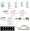
Figure 1
Figure 1
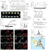
Characterization of AKAP-Lbc–KSR-1 interactions. (a) Lysates from HEK293 cells transfected with empty vector or a plasmid encoding Flag-AKAP-Lbc were subject to immunoprecipitation (IP) with anti-Flag antibodies. Proteins were resolved by SDS–PAGE and Coomassie staining, and identified by MS/MS spectrometry. (b) Lysates from HEK293 cells expressing HA–KSR-1, and transfected with control vector or vector encoding Flag–AKAP-Lbc, were immunoprecipitated using anti-Flag and proteins were identified by immunoblotting. (c) NIH3T3 cells were transfected with control vectors or vectors encoding HA–KSR-1. Cell lysates were immunoprecipitated with anti-HA, and indicated proteins were identified by immunoblotting. (d) NIH3T3 lysate and pre-immune or anti-AKAP-Lbc immunoprecipitates were immunoblotted with antibodies against KSR-1 (top) and AKAP-Lbc (bottom). (e) NIH3T3 lysate and control immunoglobulin G (IgG) or anti-AKAP-Lbc immunoprecipitates were immunoblotted with antibodies against the indicated proteins. (f) Schematic representation of AKAP-Lbc fragments used to construct GST fusion proteins for the pulldown experiments. Shaded fragment indicates KSR-1-binding fragment, as determined in g and h. (g) GST–AKAP-Lbc fragments were used as bait in pulldown experiments using HEK293 lysates. KSR-1 binding was detected by immunoblot (top). GST fusion proteins were resolved by SDS–PAGE and Ponceau S staining (bottom). (h) GST–AKAP-Lbc fragments were used as bait in pulldown experiments with in vitro-translated recombinant KSR-1. KSR-1 binding was detected by immunoblot (top). GST-fusion proteins were resolved by SDS–PAGE and Ponceau S staining (bottom). (i) Schematic representation of KSR-1 fragments used in j. Shaded fragment indicates AKAP-Lbc-binding fragment as determined in j. (j) Pyo-tagged KSR-1 fragments were co-expressed with Flag–AKAP-Lbc in HEK293 cells. Complexes were immunoprecipitated with anti-Pyo. Proteins were detected by immunoblotting with antibodies against the indicated proteins. (k) Schematic representation of EKAR, a FRET-based reporter for ERK activity. (l) Time-lapse microscopy images of FRET signals in HEK293 cells expressing EKAR alone (bottom), or with AKAP-Lbc–mCherry (top), at indicated times after addition of EGF (min). Scale bars, 10 μm. (m) Quantification of normalized YFP/CFP ratio from a FRET experiment as performed in l. Black and white bars indicate addition of EGF and UO126, respectively. Uncropped images of blots are shown in Supplementary Information, Fig. S7.
Figure 2
Figure 2
AKAP-Lbc anchors RAF. (a) Lysates from HEK293 cells expressing B-Raf and either GFP (left) or AKAP-Lbc–GFP (right) were immunoprecipitated with anti-GFP. The indicated proteins were identified by immunoblotting with antibodies against the indicated proteins. Bottom two panels indicate immunoblot of input lysate. (b) NIH3T3 lysate and pre-immune or anti-AKAP-Lbc immunoprecipitates were immunoblotted with antibodies against B-Raf (top) and AKAP-Lbc (bottom). (c) Schematic representation of the AKAP-Lbc fragments used to construct GST fusion proteins for the pulldown experiments. Shaded area indicates B-RAF binding fragments, as determined in d. (d) GST–AKAP-Lbc fragments were used as bait in pulldown experiments using in vitro-translated B-Raf. B-Raf binding was detected by immunoblot (top). GST-fusion proteins were resolved by SDS–Page and Ponceau S staining (bottom). (e) Cells were transfected, and lysates were immunoprecipitated, as in a. The immunoprecipitated complexes were then used in a kinase assay, with kinase-inactive GST–MEK1 as a substrate. Top: immunoblot of kinase assay, using antibodies against the indicated proteins. MEK1 phosphorylation was assessed with antibodies specific to MEK phosphorylated at Ser 218 and Ser 222. Bottom: the phosphorylated MEK-1 band was quantified by densitometry and normalized to the control cells. Data are means ± s.e.m. A.U.; arbitrary units. (f) HEK293 cells were transfected with vectors encoding HA–MEK1 and then co-transfected with vectors encoding AKAP-Lbc–GFP and HA–KSR-1, or empty vector controls, as indicated. Cell lysates were immunoprecipitated with anti-GFP, resolved by SDS-PAGE and immunoblotted with antibodies against MEK1, AKAP-Lbc or KSR-1, as indicated. MEK1 levels in lysates were confirmed by immunoblotting (bottom). Uncropped images of blots are shown in Supplementary Information, Fig. S7.
Figure 3
Figure 3
AKAP-Lbc enhances signal relay through the ERK kinase cascade. (a) HEK293 cells were transfected with vectors encoding HA–MEK1 and Flag–B-Raf along with increasing amounts of plasmid encoding AKAP-Lbc–GFP. Top: immunoblot of cell lysates used to detect the indicated proteins. Bottom: quantification of the phosphorylated MEK1 band (relative to total MEK) by densitometry (data are means ± s.e.m.). (bg) NIH3T3 cells were transfected with a vector encoding AKAP-Lbc–GFP (right) or control vector (left). Cells were fixed and immunostained with antibodies against phosphorylated ERK1/2 (b, c), or GFP fluorescence was observed (d, e). f is a merge of b and d, and g is a merge of c and e; in both DAPI was used as a nuclear marker. Scale bars, 20 μm. (h) Time-lapse microscopy images of FRET signals in HEK293 cells expressing EKAR and transfected with control siRNA oligonucleotides (top) or siRNA oligonucleotides against AKAP-Lbc, at indicated times after addition of EGF (min). Scale bars, 20 _m. (i) Quantification of normalized YFP/CFP ratio from a FRET experiment as performed in h. Black bar indicates addition of EGF. Data are means ± s.e.m. Inset: immunoblot of lysates from cells transfected with control or AKAP-Lbc siRNA. (j) HEK293 cells were transfected with vectors expressing EKAR and AKAP-Lbc–mCherry. Cells were pre-treated with Ht31 peptide (red) or vehicle as a control (black) before treatment with EGF and FRET recording. Normalized YFP/CFP ratios are indicated. Data are means ± s.e.m. Uncropped images of blots are shown in Supplementary Information, Fig. S7.
Figure 4
Figure 4
PKA phosphorylation of KSR-1. (a) A plasmid encoding KSR-1 was transfected into HEK293 cells, and cells were treated with forskolin and IBMX, and H-89, as indicated. Top: cell lysates were immunoblotted with antibodies against the indicated proteins. Bottom: ERK1/2 activation was measured by quantification of the phosphorylated ERK1/2 bands by densitometry. Values were normalized to total ERK1/2 levels. Data are means ± s.e.m. (b) Lysates from HEK293 cells expressing HA–KSR-1 were immunopreciptated with anti-HA. Top: autoradiograph of immunoprecipitated complexes incubated with [γ-32P] ATP. Immunoprecipitates were also incubated with cAMP and PKI (to block PKA activity) as indicated. Loading controls are also shown. Bottom: bands from the autoradiograph were quantified by densitometry. Data are means ± s.e.m. (c) HA–KSR-1 was co-expressed with AKAP-Lbc or AKAP-LbcΔPKA in HEK293 cells. Cell lysates were immunoprecipitated with anti-HA. Top: autoradiograph of immunocomplexes incubated with [γ-32P]ATP, and with cAMP and PKI as indicated. Loading controls are shown. Bottom: bands from the autoradiograph were quantified by densitometry. Data are means ± s.e.m. (d) Sequence alignment of a conserved consensus PKA phosphorylation site in KSR-1. The target serine is indicated. (e) Lysates of cells expressing wild-type (WT) KSR-1 or a S838A mutant were immunoprecipitated with anti-HA. Top: autoradiograph of immunocomplexes incubated with [γ-32P]ATP, and with PKA and PKI as indicated. Loading controls are shown. Bottom: bands from the autoradiograph were quantified by densitometry. Data are means ± s.e.m. Uncropped images of blots are shown in Supplementary Information, Fig. S7.
Figure 5
Figure 5
Phosphorylation of KSR-1 on Ser 838 controls ERK1/2 signalling. (a) KSR-1 immunoprecipitates from KSR-1−/− mouse embryonic fibroblasts (MEFs) and MEFs expressing wild-type KSR-1 were screened for AKAP-Lbc, PKA RII and KSR-1 by immunoblotting. (b) Time-course of EGF-stimulated ERK1/2 activity in KSR-1−/− MEFs and MEFs expressing wild-type KSR-1 or KSR-1S838A. Starved cells were treated with EGF for the indicated times. ERK activation was assessed by immunoblotting for phosphorylated ERK1/2, ERK1/2, KSR-1 and AKAP-Lbc. (c) Quantification of phosphorylated ERK1/2 bands by densitometry, from experiment carried out as in b (data are means ± s.e.m.). (d) Time-lapse microscopy images of FRET signals in HEK293 cells expressing EKAR, and AKAP-Lbc and wild-type KSR-1 (top), or AKAP-Lbc and KSR-1S838A (bottom), at indicated times after addition of EGF (min). Scale bars, 20 μm. (e) Quantification of YFP/CFP ratios for experiment performed as in d. Black bar indicates addition of EGF. Data are means ± s.e.m. (f) HEK293 cells expressing wild-type KSR-1 or KSR-1S838A were starved and stimulated with forskolin/IBMX, as indicated. ERK activation was measured by immunoblotting of the cell lysates. (g) Quantification of phosphorylated ERK1/2 bands by densitometry from experiments performed in f. Data are normalized to the density of the total ERK1/2 bands. Data are means ± s.e.m. (asterisk indicates P < 0.05, paired t-test). (h) KSR-1−/− MEFs and MEFs expressing wild-type or KSR-1S838A were treated with forskolin and IBMX, and H-89, as indicated. ERK activation was assessed by immunoblotting for phosphorylated ERK1/2, ERK1/2, KSR-1 and AKAP-Lbc. (i) Quantification of phosphorylated ERK1/2 bands by densitometry from experiments performed in h. Data are normalized to the density of the total ERK1/2 bands. Data are means ± s.e.m. (asterisk indicates P < 0.001, ANOVA). (j–u) Immunofluorescence microscopy analysis of ERK activation. KSR-1−/− MEFs (jm), and MEFs expressing KSR-1 (nq) or KSR-1S838A (ru) were starved, treated with forskolin/IBMX for 10 min, fixed, and immunostained for phosphorylated ERK1/2 and total ERK1/2, as indicated. Scale bars, 20 μm. (v) Schematic representation of the AKAP-Lbc–KSR-1 core unit directing growth factor and cAMP signals through the ERK signalling network. Uncropped images of blots are shown in Supplementary Information, Fig. S7.

Similar articles

Cited by

  • UCR1C is a novel activator of phosphodiesterase 4 (PDE4) long isoforms and attenuates cardiomyocyte hypertrophy.
    Wang L, Burmeister BT, Johnson KR, Baillie GS, Karginov AV, Skidgel RA, O'Bryan JP, Carnegie GK. Wang L, et al. Cell Signal. 2015 May;27(5):908-22. doi: 10.1016/j.cellsig.2015.02.003. Epub 2015 Feb 12. Cell Signal. 2015. PMID: 25683917 Free PMC article.
  • The MLL recombinome of acute leukemias in 2013.
    Meyer C, Hofmann J, Burmeister T, Gröger D, Park TS, Emerenciano M, Pombo de Oliveira M, Renneville A, Villarese P, Macintyre E, Cavé H, Clappier E, Mass-Malo K, Zuna J, Trka J, De Braekeleer E, De Braekeleer M, Oh SH, Tsaur G, Fechina L, van der Velden VH, van Dongen JJ, Delabesse E, Binato R, Silva ML, Kustanovich A, Aleinikova O, Harris MH, Lund-Aho T, Juvonen V, Heidenreich O, Vormoor J, Choi WW, Jarosova M, Kolenova A, Bueno C, Menendez P, Wehner S, Eckert C, Talmant P, Tondeur S, Lippert E, Launay E, Henry C, Ballerini P, Lapillone H, Callanan MB, Cayuela JM, Herbaux C, Cazzaniga G, Kakadiya PM, Bohlander S, Ahlmann M, Choi JR, Gameiro P, Lee DS, Krauter J, Cornillet-Lefebvre P, Te Kronnie G, Schäfer BW, Kubetzko S, Alonso CN, zur Stadt U, Sutton R, Venn NC, Izraeli S, Trakhtenbrot L, Madsen HO, Archer P, Hancock J, Cerveira N, Teixeira MR, Lo Nigro L, Möricke A, Stanulla M, Schrappe M, Sedék L, Szczepański T, Zwaan CM, Coenen EA, van den Heuvel-Eibrink MM, Strehl S, Dworzak M, Panzer-Grümayer R, Dingermann T, Klingebiel T, Marschalek R. Meyer C, et al. Leukemia. 2013 Nov;27(11):2165-76. doi: 10.1038/leu.2013.135. Epub 2013 Apr 30. Leukemia. 2013. PMID: 23628958 Free PMC article.
  • KRAS mutation: from undruggable to druggable in cancer.
    Huang L, Guo Z, Wang F, Fu L. Huang L, et al. Signal Transduct Target Ther. 2021 Nov 15;6(1):386. doi: 10.1038/s41392-021-00780-4. Signal Transduct Target Ther. 2021. PMID: 34776511 Free PMC article. Review.
  • The crystal structure of the RhoA-AKAP-Lbc DH-PH domain complex.
    Abdul Azeez KR, Knapp S, Fernandes JM, Klussmann E, Elkins JM. Abdul Azeez KR, et al. Biochem J. 2014 Dec 1;464(2):231-9. doi: 10.1042/BJ20140606. Biochem J. 2014. PMID: 25186459 Free PMC article.
  • Analysis of RAS and drug induced homo- and heterodimerization of RAF and KSR1 proteins in living cells using split Nanoluc luciferase.
    Rohrer L, Spohr C, Beha C, Griffin R, Braun S, Halbach S, Brummer T. Rohrer L, et al. Cell Commun Signal. 2023 Jun 14;21(1):136. doi: 10.1186/s12964-023-01146-9. Cell Commun Signal. 2023. PMID: 37316874 Free PMC article.

References

    1. Raman M, Chen W, Cobb MH. Differential regulation and properties of MAPKs. Oncogene. 2007;26:3100–3112. - PubMed
    1. Mansour SJ, et al. Transformation of mammalian cells by constitutively active MAP kinase kinase. Science. 1994;265:966–970. - PubMed
    1. Wan PT, et al. Mechanism of activation of the RAF-ERK signaling pathway by oncogenic mutations of B-RAF. Cell. 2004;116:855–867. - PubMed
    1. Wilhelm SM, et al. Preclinical overview of sorafenib, a multikinase inhibitor that targets both Raf and VEGF and PDGF receptor tyrosine kinase signaling. Mol Cancer Ther. 2008;7:3129–3140. - PubMed
    1. Tsai J, et al. Discovery of a selective inhibitor of oncogenic B-Raf kinase with potent antimelanoma activity. Proc Natl Acad Sci USA. 2008;105:3041–3046. - PMC - PubMed

Publication types

MeSH terms