. 2010 Dec;17(12):1478-85.
doi: 10.1038/nsmb.1957. Epub 2010 Nov 21.

Mre11-Rad50-Xrs2 and Sae2 promote 5' strand resection of DNA double-strand breaks

Affiliations

Mre11-Rad50-Xrs2 and Sae2 promote 5' strand resection of DNA double-strand breaks

Matthew L Nicolette et al. Nat Struct Mol Biol. 2010 Dec.

Abstract

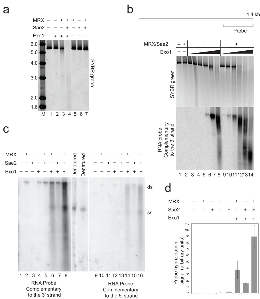
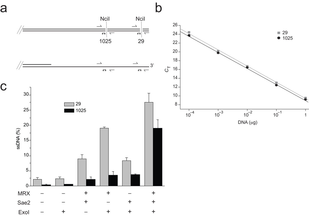
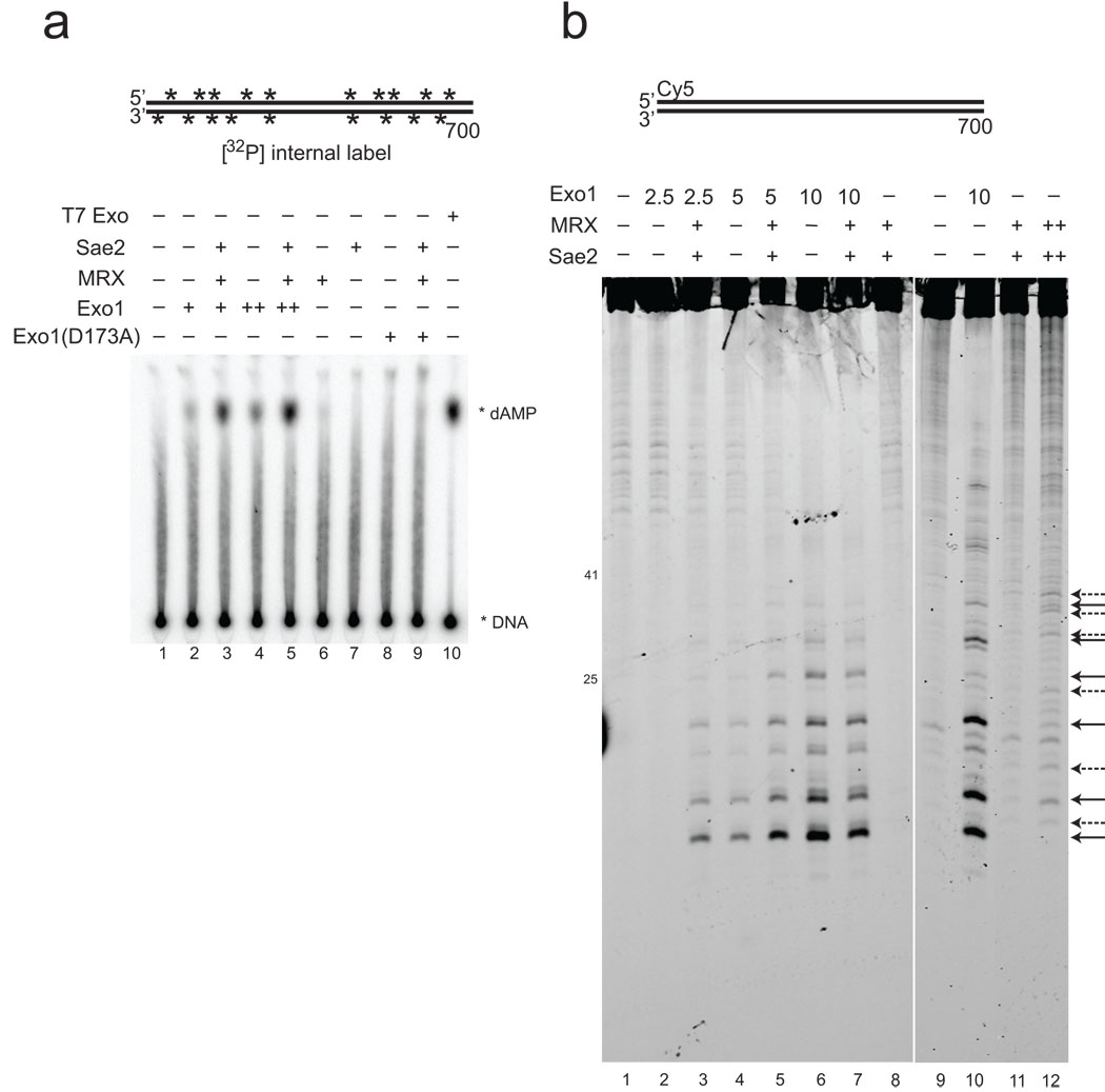
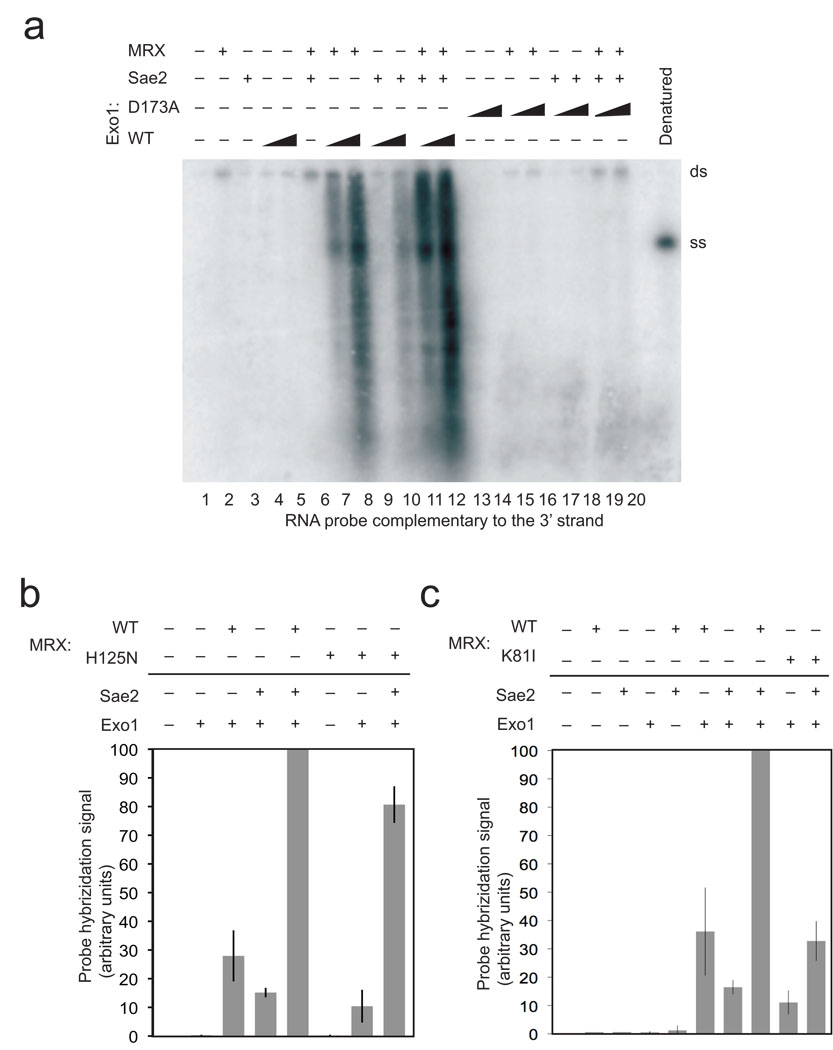
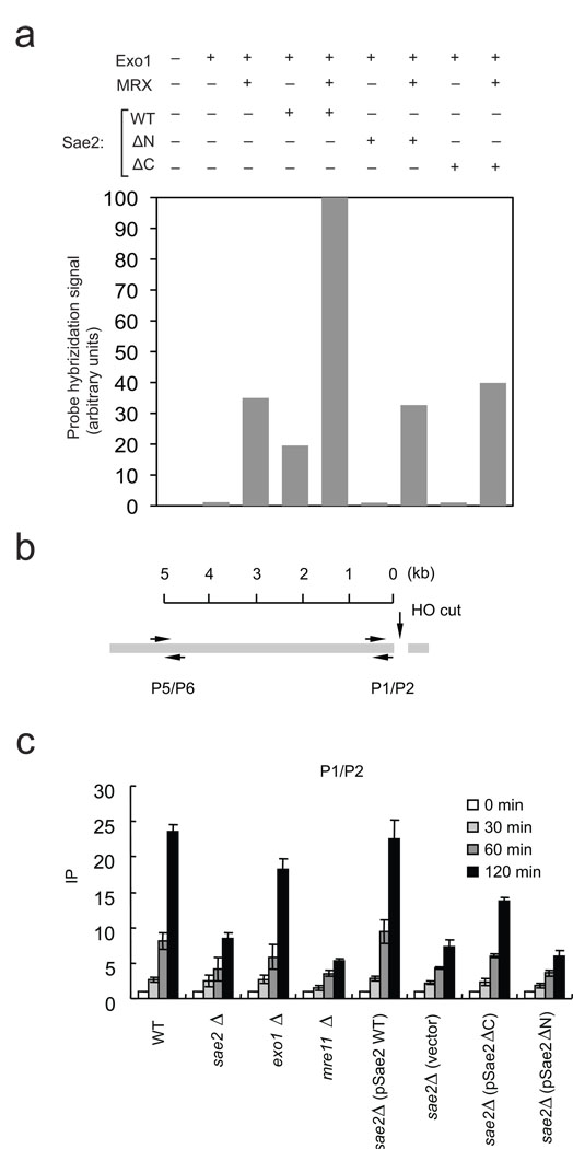
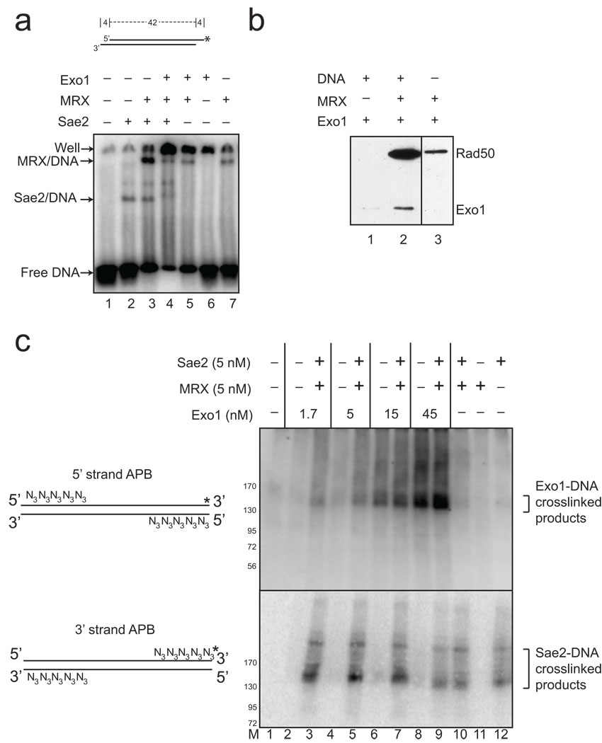
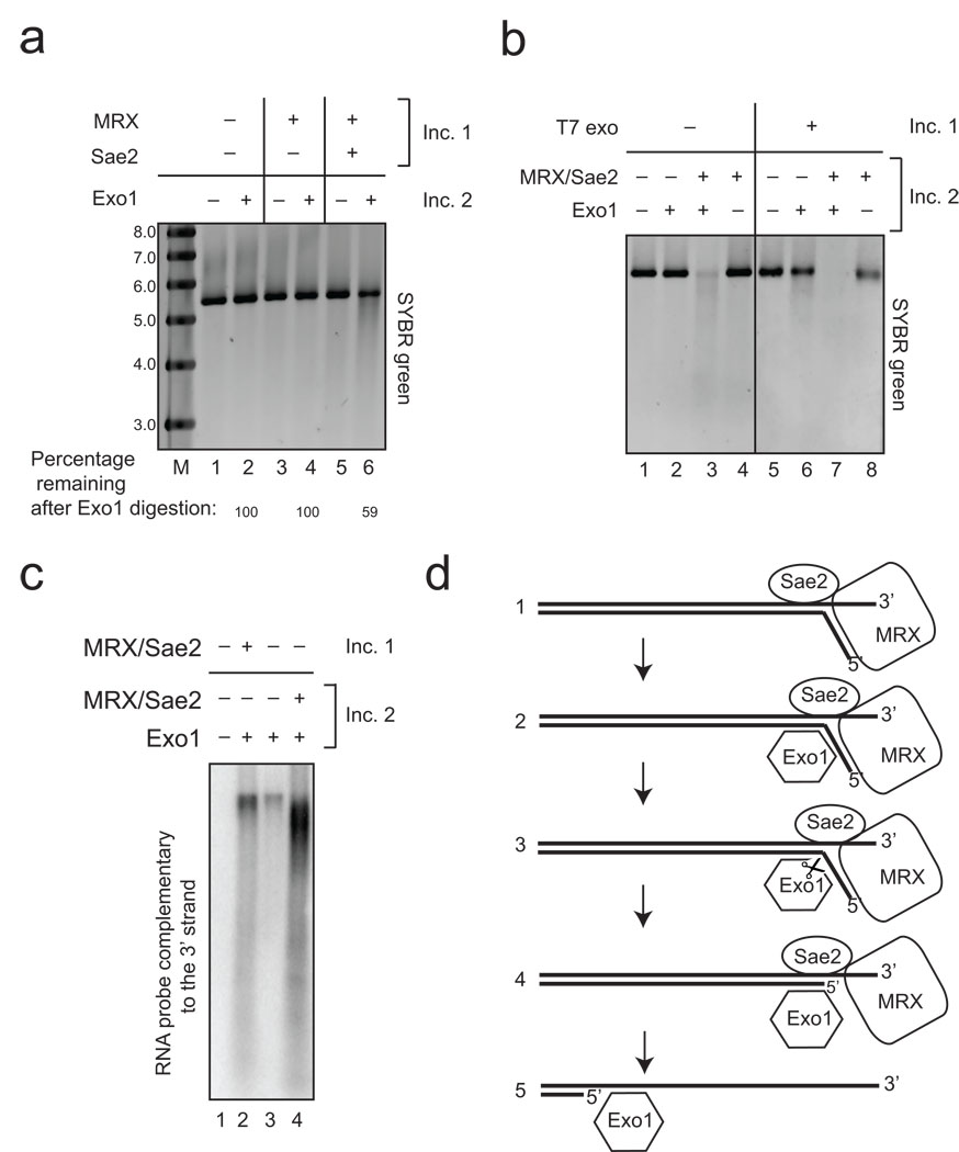
The repair of DNA double-strand breaks (DSBs) by homologous recombination is essential for genomic stability. The first step in this process is resection of 5' strands to generate 3' single-stranded DNA intermediates. Efficient resection in budding yeast requires the Mre11-Rad50-Xrs2 (MRX) complex and the Sae2 protein, although the role of MRX has been unclear because Mre11 paradoxically has 3'→5' exonuclease activity in vitro. Here we reconstitute resection with purified MRX, Sae2 and Exo1 proteins and show that degradation of the 5' strand is catalyzed by Exo1 yet completely dependent on MRX and Sae2 when Exo1 levels are limiting. This stimulation is mainly caused by cooperative binding of DNA substrates by Exo1, MRX and Sae2. This work establishes the direct role of MRX and Sae2 in promoting the resection of 5' strands in DNA DSB repair.


Figures

Figure 1
Figure 1. 5’ strand degradation at a DNA break by Exo1 is promoted by Mre11–Rad50–Xrs2 and Sae2
(a) 5.6 kb double-stranded DNA (pTP407) linearized with BseRI (0.3 nM) was incubated with Exo1 (0.5 nM), MRX (5 nM), and Sae2 (5 nM) for 60 min. at 30°C. The native agarose gel containing the reactions is shown after visualization of the DNA with SYBR green. Lane marked "M" contains MW markers as indicated (kb). (b) Resection assays were performed with a 4.4 kb plasmid DNA substrate (pNO1) linearized with SphI and analyzed as in (a) with SYBR green staining (top panel), and with non-denaturing Southern hybridization with a strand-specific RNA probe for the 3' strand at one end (see diagram). Reactions contained 14 nM MRX, 3.5 nM Sae2, and 0.4, 0.8, 1.6, 3.2, 6.4, and 32 nM Exo1 and were incubated for 60 min. at 30°C. (c) Resection assays were performed as in (b) with 4 nM Exo1. The reactions were split and separated in parallel in a non-denaturing gel, followed by non-denaturing Southern hybridization and probed separately for single-stranded 3’ strand (left) or 5’ strand (right) DNA adjacent to the break site. Denatured plasmid DNA was used as a marker for the single-stranded DNA “ss”, and the position of the unresected plasmid is marked as double-stranded, “ds”. (d) Quantitation of the reactions shown in (b), in addition to reactions from 4 other independent experiments, using phosphorimager analysis to quantitate the total counts in each lane. Within each experiment, the signal from the reactions containing wild-type MRX, Exo1, and Sae2 was set to 100%, with the other values shown relative to this. Error bars indicate standard deviation.
Figure 2
Figure 2. Characterization of the products of cooperative DNA resection by Exo1, MRX, and Sae2
(a) Diagram of the end of the pNO1 DNA substrate cut with SphI, with locations of NciI sites, PCR primers, and qPCR 6-FAM/TAMRA probes (asterisks). NciI digests double-stranded DNA (top) but leaves single-stranded DNA intact (bottom). (b) Standard curves using undigested DNA with the primer sets for the 29 bp (squares) and 1025 bp (circles) NciI sites. (c) qPCR results from resection assays performed as in Fig. 1B but analyzed using the qPCR technique to assess the level of DNA resection at each site: 29 nt from the end (top panel) and 1025 nt from the end (bottom panel). From each reaction, undigested aliquots were analyzed and compared with digested aliquots to obtain a ΔCT value, which was used to calculate the percentage of ssDNA in the reaction as described in the Methods. Each resection reaction was performed in triplicate and a qPCR analysis done for each; the average of these is shown in the graph with the standard error.
Figure 3
Figure 3. Digestion of linear DNA by Exo1 produces both single nucleotide and oligonucleotide products
(a) Resection assays were performed as in Fig. 1B except with 700 bp DNA substrate internally labeled with 32[P], with Exo1 (0.2, 0.4 nM), MRX (1.6 nM), and Sae2 (0.6 nM) or T7 exonuclease (1 unit per reaction). Reactions were stopped with SDS and EDTA and separated by thin layer chromatography; migration of the labeled dAMP product is indicated. T7 exonuclease was used as a positive control to generate single nucleotide products. (b) Resection assays were performed as in (a) except that the 700 bp DNA substrate was labeled on one 5' strand with Cy5. Reactions contained 2.5 (lanes 2, 3), 5 (lanes 4, 5), and 10 (lanes 6, 7, 10) nM Exo1, 7.5 nM MRX, and 1.5 nM Sae2 as indicated, with 25 nM MRX and 3 nM Sae2 in the reaction in lane 12. Arrows on the right side indicate positions of cleavage products (solid line: Exo1; dashed line: MRX–Sae2).
Figure 4
Figure 4. Mutations in Exo1 and Rad50 prevent DNA end processing
(a) Resection assays were performed and analyzed as in Fig. 1B using non-denaturing southern hybridication with a 3' strand probe and with a catalytic mutant of Exo1, D173A, as indicated. Concentrations of wild-type and mutant Exo1 were 4 nM and 8 nM. (b) Resection assays were performed as in Fig. 1B with the probe specific for the 3' strand but with MRX complexes containing the Mre11 mutant H125N. Results from 3 experiments were quantitated using phosphorimager analysis to quantitate the total counts in each lane and the average of these shown, with error bars indicating standard deviation. Within each experiment, the signal from the reactions containing wild-type MRX, Exo1, and Sae2 was the highest and set to 100%, with the signals in other lanes shown relative to this value. (c) Resection assays were performed, analyzed, and presented as in (b) but with the MR(K81I)X mutant complex. Reactions from three independent experiments were quantitated.
Figure 5
Figure 5. Mutations in Sae2 reduce the efficiency of Exo1-mediated DSB resection in vitro and in vivo
(a) Resection assays were performed, analyzed, and presented as in Fig. 4B but with the Sae2 mutants ΔC (deletion of amino acids 251–345) and ΔN (deletion of amino acids 21 −172) as indicated. Reactions from two independent experiments were quantitated and the average value is shown. (b) Schematic of the MAT locus containing an HO endonuclease cut site, and the locations of PCR primers used to assess the levels of RPA in sae2Δ strains carrying plasmids expressing a vector control, wild type, ΔN, or ΔC truncations of Sae2. mre11Δ and exo1Δ strains are also shown for comparison. Chromatin immunoprecipitation assays were performed using an anti-RPA antibody as described previously . PCR signals from a primer set that anneal 0.2 kb (P1–P2) from an HO break at different durations of HO expression were quantified and plotted in (c).
Figure 6
Figure 6. MRX and Sae2 promote Exo1 DNA binding
(a) Gel mobility shift assays were performed with wild-type MRX (2.5 nM), Sae2 (2.5 nM), and Exo1 D173A (4 nM) proteins as indicated and a 32[P]-labeled, double-stranded oligonucleotide substrate containing 4 nt 3’ overhangs on both ends. Reactions were incubated for 15 min on ice before separation on a native acrylamide gel. (b) MRX and Exo1 D173A proteins were incubated with biotinylated, blunt 100 bp duplex DNA as indicated, crosslinked with formaldehyde, and proteins bound to the DNA were isolated using streptavidin-coated magnetic beads. Bound protein were visualized by SDS-PAGE and western blotting with anti-Flag antibody for Exo1 and Rad50. (c) Protein-DNA binding assays were performed with a 90 bp blunt DNA substrate, containing 5 azide groups (N3) on the 5' ends of the 5' strands or the 3' ends of the 3' strands as shown in the diagram. Both DNA substrates were labeled with 32[P] ("*"). Proteins were incubated with the DNA substrates on ice, UV irradiated, separated by SDS-PAGE and transferred to a PVDF membrane to remove all uncrosslinked DNA before phosphorimager analysis. Migration of molecular weight markers in the gels are shown in the lane marked “M”.
Figure 7
Figure 7. MRX and Sae2 facilitate Exo1 activity through distinct "processing" and “recruitment” pathways
The resection reaction was performed as in Fig. 1A using pTP407 plasmid DNA linearized with BseRI except that the reaction was done in two stages. The first incubation (“Inc. 1”), included MRX (50 nM) and Sae2 (26 nM) as indicated but the DNA was deproteinized with SDS and proteinase K, followed by ethanol precipitation. The DNA from each reaction was incubated in a second reaction (“Inc. 2”) that included Exo1 (1 nM) as indicated. Reactions were separated by native agarose gel electrophoresis and DNA stained with SYBR green. Lane marked “M” contains molecular weight markers with sizes in kb as indicated. (b). 2-stage reactions were performed as in (a) except that T7 exonuclease (1 unit) was used in the first incubation and MRX (5 nM) and Sae2 (5 nM) were also added in the second incubation as indicated. (c) 2-stage reactions were performed as in (b) except with pNO1 plasmid DNA linearized with Sph1 and analyzed by non-denaturing southern hybridization using a probe specific for the 3' strand as in Fig. 1B. Reactions included MRX (14 nM), Sae2 (3.5 nM), and Exo1 (4 nM) as indicated. Lane marked “M” shows migration of molecular weight markers with sizes in kb as indicated. (d) Working model for association and cleavage of DNA ends by MRX, Sae2, and Exo1. We hypothesize a DNA-unwinding step followed by inefficient MRX–Sae2 cleavage, Exo1 recruitment, and further excision catalyzed by Exo1 (see text for details).

Similar articles

Cited by

References

    1. San Filippo J, Sung P, Klein H. Mechanism of eukaryotic homologous recombination. Annu Rev Biochem. 2008;77:229–257. - PubMed
    1. Aylon Y, Liefshitz B, Kupiec M. The CDK regulates repair of double-strand breaks by homologous recombination during the cell cycle. EMBO J. 2004;23:4868–4875. - PMC - PubMed
    1. Ira G, et al. DNA end resection, homologous recombination and DNA damage checkpoint activation require CDK1. Nature. 2004;431:1011–1017. - PMC - PubMed
    1. Wyman C, Kanaar R. DNA double-strand break repair: all's well that ends well. Annu Rev Genet. 2006;40:363–383. - PubMed
    1. Krogh BO, Symington LS. Recombination proteins in yeast. Annu Rev Genet. 2004;38:233–271. - PubMed

Publication types

MeSH terms