. 2010 Dec;16(12):1450-5.
doi: 10.1038/nm.2265. Epub 2010 Nov 21.

Invasive three-dimensional organotypic neoplasia from multiple normal human epithelia

Affiliations

Invasive three-dimensional organotypic neoplasia from multiple normal human epithelia

Todd W Ridky et al. Nat Med. 2010 Dec.

Abstract

Refined cancer models are required if researchers are to assess the burgeoning number of potential targets for cancer therapeutics in a clinically relevant context that allows a fast turnaround. Here we use tumor-associated genetic pathways to transform primary human epithelial cells from the epidermis, oropharynx, esophagus and cervix into genetically defined tumors in a human three-dimensional (3D) tissue environment that incorporates cell-populated stroma and intact basement membrane. These engineered organotypic tissues recapitulated natural features of tumor progression, including epithelial invasion through basement membrane, a complex process that is necessary for biological malignancy in 90% of human cancers. Invasion was rapid and was potentiated by stromal cells. Oncogenic signals in 3D tissue, but not 2D culture, resembled gene expression profiles from spontaneous human cancers. We screened 3D organotypic neoplasia with well-characterized signaling pathway inhibitors to distill a clinically faithful cancer gene signature. Multitissue 3D human tissue cancer models may provide an efficient and relevant complement to current approaches to characterizing cancer progression.


Figures
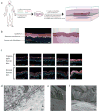
Figure 1
Figure 1. 3-D organotypic human epithelial tissue
(a) Experimental approach. (b) Normal stratified squamous epithelium and stromal architecture of regenerated human skin. Immunofluorescence image (left) highlighting epidermis (orange=pan-keratin) established on an intact basement membrane (BM) (green=type VII collagen BM marker), with fibroblast-populated dermis below. The corresponding hemotoxylin and eosin stained section is shown (right); scale bar=100μm. (c) Normal differentiation and BM protein distribution in organotypic and native human epidermal tissue; scale bar=100μm. (d) Transmission electron microscopy (EM) reveals a continuous intact BM with hallmarks of native tissue. Lamina densa (open black arrow), lamina lucida with anchoring filaments (open white arrow), anchoring fibrils (closed gray arrow), hemidesmosomes (closed white arrows), and dermal type I collagen (closed black arrows). (e) Scanning EM of the surface of organotypic human skin tissue at the edge of multilayered epidermal keratinocytes (white arrow) regenerated on intact native BM (black arrow). (f) Scanning EM of tissue from (e) cut to expose the epidermal-dermal interface with collagen fibers on BM surface (black arrow), and basal keratinocytes (white arrow; note prominent appearance of keratin intermediate filaments). Dots outline individual basal cells sitting on the BM.
Figure 2
Figure 2. Organotypic neoplasia from multiple stratified epithelia
Ras-driven invasive tissue compared with LacZ-expressing control. Note invasion of epithelial cells derived from four different tissues (orange=keratin) through the BM (green=type VII collagen) into the surrounding stroma. In contrast, control tissues respect the BM; scale bar=100μm.
Figure 3
Figure 3. Stromal cells potentiate invasion in organotypic neoplasia
(a) Normal control keratinocytes respecting epithelial boundary delimited by the basement membrane. (b,c) Invasive Ras-expressing keratinocytes invading into stroma. (d) Higher magnification of early epidermal keratinocyte invasion. Note penetration by epithelial cells through disrupted BM; scale bar=10μm. (e,f) Invasion is potentiated by the presence of stromal fibroblasts. Compared to control stroma lacking fibroblasts (e), Ras-driven organotypic epidermal neoplasia displays significantly deeper invasion in the presence of living fibroblasts (f); scale bar=100μm. (g) Invasion depth from the overlying basement membrane (collagen VII staining) to the deepest keratin positive cell directly in the stroma was measured. Depth is an average of 6 representative measurements at different points equally spaced across the section.
Figure 4
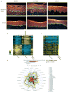
Figure 4. Analysis of organotypic neoplasia
(a) Effects of the PI3K inhibitor, LY294002, and the Mek and Erk inhibitor, U0126, on organotypic epidermal neoplasia; scale bar=100μm. (b) Expression data from Ras-driven organotypic epidermal neoplasia compared to normal epidermal control tissues (n=8 biologic replicates each) identifies 483 genes differentially expressed (≥2-fold change, FDR<0.03); reversion by U0126 (2 replicates) shown at right. Sample names are listed below the heat map. Cells used for this experiment were derived from 4 different donors (A,B,C,D) and replicate tissues were generated from each cell population. (c) Correlation of relative gene expression in Ras-driven organotypic neoplasia with relative gene expression in spontaneously occurring human head and neck SCC. The heat map displays the expression of the U0126 reverted genes in the tumor specimens relative to matched patient normal. The right column represents the relative expression of the gene set in organotypic neoplasia. (d) Pearson correlations between gene expression changes in spontaneous human SCC tumors (radially arrayed around edge of figure) and 5 different model systems (concentric plots within figure). Thicker colored lines represent median (or mean) values for each category.

References

    1. Stratton MR, Campbell PJ, Futreal PA. The cancer genome. Nature. 2009;458:719–724. - PMC - PubMed
    1. Campbell PJ, et al. Identification of somatically acquired rearrangements in cancer using genome-wide massively parallel paired-end sequencing. Nat Genet. 2008;40:722–729. - PMC - PubMed
    1. Chin L, Gray JW. Translating insights from the cancer genome into clinical practice. Nature. 2008;452:553–563. - PMC - PubMed
    1. Rangarajan A, Hong SJ, Gifford A, Weinberg RA. Species- and cell typespecific requirements for cellular transformation. Cancer Cell. 2004;6:171–183. - PubMed
    1. Hamad NM, et al. Distinct requirements for Ras oncogenesis in human versus mouse cells. Genes Dev. 2002;16:2045–2057. - PMC - PubMed

Publication types